Eye Notes Parts of sight sense Eyes Accessory

-thinnest skin •")

 • Regulate amount of light entering the eye • Control")
 • Lens-behind the iris and pupil – Held in place by")


 and retinal in")
 w The wavelength of light determines the color perceived from it;")

- Slides: 19
Eye Notes
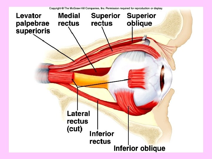
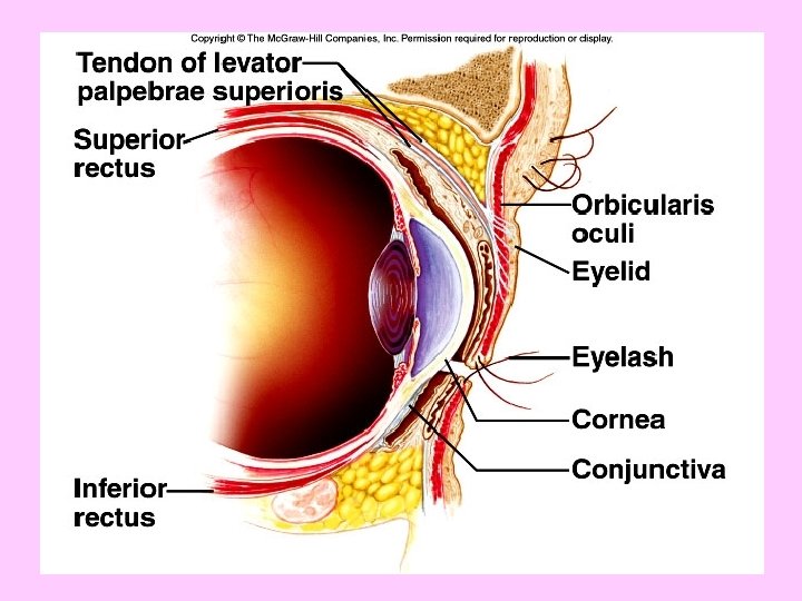
Parts of sight sense • Eyes • Accessory organs – Eyelids (palpebrae)-thinnest skin • Protect eye – Canthus-corners of the eye – Eyelashes-hairs that prevent particles from getting into the eye – Conjuctiva-membrane inside eyelids (prevent eyelids from sticking together) • Conjunctivitis-pink eye – Lacrimal apparatus-has a gland that produces tears/pink tissue in corner of eye • Tears have lysozymes-antibacterial enzyme – 6 muscles
Parts of outer eye Blindness-loss of transparency of cornea Can receive a donor cornea/no blood involved • Cornea-transparent window of the eye – Focus entering light – Covers colored portion of the eye – Limited repair • Sclera-white portion of eye – Made of collagen and elastin – Protect eye and attaches muscles • Optic nerve Parts of fibrous tunic -Mechanical support and physical protection -Important for focus
Vascular Tunic (middle eye) • Regulate amount of light entering the eye • Control shape of lens (an essential part of the focusing process • Choroid coat – Vascular and nutritive – Joined to sclera – Melanocytes (pigments)-absorb excess light and keep inside eye dark (tons of pigments) • Ciliary body-forms a ring around the eye • Composed of muscles and ligaments • Holds lens in position
Middle Eye (cont) • Lens-behind the iris and pupil – Held in place by suspensory ligaments – Ciliary muscles and ligaments help to change its shape in order to focus • Accomodation – Relaxation creates flat shape to see distance – Contraction creates convex shape to see close • Cataracts-lens become cloudy and opaque – Can cause blindness
Iris-part of middle eye • Thin diaphragm of connective tissue and smooth muscle • Colored portion of eye – Thickness and # pigments determines eye color – More is black, brown colors; less is blue and gray • Adjusts the amount of light that enters the pupil – The pupil is the opening at the center of the iris. – Dark part of eye • Aqueous humor-fluid between cornea and lens – Provides nourishment and maintains shape
Smooth Muscle Role • Regulates light by regulating pupil size – Contract-small size so less light • During bright light – Relax-big size so more light • During dim light
• http: //www. youtube. com/watch? v=0 Hz. Wml d. LDHI&feature=related
Glaucoma • Rate of aqueous humor formation is more than its rate of removal • Builds pressure on the eye – Blood vessels shut-rob cells of nutrients • Cells die and may cause blindness
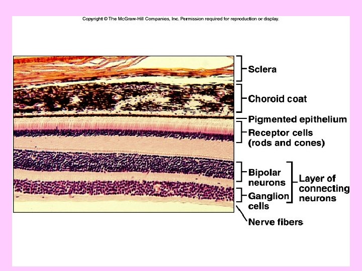
Inner Eye Parts • Retina – Photoreceptors – Thin and delicate – Contains a depression called fovea centralis – This produces the sharpest vision • Optic Disc – Where nerve fibers leave the eye and join the optic nerve – Lacks receptor cells-known as blind spot – Vitreous humor-liquid that fills the posterior cavity • Floaters-when clumps form in this liquid
Light Refraction • Refraction-bending of light waves • Convex surface of cornea and the lens refracts light and converges the rays onto the retina – Image is upside down and reverse • Visual cortex interprets this image correctly
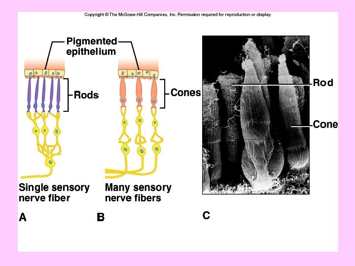
Visual Receptors • Rods – Long and thin – Provide black and white vision – More sensitive to light – Provide vision in dim light • Cones – Short and blunt – Color vision – Sharp image – To see something in detail, a person moves the eyes so the image falls on the fovea centralis (has tons of cones) – Cone numbers decrease as you move away from fovea centralis
Visual pigments w Rhodopsin-light sensitive pigment w Turns into opsin (protein) and retinal in the presence of light w This break down triggers an enzyme to change the cell membrane of the rod cell w Change causes a nerve impulse to be sent w Nerve impulses travel away from the retina and are interpreted as vision. w The light-sensitive pigments in cones are also proteins; there are three types, each containing a different visual pigment.
Visual Pigments (cont) w The wavelength of light determines the color perceived from it; each of the three pigments is sensitive to different wavelengths of light. w The color perceived depends upon which sets of cones the light stimulates: if all three sets are stimulated, the color is white; if none are stimulated, the color is black. Colorblindness-lack of cone pigments
• Nearsightedness-myopia – Concave lens • Farsightedness-hyperopia – Convex lens • Ophthalmoscope-used to examine the interior of the eye