
Eye Histology BY Dr Zahid Sarfaraz Khan AP
Eye Histology BY Dr. Zahid Sarfaraz Khan A/P ANATOMY KGMC
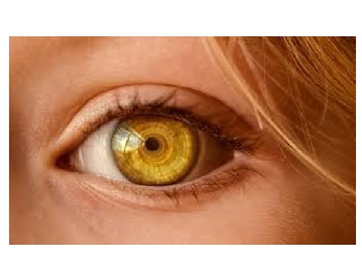
Eye The eye is a complex and highly developed photosensitive organ. That analyses: Ø The form Ø Intensity Ø And color of light reflected from objects, providing the sense of sight. ØThe eyes are located in protective areas of the skull, the orbits, which also contain cushions of adipose tissue.
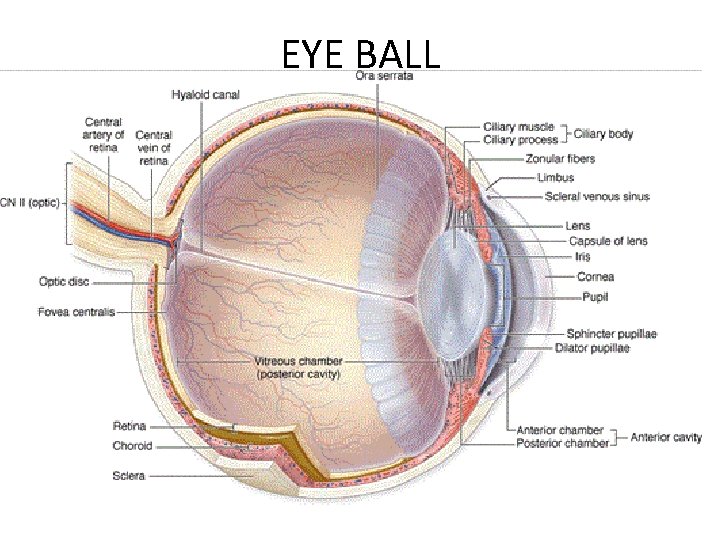
EYE BALL
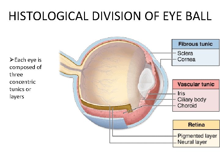
HISTOLOGICAL DIVISION OF EYE BALL ØEach eye is composed of three concentric tunics or layers
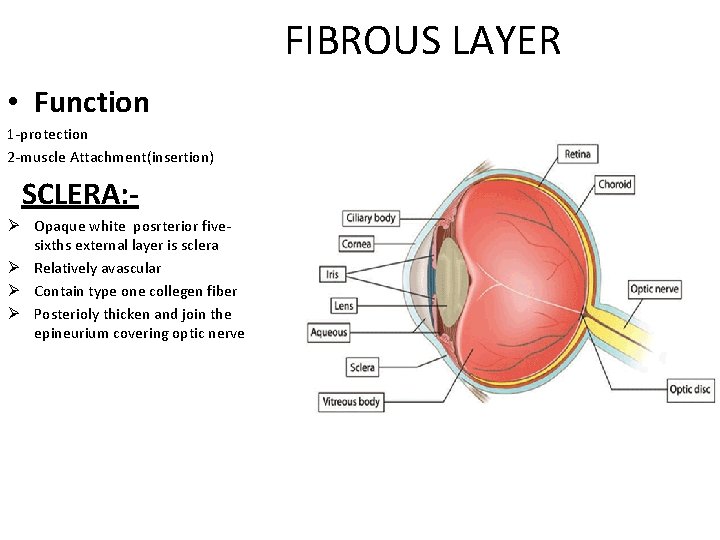
FIBROUS LAYER • Function 1 -protection 2 -muscle Attachment(insertion) SCLERA: Ø Opaque white posrterior fivesixths external layer is sclera Ø Relatively avascular Ø Contain type one collegen fiber Ø Posterioly thicken and join the epineurium covering optic nerve
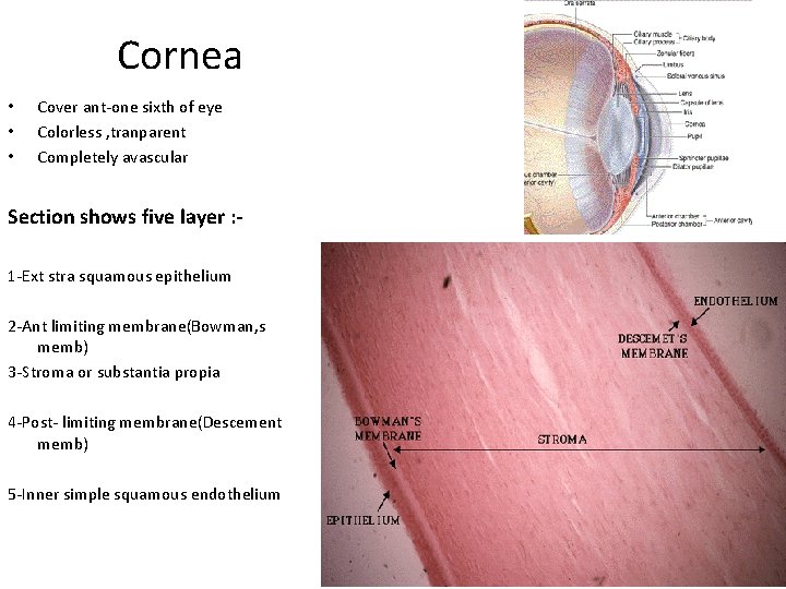
Cornea • • • Cover ant-one sixth of eye Colorless , tranparent Completely avascular Section shows five layer : 1 -Ext stra squamous epithelium 2 -Ant limiting membrane(Bowman, s memb) 3 -Stroma or substantia propia 4 -Post- limiting membrane(Descement memb) 5 -Inner simple squamous endothelium
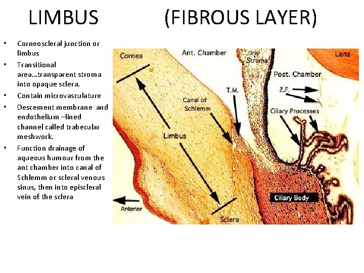
LIMBUS (FIBROUS LAYER) • • • Corneoscleral junction or limbus Transitional area…transparent stroma into opaque sclera. Contain microvasculature Descement membrane and endothelium –lined channel called trabecular meshwork. Function drainage of aqueous humour from the ant chamber into canal of Schlemm or scleral venous sinus, then into episcleral vein of the sclera
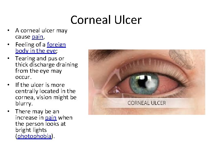
Corneal Ulcer • A corneal ulcer may cause pain, • Feeling of a foreign body in the eye; • Tearing and pus or thick discharge draining from the eye may occur. • If the ulcer is more centrally located in the cornea, vision might be blurry. • There may be an increase in pain when the person looks at bright lights (photophobia).
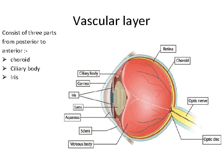
Vascular layer Consist of three parts from posterior to anterior : Ø choroid Ø Ciliary body Ø Iris
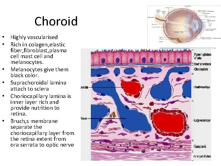
Choroid • Highly vascularised • Rich in colagen, elastic fiber, fibroblast, plasma cell mast cell and melanocytes. • Melanocytes give them black color. • Suprachoroidal lamina attach to sclera • Choriocapillary lamina is inner layer rich and provide nutrition to retina. • Bruch, s membrane separate the choriocapillary layer from the retina extent from ora serrata to optic nerve
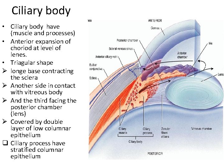
Ciliary body • Ciliary body have (muscle and processes) • Anterior expansion of choriod at level of lenes. • Triagular shape Ø longe base contracting the sclera Ø Another side in contact with vitreous body Ø And the third facing the posterior chamber (lens) Ø Covered by double layer of low columnar epithelium q Ciliary process have stratified columnar epithelium
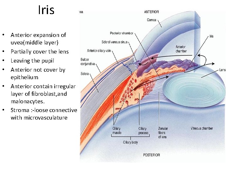
Iris • Anterior expansion of uvea(middle layer) • Partially cover the lens • Leaving the pupil • Anterior not cover by epithelium • Anterior contain irregular layer of fibroblast, and malonacytes. • Stroma : -loose connective with microvasculature
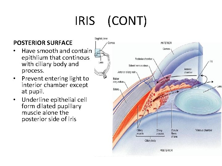
IRIS (CONT) POSTERIOR SURFACE • Have smooth and contain epithlium that continous with ciliary body and process. • Prevent entering light to interior chamber except at pupil. • Underline epithelial cell form dilated pupillary muscle alone the posterior side of iris
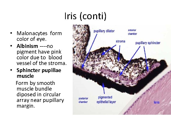
Iris (conti) • Malonacytes form color of eye. • Albinism ----no pigment have pink color due to blood vessel of the stroma. • Sphincter pupillae muscle Form by smooth muscle bundle diposed in circular array near pupillary margin.
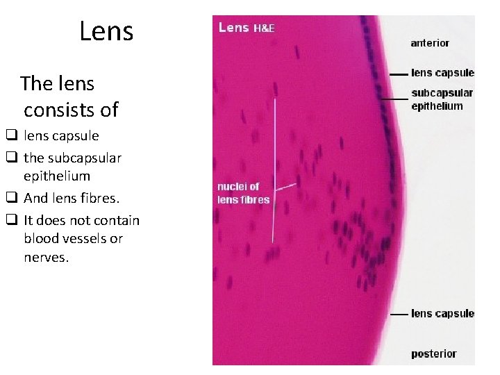
Lens The lens consists of q lens capsule q the subcapsular epithelium q And lens fibres. q It does not contain blood vessels or nerves.
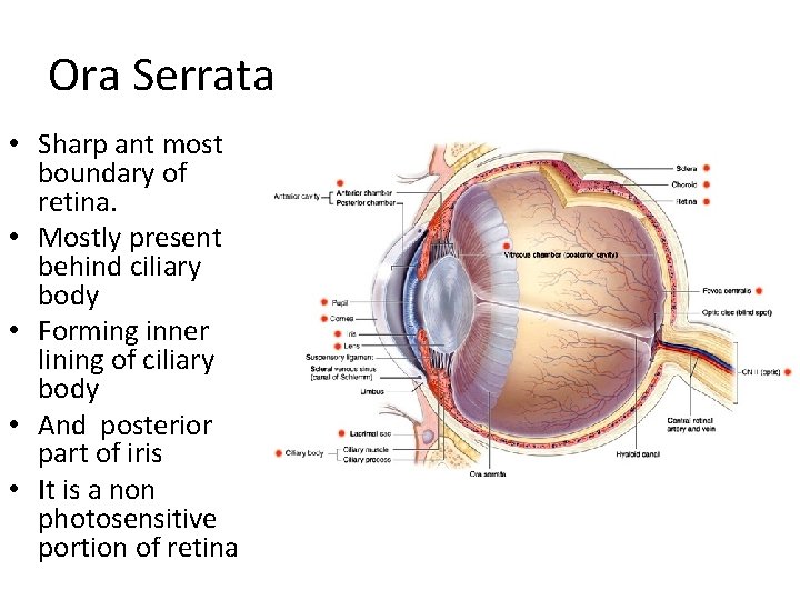
Ora Serrata • Sharp ant most boundary of retina. • Mostly present behind ciliary body • Forming inner lining of ciliary body • And posterior part of iris • It is a non photosensitive portion of retina
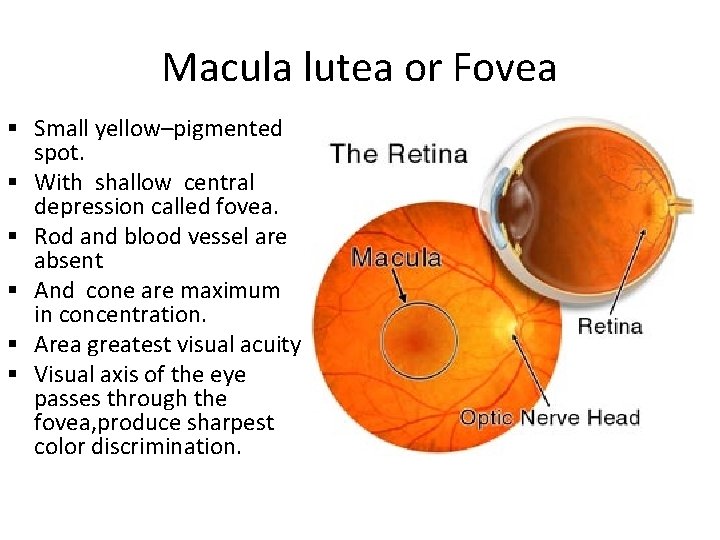
Macula lutea or Fovea § Small yellow–pigmented spot. § With shallow central depression called fovea. § Rod and blood vessel are absent § And cone are maximum in concentration. § Area greatest visual acuity § Visual axis of the eye passes through the fovea, produce sharpest color discrimination.
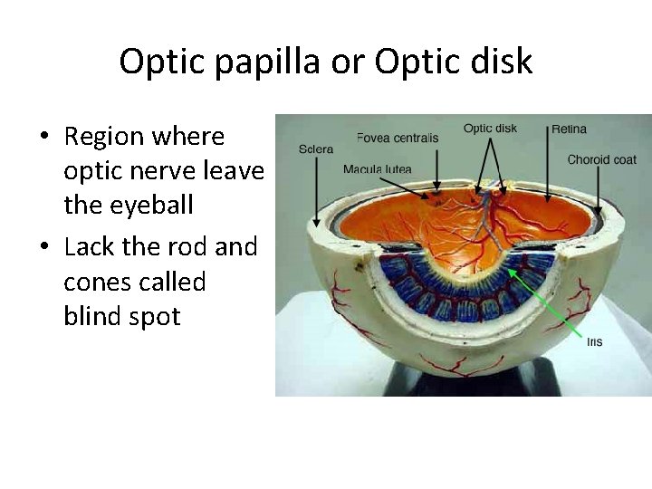
Optic papilla or Optic disk • Region where optic nerve leave the eyeball • Lack the rod and cones called blind spot

Retinal organization … • The retina is made of several cell layers: • Outermost layer —pigmented epithelium cells • Basement membrane of P. E. cells forms the inner most layer of glassy (Bruch, s) membrane of choriod. • The cubiodal P. E. CELLS contain melanin pigments granules in their cytoplasm.
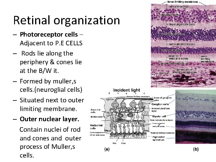
Retinal organization – Photoreceptor cells – Adjacent to P. E CELLS – Rods lie along the periphery & cones lie at the B/W it. – Formed by muller, s cells. (neuroglial cells) – Situated next to outer limiting membrane. – Outer nuclear layer. Contain nuclei of rod and cones and outer process of Muller, s cells.
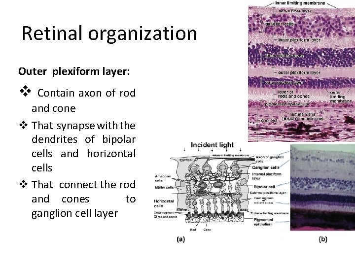
Retinal organization Outer plexiform layer: v Contain axon of rod and cone v That synapse with the dendrites of bipolar cells and horizontal cells v That connect the rod and cones to ganglion cell layer
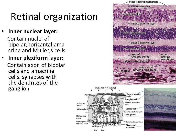
Retinal organization • Inner nuclear layer: Contain nuclei of bipolar, horizantal, ama crine and Muller, s cells. • Inner plexiform layer: Contain axon of bipolar cells and amacrine cells. synapses with the dendrites of the ganglion
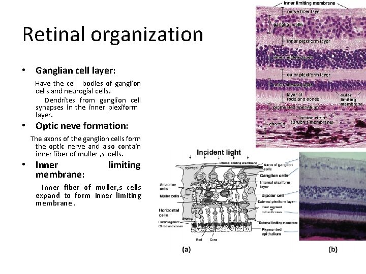
Retinal organization • Ganglian cell layer: Have the cell bodies of ganglion cells and neurogial cells. Dendrites from ganglion cell synapses in the inner plexiform layer. • Optic neve formation: The axons of the ganglion cells form the optic nerve and also contain inner fiber of muller , s cells. • Inner membrane: limiting Inner fiber of muller, s cells expand to form inner limiting membrane.
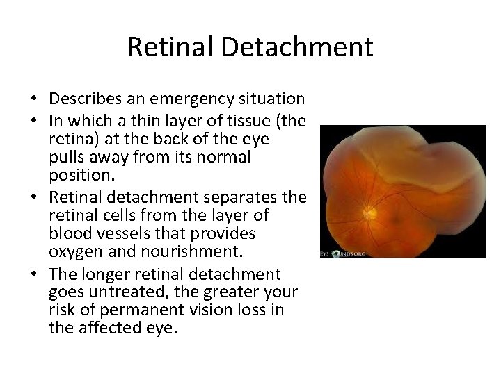
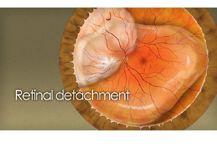
Retinal Detachment • Describes an emergency situation • In which a thin layer of tissue (the retina) at the back of the eye pulls away from its normal position. • Retinal detachment separates the retinal cells from the layer of blood vessels that provides oxygen and nourishment. • The longer retinal detachment goes untreated, the greater your risk of permanent vision loss in the affected eye.
Symptom Retinal detachment • Itself is painless. • But warning signs almost always appear before it occurs or has advanced, such as: • The sudden appearance of many floaters — tiny specks that seem to drift through your field of vision • Flashes of light in one or both eyes • Blurred vision • Gradually reduced side (peripheral) vision • A curtain-like shadow over your visual field
How Retinal Detachment Occurs • when the gel-like material (vitreous) leaks through a retinal hole or tear and collects underneath the retina. • Aging or retinal disorders can cause the retina to thin. • Fluid inside the vitreous then finds its way through the tear and collects under the retina, peeling it away from the underlying tissues. • The areas where the retina is detached lose this blood supply and stop working, so you lose vision.

Thank you
- Slides: 32