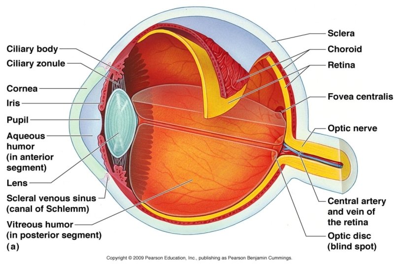
Eye Anatomy Eye Structure enclosed and cushioned by

Eye Anatomy

Eye Structure • enclosed and cushioned by fat and walls of bony orbit

• composed of 3 layers = tunics • filled with fluid = humors – to maintain shape • lens – for focusing – divides eye into anterior and posterior chambers

3 Layers: Tunics

1. Fibrous Tunic • outermost • 2 parts 1. sclera “white of the eye” – Tough – Anchors muscles

1. Fibrous tunic • 2 parts 1. Sclera 2. Cornea “clear window” – lacks blood vessels – easy to transplant – nutrition via diffusion from aqueous humor fluid behind
 § vascular §")
2. Vascular Tunic • 3 regions • middle 1. choroid (coat) § vascular § pigmented § posterior part § absorbs light and prevents scattering

2. Vascular Tunic 3 regions 1. choroid 2. ciliary body § encircles lens § made of smooth muscle - ciliary muscles § controls lens shape

2. Vascular Tunic 3 regions 1. choroid 2. ciliary body § folds secrete aqueous humor - ciliary processes § suspensory ligaments hold lens up

2. Vascular Tunic 3. iris § visible colored area § contain melanin § amount and distribution = different colors

2. Vascular Tunic 3. iris § made of muscles that control pupil size - regulating light § pupil is hole in center of iris

• pupillary reflex also responds to interests or emotions boredom or repugnant material = constriction of pupil appealing subject matter, problem solving = dilates

3. Sensory tunic = retina 2 layers 1. outer is pigmented to absorb light 2. inner layer § transparent § photoreceptor neurons (rods and cones)

Detached Retina • separation of pigmented from transparent Warning signs include: • seeing many new flashing lights • showers of many floaters • blurred vision • a curtain-like blockage

3. Sensory tunic = retina Fovea • optic disc § where optic nerve leaves eye § “blind spot” § off center § optic nerve has blood vessels in center

Photoreceptors Rods § dim light § black and white just outside fovea § peripheral vision

Cones Photoreceptors § bright light § color vision Optic Disc § concentrated in center fovea centralis
 light → ganglion cells → bipolar neurons")
nervous flow is opposite (→ = light) light → ganglion cells → bipolar neurons → photoreceptors→ back up and to optic nerve

Chambers and Humors

Posterior Chamber with Vitreous Humor • transmits light • supports back surface of lens • supports eye shape • vitreous humor § thick and clear § lasts a lifetime

Anterior Chambers with Aqueous Humor • chamber between cornea and lens • formed in ciliary processes • watery • constantly produced and drained via canals of Schlemm

Glaucoma • if canals are blocked • unable to drain
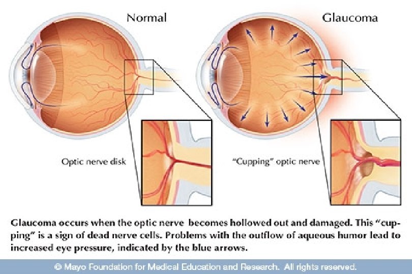
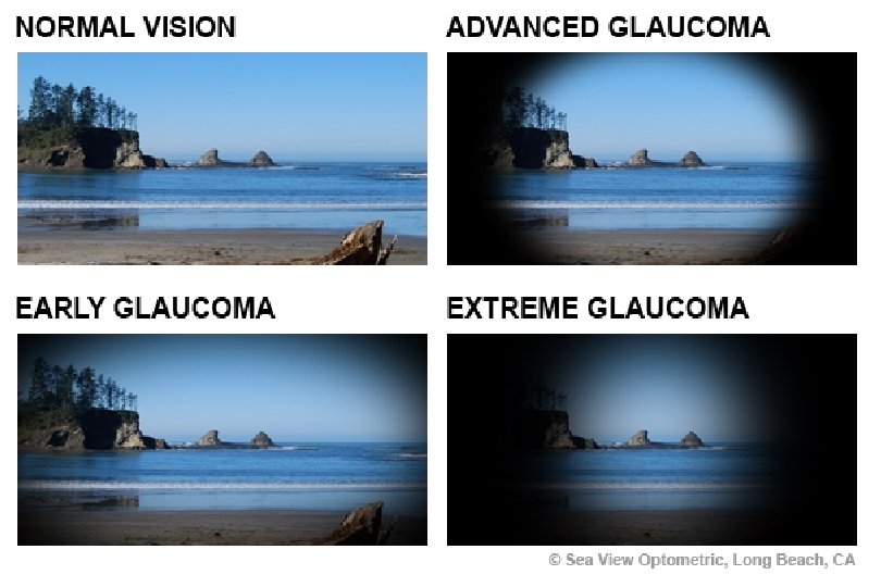

Glaucoma • if canals are blocked • unable to drain • = nerve cells gradually destroyed by pressure • treated with drugs

Lens

Lens • • biconvex transparent flexible becomes less elastic with age

Cataracts • lens clouding
- Slides: 29