Extrahepatic biliary apparatus Dr Shivarama Bhat functions Collects

Extrahepatic biliary apparatus Dr Shivarama Bhat

functions Collects from liver Stores in gall bladder Transmits to 2 nd part of duodenum

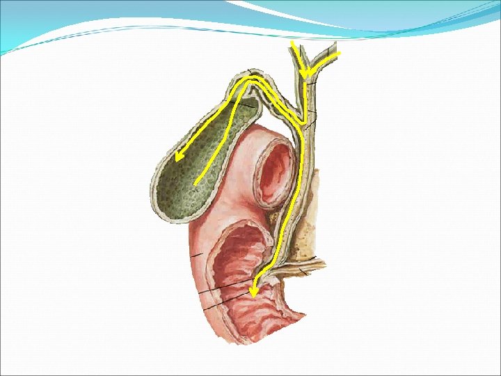
Apparatus Consists of Left and right hepatic ducts Common hepatic duct Gallbladder Cystic duct Common bile duct

Intrahepatic part
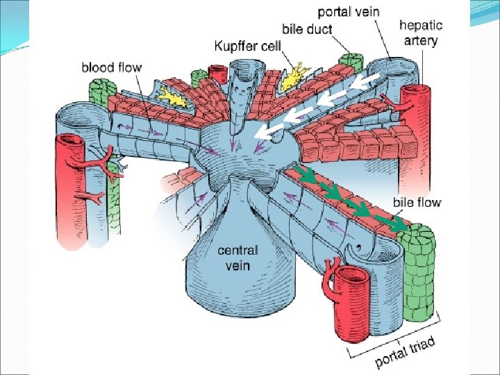

Hepatic ducts Right & left Porta hepatisemergence Behind forwards: ü Portal vein ü Hepatic artery ü Bile duct

Common hepatic duct Right & left hepatic ducts Right end of porta hepatis 3 cm +cystic duct(right)=bile duct

accessory hepatic duct 15% Right lobe of liver Terminate in

THE GALLBLADDER

Gall bladder Pear-shaped, hollow structure situated in fossa for gall bladder On inferior surface of liver Extending from right end of porta hepatis to inferior border of liver

Measurements: 7 -10 cm long ~ 3 cm diameter 30 – 50 cc volume

Parts 3 parts • Fundus of gallbladder Surface projection: at the junction of right midclavicular line and right costal arch • Body of gallbladder • Neck of gallbladder Narrow upper end

Fundus of GB: may be palpated in angle between lateral border of right rectus abdominis and 9 th costal margin surrounded by peritoneum Anteriorly – ant abdominal wall Posteriorly – transverse colon

Body of Gallbladder Lies in gall bladder fossa Upper end continous with neck at right end of porta hepatis Superior surface devoid of peritoneum Inferior surface covered with peritoneum related to transverse colon & duodenum
")
Neck Situated near right end of porta hepatis Antero-superiorly→postero-inferiorly continuous with cystic duct (constriction) Attached to liver by loose (areolar) connective tissue –cystic vessels Inferiorly – 1 st part of duodenum Mucous membrane-folded spirally

Hartmann’s pouch Dilated posteromedial wall of neck Directed downwards and backwards Normal variation Gall stones may lodge in it pathological

Cystic duct 3 -4 cm long Extends from neck of gallbladder to common hepatic duct Joins with common hepatic duct inferior to porta hepatis Downwards, backwards, to left superior and posterior to pylorus of stomach Spiral valve may extend into neck of gallbladder: 5 -12 cresecentic folds

Bile duct Formed by union of cystic and common hepatic duct 7. 5 cm long Narrow tube, 6 mm diameter

Course Downwards & backwards Deep to pyloric sphincter 3 parts supraduodenal: through lesser omentum retroduodenal: behind 1 st part of duodenum infraduodenal: behind /embedded in head of pancreas

Supraduodenal part

Retorduodenal part

Infraduodenal part

Intraduodenal segment Enters the wall of descending part of duodenum obliquely where joins the pancreatic duct to form the hepatopancreatic ampulla /Ampulla of Vater opens at the major duodenal papilla 8 -10 cm distal to pylorus

Sphincter of Oddi Spincter of bile duct/choledochus /Boyden Sphincter of ampullae Sphincter pancreaticus

Sphincter of Oddi

liver common hepatic duct cystic duct Triangle of Calot Boundaries: Content: cystic artery

BLOOD SUPPLY OF BILE DUCT: Cystic artery Hepatic artery Posterior superior pancreaticoduodenal

Right hepatic artery Cystic artery Left hepatic artery Cystic artery Ventral br. hepatic artery dorsal br. common hepatic artery

Cystic artery variation

Venous drainage Superior surface of gall bladder drains veins entering through liver into the hepatic veins. Rest of gall bladder-cystic veins lower part of the bile duct drains into the portal vein.

LYMPHATIC DRAINAGE Lymphatics from the gall bladder cystic duct, hepatic duct and upper part of the bile duct pass to the cystic node , these are the most constant members of the upper hepatic nodes. the node of the anterior border of the epiploic foramen The lower part of the bile duct drains into the lower hepatic and the upper pancreaticosplenic nodes.

NERVE SUPPLY The cystic plexus of nerves, supplying the territory of the cystic artery derived from the hepatic plexus, which receives fibres from coeliac plexus, left and right vagus right phrenic nerves. The nerve plexus supplies the lower part of the bile duct over the superior pancreaticoduodenal artery.

NERVE SUPPLY Parasympathetic nerves are motor to musculature of the gall bladder and bile ducts, but inhibitory to the sphincters of the bile duct. Gall bladder pain via vagus is referred to stomach. Sympathetic nerves (T 7 -9) are vasomotor and motor to sphincters. Pain via sympathetic nerves is referred to the inferior angle of the scapula. Pain via the phrenic nerve is referred to the right shoulder

Functions of Gall bladder Storage of bile Absorption of water and concentration of bile 10 times

Functions of Gall bladder Bile salt: cholesterol solvent Bile cholesterol compound absorbed inflammed Bile salt absorbed Cholesterol ppt

Applied anatomy ANOMALIES OF THE GALL BLADDER ANOMALIES OF THE DUCTS ANOMALIES OF BLOOD VESSELS

Anomalies of gall bladder

ANOMALIES OF THE DUCTS

Cholecystogram After meal Oral cholecystogram

Gallbladder Disease Cholecystitis - Normal US Murphy’s sign Acute cholecystitis

Cholelithiasis GB shows likely sites of stone formation/deposition magnetic resonance cholangiopancreatography

Gall stones

Courvoisier’s law Malignant growth of head of pancreas Distended GB Stone in GB Fribrotic non-distensible GB

Biliary colic

Biliary fistula May occur due to acute cholecystitis with obstuction of neck of GB

Obstructive jaundice/post hepatic jaundice common causes gallstones in the common bile duct pancreatic cancer in the head of the pancreas.
- Slides: 47