Extraembryonic Membranes Meaning of Extraembryonic Structures in Chick

Extra-embryonic Membranes

Meaning of Extraembryonic Structures in Chick Embryo: The embryo of chick possesses four extra embryonic or foetal membranes: namely, the yolk sac, the allantois, the amnion and the serosa or chorion. In amphibian embryo, the yolk sac and the allantois are present in rudimentary condition. The amnion and the chorion are developed in the reptilian embryo for the first time in evolutionary history of the vertebrates. In birds these two structures are retained while in mam mals these are also present in a modified form. All the extra embryonic mem branes are discarded at hatching while the yolk sac is incorporated into the small intestine. As development goes on, the closely set ectoderm and somatopleure (somatic mesoderm) as well as the endoderm and splanchnopleure (splanchnic mesoderm) extend into the extraembryonic area. The developing embryo becomes located at the central area of the blastodisc.

Kinds of Extra-Embryonic Membranes: Four sets of extra-embryonic membranes are common to the embryos of all terrestrial vertebrates including chick. These are i. Amnion: The amnion is a thin mem brane which eventually encloses the entire developing embryo in a fluid filled sac. Reptiles, birds and mammals possess ing this amnion are often called amniotes, while fishes and amphibians, lacking it, are collectively called anamniotes. ii. Yolk Sac: It is the most primitive struc ture containing network of blood vessels and encloses the yolk of the egg. A yolk sac is also present in those fishes which have megalecithal eggs. Despite the lack of stored yolk in mammalian eggs (except in prototherians), the yolk sac has been preserved, as it serves many impor tant secondary functions.

Kinds of Extra-Embryonic Membranes: iii. Allantois: Allantois is a large sac like structure in reptiles and birds, while its role in mammals varies with the efficiency of the interchange that takes place at the foetal maternal inter face. Allantois in human has been reduced to a mere ves tige which contributes only as a well developed vascular network to the high ly efficient placenta. iv. Chorion (Serosa): Chorion is a very thin membrane and it covers the embryo and other extra embryonic membranes. It is formed by the fusion of the amniotic folds over the embryo. All these extra embryonic membranes are composite structures as they involve two germ layers. q The amnion and chorion are made up of extra embryonic ectoderm and somatic layer of mesoderm, while the yolk sac and allantois are composed of extra embryonic endoderm and splanchnic layer of mesoderm.

Development of Extra-Embryonic Membranes: The somatic layer of mesoderm and ectoderm are collectively known as somatopleure, while the splanch nic layer of mesoderm along with endoderm forms the splanchnopleure. At the time of development of the avian blastoderm, the somatopleure and splanchnopleure gradu ally spread peripherally over the yolk mass, far beyond the area where the body of the embryo is taking form. Shortly, the embryo proper begins to be undercut by a series of body folds that serve to delimit the embryo nic regions from the more peripheral extra embryonic somatopleure and splanchno pleure. After the formation of the body folds, the somatopleure and splanchno pleure of chick develop into the four extra embryonic membranes

i. Development of Yolk Sac: The yolk sac is the first extra embryonic membrane to make its appearance. As the early blastoderm expands, the extra embryonic splanchno pleure continues to spread over the yolk mass and eventually encloses the yolk com pletely to form the yolk sac is connected with the digestive tract by the yolk stalk, the yolky food reserves are not transmitted to the embryo by this route. Rather, the digestion of the yolk is done by the endodermal lining of the yolk sac through the mediation of appro priate enzymes. The entire yolk is not completely absorbed during embryonic life. the remains of the yolk sac are enclosed within the body walls of the embryo. During the first 6 days after hatch ing, the resorption of the remaining part of the yolk sac and yolk gets completed. This remaining yolk reserves are vital to the newly hatched chick


ii. Development of Amnion and Chorion: The amnion and chorion are deve loped simultaneously and both are derived from the extra embryonic somatopleure. At about the 30 th hour of incubation, the head of the embryo sinks into the yolk and at the same time the extra embryonic somatopleure is elevated over the embryo. The initial elevation is over the head end of the embryo, producing a double somatopleuric hood, called the cephalic amniotic fold. As the cephalic amniotic fold gradually extends backward, towards the tail region, its caudally extending side limbs called lateral amnio tic folds arch over the embryo from each side to be joined finally by a similar fold or eleva tion from the tail region called the caudal amniotic fold. The fusion of the amniotic folds results in the formation of two sac like membranes and two cavities. The inner somatopleuric membrane becomes the amnion and the outer one, the chorion. The cavity between the amnion and the embryo is called the amniotic cavity and is lined by ectoderm. The cavity lying between the amnion and chorion is called the chorionic cavity and is lined by the mesoderm.
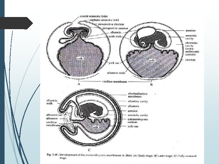

iii. Development of Allantois: The allantois first appears late in the 3 rd day of incubation. It bulges out as a ventral out growth of the endodermal hindgut. The outgrowth consists of an inner layer of endoderm and an outer layer of splanchnic mesoderm. The allantois enlarges very rapidly from the fourth day to the tenth day of incubation. It penetrates into the extra embryonic coelom, into the space between the yolk sac, the amnion and the chorion The base of the allantois remains connected with the hindgut of the embryo by means of a narrow allantoic stalk. Soon, the mesodermal layer of the allantois becomes fused with the adja cent mesodermal layer of the chorion to form a single mesodermal layer called chorioal lantoic membrane.


Functions of the Extra-Embryonic Membranes: i. Functions of Yolk Sac: The yolk sac which spreads over the large amount of yolk, serves as the digestive and absorptive organ by which the yolk is made available for the growing embryo. It functions as the first respiratory organ. It acts as a haemopoetic organ like the liver. The yolk sac also serves as the place of origin of blood cells, at later stages of development. ii. Functions of Amnion and Chorion: The amnion serves as a protective organ where the embryo is saved from the danger of desiccation. The amniotic fluid acts as an efficient shock absorber and thus, protects the soft, collapsible and almost skeletons early chick embryo from mechanical shocks. As the amnion isolates the embryo from the egg shell, it thus protects it from adhesion to the shell or from friction against it. The mesoderm of amnion, during later developmental stages, form muscle cells which contract rhythmically, thus rocking the embryo within the amniotic fluid. This rock ing prevents the adhesion of amnion to the different embryonic membranes. It also helps in preventing the stagnation of blood in the vessels, a condition that might tend to occur on account of pressure from growing organs.

Functions of the Extra-Embryonic Membranes: The chorion at later developmental stage joins with the allantois to serve as a nutritional and respiratory organ. iii. Functions of Allantois: Allantois acts as a reservoir for the secretions (excretory wastes) coming from the developing excretory organs. During early stages of development the chick excretes mostly urea, but later it becomes chiefly uric acid. This change is significant as urea is a relatively soluble substance and would require large amount of water to keep it at nontoxic level. Uric acid is relatively insoluble and can be stored without any ill effects. The chorioallantoic membrane acts as a respiratory surface for the embryo. Thus, the yolk sac, amnion, chorion and allantois can be regarded as an adaptation for the egg and embryo to carry on its develop ment on dry land.
- Slides: 13