Extracellular Matrix Animal tissue is not only composed


 Glycosaminoglycan • It is a long, un branched linear polysaccharide chains and consists")
 Fibrous Protein: A. Structural Fibrous Protein: • (a) Collagen: It is a hydrophobic")

 Fibronectin: Fibronectin is a glycoprotein. It is made")


 Specialised Extracellular Matrix Basal Laminae: • Basal lamina is a continuous thin mat")

- Slides: 15
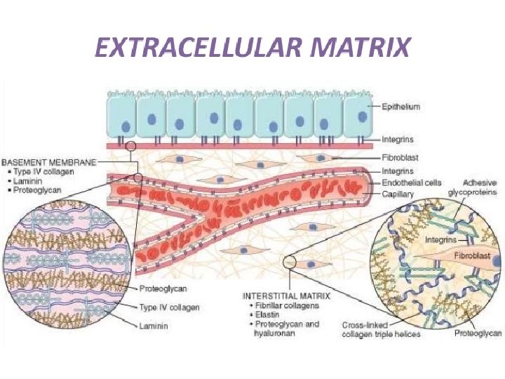
Extracellular Matrix Animal tissue is not only composed of cells but also contains many types of extracellular space or intercellular space. These spaces are again filled up by many types of macro molecules constituting the extracellular matrix. The macromolecules that constitute the extracellular matrix are mainly secreted locally by the cells. In most of the connective tissues the macromolecules are secreted by fibroblast (Fig. 4. 9). In some specialised connective tissues, such as cartilage and bone, they are secreted by chondroblasts and osteoblasts, re spectively.
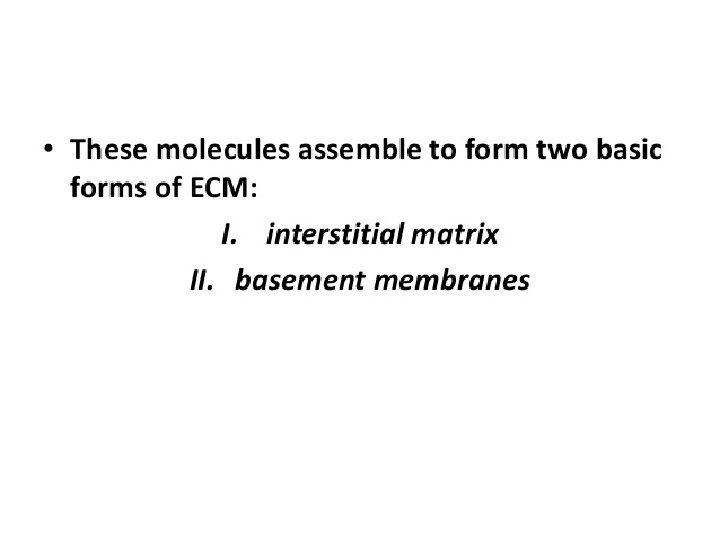
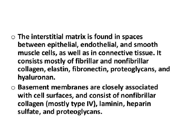
Types of Extracellular Matrix: • The extracellular matrix is made of three main types of extracellular macromolecules: i) Polysaccharide glycosaminoglycan’s (com monly known as muco polysaccharides) or GAGs which are usually linked covalently to proteins in the form of proteoglycans (ii) Fibrous proteins of two functional types: (a) Mainly structural (e. g. , collagen and elastin) and (b) Mainly adhesive (e. g. , fibronectin and laminin) (iii) Specialised extracellular matrix or basal lamina.
(i) Glycosaminoglycan • It is a long, un branched linear polysaccharide chains and consists of repeating disaccharide units in which one of two sugars is always either N acetyl glucosamine or N acetylgalactosamine. Hence it is named glycosaminoglycan. There are four main classes of glycosaminoglycan’s: (i) Hyaluronic acid, (ii) Chondroitin sulfate and dermatan sulfate, (iii) Heparan sulfate and heparin, and (iv) Keratan sulfate.
(ii) Fibrous Protein: A. Structural Fibrous Protein: • (a) Collagen: It is a hydrophobic protein. This protein is found in all multicellular animals and is secreted mainly by connective tissue cells. • The basic molecular unit of collagen is tropocollagen or pro collagen which is 300 nm in length and 1. 5 nm wide. It is made of three polypeptide chains that are coiled together to form a triple helical structure. • The amino acid composition of the polypeptide chain of collagen is very simple; they have a large amount of proline and many of the proline and lysine residues are hydroxylated. So far, about 20 distinct a chains of collagen have been identified. These are encoded by separate genes.
• These are types I, III, IV, and V. Types I, III, and V are fibrillar collagens, while type IV is non fibrillar. (b) Elastin: Elastin is a fibrillar cross linked, random coil, hydrophobic, non glycosylated protein that gives the elasticity of the tissues—such as skin, blood vessels and lungs—in order to function. This protein is rich in proline and glycine and contains little amount of hydroxyproline and hydroxyserine. It is secreted into the extracellular space and forms an extensive cross linked network of fibres and sheets that can stretch and recoil like a rubber band imparts the elasticity to the extracellular matrix. Elastin fibre also contains a glycoprotein which is distributed as micro fibrils on the elastin fibre surface.
B. Adhesive Fibrous Protein: • (a) Fibronectin: Fibronectin is a glycoprotein. It is made of two polypeptide chains which are similar but not identical. The two polypeptides are joined by two disulfide bonds near the carboxyl terminus. Each chain is folded into a series of globu lar domains joined by a flexible polypeptide segments. • Individual domains are specialised for binding to a particular molecule or to a cell. For example, one domain binds to collagen, another to heparin, another to specific receptors on the surface of various types of cells, and so on. In this way fibronectin builds up the close organisation of the matrix and help cells attach to it.
Fibronectin occurs in three forms: § Soluble Dineric Form § Oligomers of Fibronectin § Highly Insoluble Fibrillar Fibronectin (b) Lamina: Laminin is an adhesive glycoprotein. It is secreted specially by epithelial cells. This protein is a major part of all basal laminae. It binds the epithelial cells to type IV collagen of basal Lamina. Laminin is composed of three multi-domain polypeptide chains, such as A chain, B 1 chain and B 2 chain.
• Laminin is the first extracellular matrix pro tein to appear in the embryo. • In the kidney it acts a major barrier to filtration. When this protein deposits in the glomerular basement membrane, antibodies are produced against laminin and severely affect the kidney func tions. • Laminin is increased in basement mem branes of diabetic patients.
(iii) Specialised Extracellular Matrix Basal Laminae: • Basal lamina is a continuous thin mat or sheet like specialised extracellular structure that un derlies all epithelial cells • Individual muscle cells, fat cells, Schwann cells are wrapped by basal lamina. • The basal lamina separate these cells from the connective tissue. • Basal lamina is also able to determine cell polarity, influence cell metabolism, organise the proteins in neighbouring plasma membrane, induces cell differentiation and also facilitate cell migration.
• The macromolecules that comprise the basal lamina are synthesised by the cells that sit on it. • The precise composition of basal lamina varies from tissue to tissue but, in general, it is made of huge quantity of type IV collagen, together with proteoglycan — primarily heparan sulfate and some glycoproteins like laminin and enlactin. • In cross sectional view, most of the basal lamina consists of two distinct layers—an electron lucent layer, i. e. , lamina lucida or rara, which remains in close contact with plasma membrane of the epithelial cells that sit on it; and an electron dense layer, or lamina densa, that is present just below the lamina lucida.