Extra Cellular Matrix Figure 19 43 The structure


. A) A model of")
 Three polypeptide chains coil around")

 a low-power view")
 yellow")


 • Cadherins: cell-cell junctions • Integrins: cell- matrix junctions •")


 A) Electron micrograph of a gap")


 • hyaluronic acid (joints, eyeballs…) • chondroitin sulfate (cartillage,")


- Slides: 61
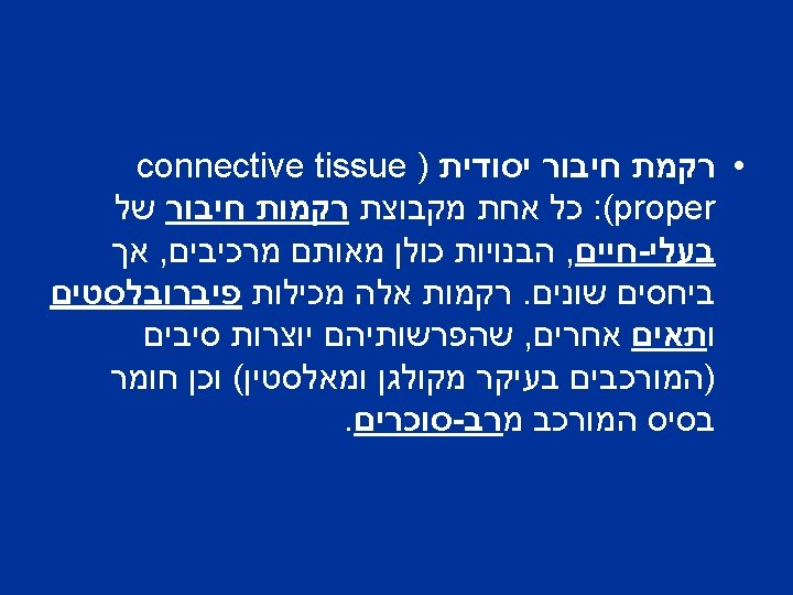
Extra Cellular Matrix
Figure 19 -43. The structure of a typical collagen molecule). A) A model of part of a single collagen a chain in which each amino acid is represented by a sphere. The chain is about 1000 amino acids long. It is arranged as a lefthanded helix, with three amino acids per turn and with glycine as every third amino acid. Therefore, an a chain is composed of a series of triplet Gly. X-Y sequences, in which X and Y can be any amino acid (although X is commonly proline and Y is commonly hydroxyproline). (B) A model of part of a collagen molecule in which three a chains, each shown in a different color, are wrapped around one another to form a triplestranded helical rod. Glycine is the only amino acid small enough to occupy the crowded interior of the triple helix. Only a short length of the molecule is shown; the entire molecule is 300 nm long. (From model by B. L. Trus (.
Collagen structure Figure 12. 52. Structure of collagen A) Three polypeptide chains coil around one another in a characteristic triple helix structure. B) The amino acid sequence of a collagen triple helix domain consists of Gly-X-Y repeats, in which X is frequently proline and Y is frequently hydroxyproline Hyp.
Figure 19 -52. Stretching a network of elastin molecules. The molecules are joined together by covalent bonds) red (to generate a cross -linked network. In this model, each elastin molecule in the network can expand contract as a random coil, so that the entire assembly can stretch and recoil like a rubber band.
Figure 19 -51. Elastic fibers. These scanning electron micrographs show (A) a low-power view of a segment of a dog's aorta and (B) a high-power view of the dense network of longitudinally oriented elastic fibers in the outer layer of the same blood vessel. All the other components have been digested away with enzymes and formic acid. (From K. S. Haas, S. J. Phillips, A. J. Comerota, and J. W. White , Anat. Rec ©. 1991 , 96 230: 86. Wiley-Liss, Inc (.
Figure 19 -55. Three ways in which basal laminae are organized. Basal laminae) yellow (surround certain cells (such as skeletal muscle cells), underlie epithelia, and are interposed between two cell sheets (as in the kidney glomerulus). Note that, in the kidney glomerulus, both cell sheets have gaps in them, so that the basal lamina serves as the permeability barrier determining which molecules will pass into the urine from the blood.
קשרים בין תאים Anchoring junctions • Tight junctions • Gap junctions •
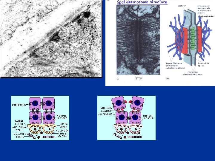
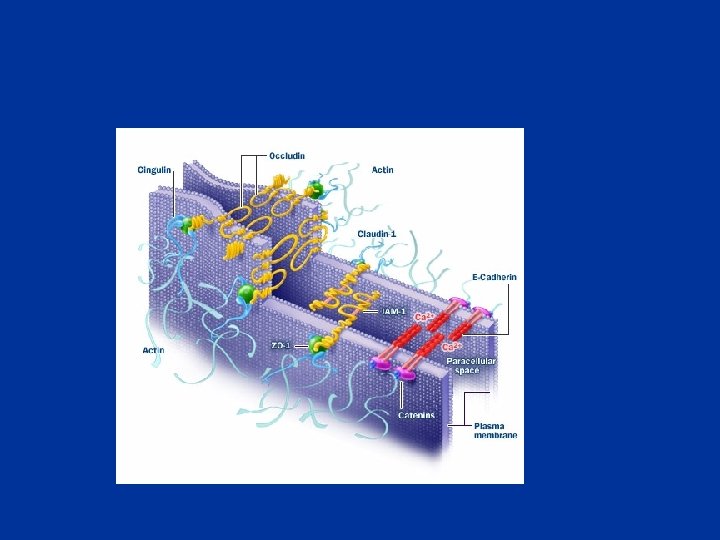
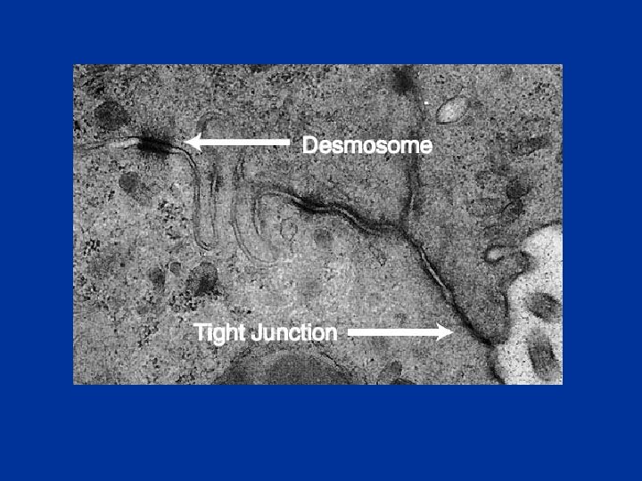
Anchoring junctions Figure 12. 64. Stable cell-cell junctions mediated by the cadherins Homophilic interactions between cadherins mediate two types of stable cell-cell adhesions. In adherens junctions, the cadherins are linked to bundles of actin filaments via the catenins (see Figure 11. 14. (In desmosomes, desmoplakin links members of the cadherin superfamily (desmogleins and desmocollins) to intermediate filaments
Anchoring junctions Cell-cell Adherent j. desmosomes cadherins Cell-matrix Focal adhesion hemidesmosme integrin
Cell adhesion molecules (CAMs) • Cadherins: cell-cell junctions • Integrins: cell- matrix junctions • Selectins: cell- cell adhesion (blood cells bind to blood vessel)
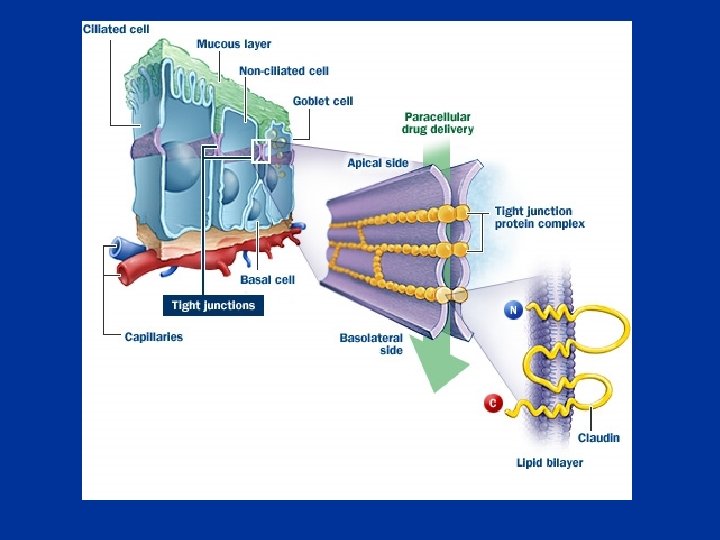
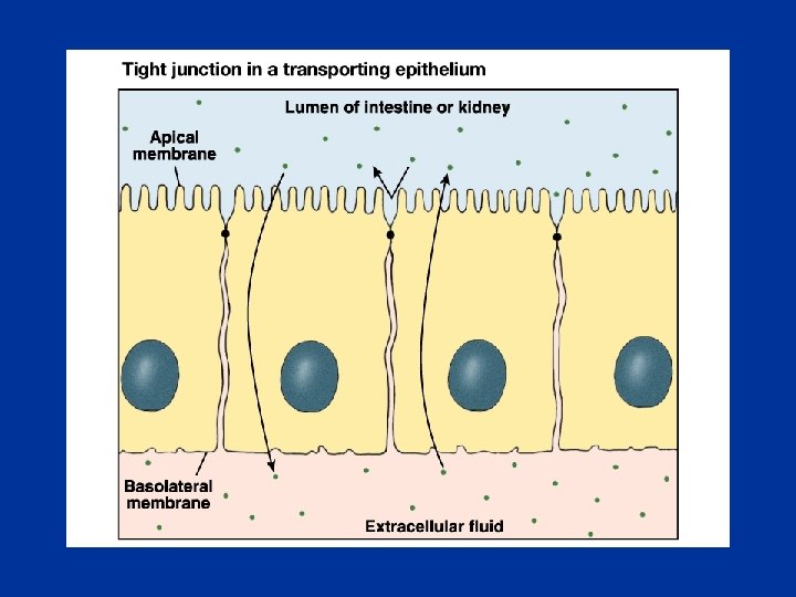
Tight junctions
Figure 12. 65. Tight junctions are formed by interactions between strands of transmembrane proteins (occludin and claudins) on adjacent cells.
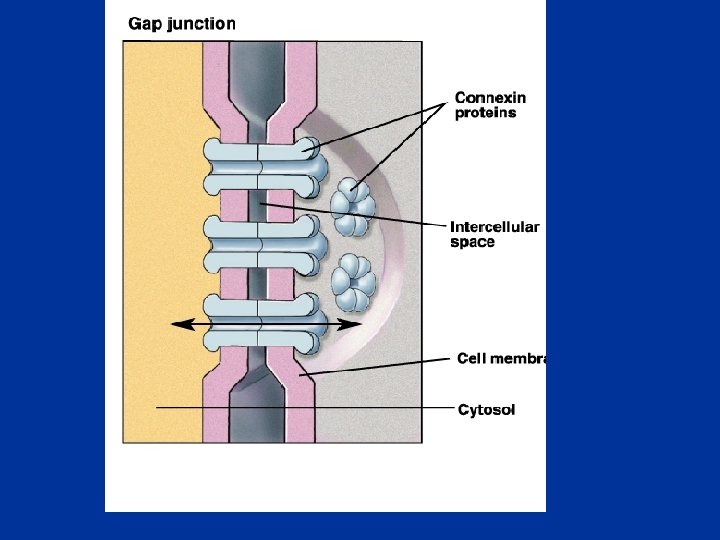
Gap junctions Figure 12. 66. Gap junctions) A) Electron micrograph of a gap junction (arrows) between two liver cells. (B) Gap junctions consist of assemblies of six connexins, which form open channels through the plasma membranes of adjacent cells. (A, Don Fawcett and R. Wood/Photo Researchers, Inc (.
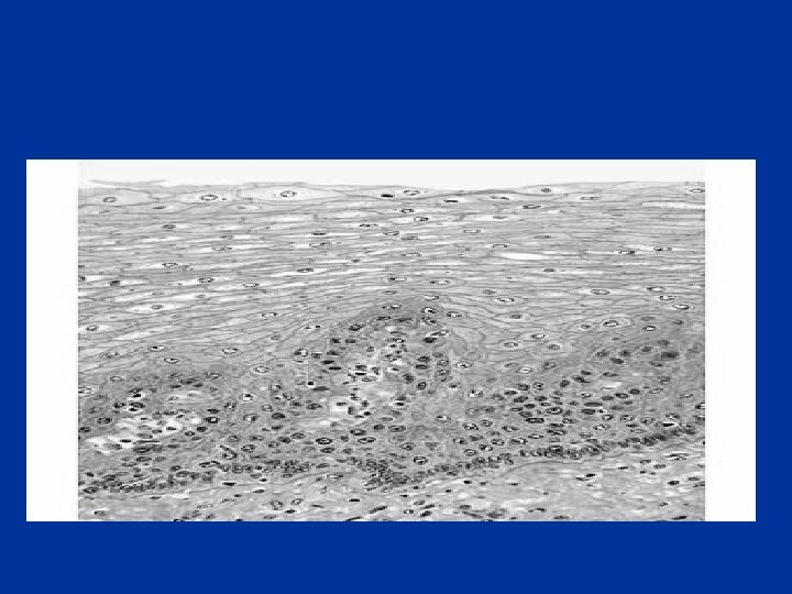
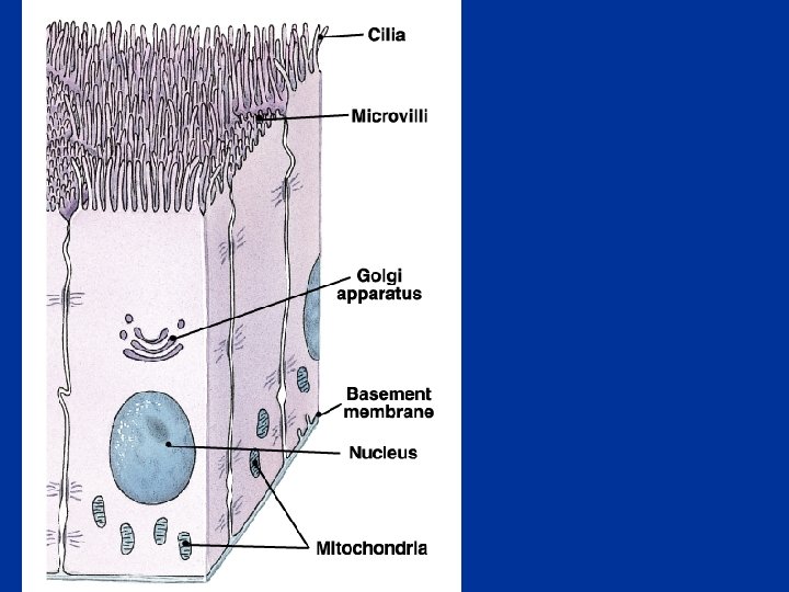
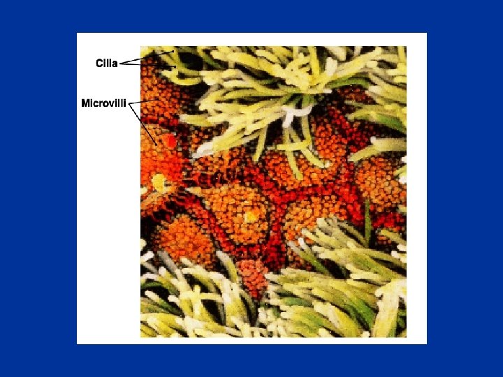
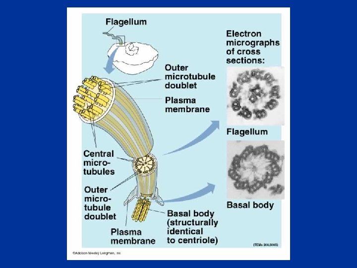
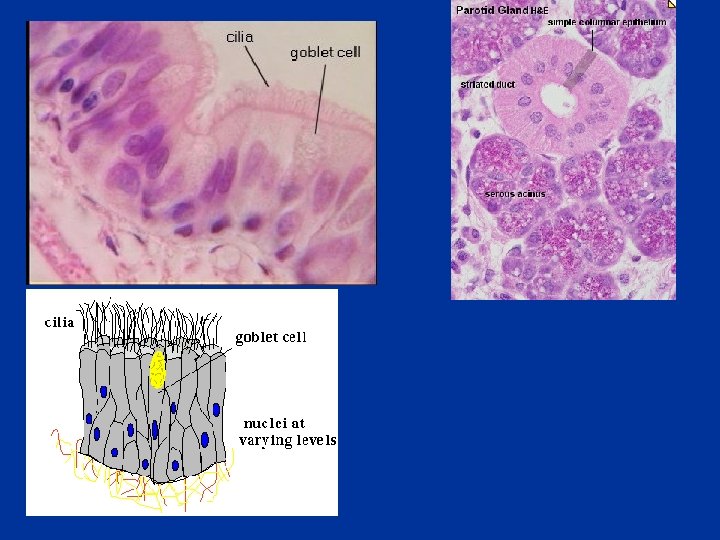
אפיתל • • • exchange Transporting Ciliated Protective Secretory • • • Simple Stratified Transition Squamous Cuboidal columnar
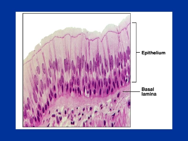
Simple epithelia
Stratified/ transition epithelia
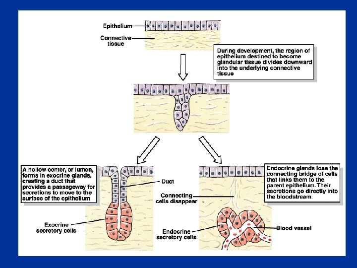
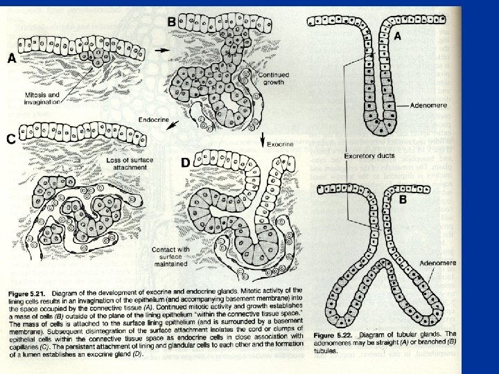
Unicellular gland
Multicellular gland
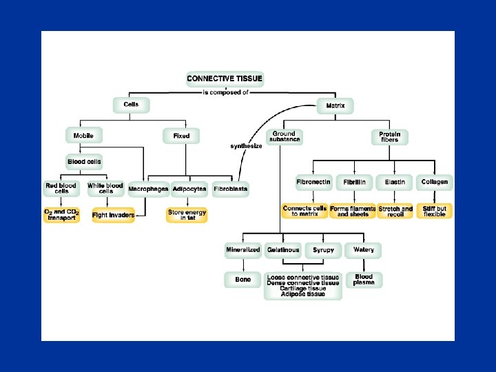
Ground substance • Glycosaminoglycans (polysacharides) • hyaluronic acid (joints, eyeballs…) • chondroitin sulfate (cartillage, bone, skin, blood vessels…) • Dermatan sulfate • Keratan sulfate
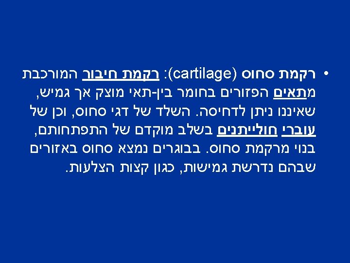
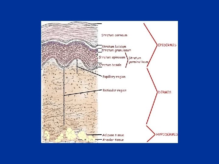
• Loose connective tissue: Adipose tissue Reticular connective tissue • Dense connective tissue: tendons, ligaments, heart valves, dermis, perichondrium, periosteum, • Cartilage • Bone • Liquid connective tissue
Loose connective tissue Areolar connective tissue
Figure 19 -34. The connective tissue underlying an epithelium. This tissue contains a variety of cells and extracellular matrix components. The predominant cell type is the fibroblast, which secretes abundant extracellular matrix.
Dense connective tissue
Bone