Experiment Objectives Preparing Staining and Observing Gbanding human


Experiment Objectives • Preparing, Staining and Observing Gbanding human chromosomes • Develop an understanding of karyotyping and the association of various chromosomal abnormalities to diseases.

Introduction • Chromosomes are composed of double-stranded DNA associated with specific proteins. • The nuclei of normal human somatic cells each contain 23 pairs of chromosomes. • 1 set came from the mother “maternal ”, 1 set came from the father ” paternal ”. • During metaphase, chromosomes become condensed and stain intensely with basic dyes. Mazen Zaharna Molecular Biology 1/2009
 chromosomes,")
Human Chromosomes • 22 of these sets are called autosomes (or “self chromosomes”) chromosomes, are numbered from 1 to 22 approximating decreasing size order. • 1 set are the sex chromosomes • A female carries two X chromosomes (XX) • A male carries an X chromosome and a Y chromosome (XY)

Why do scientists look at chromosomes? • Scientists can diagnose or predict genetic disorders by looking at chromosomes. • This kind of analysis is used in prenatal testing and in diagnosing certain disorders, such as üDown syndrome, üor in diagnosing a specific types of leukemia.

Chromosome abnormalities • Chromosome abnormalities can be ünumerical, as in the presence of • extra • or missing chromosomes , üStructural as in translocations, inversions, large scale deletions or duplications.
")
Chromosomal Abnormalities • Alterations in chromosome number. ü Euploid - normal set (2 n) ü Polyploidy – extra set of the entire genome. (3 n, 4 n etc) ü Aneuploidy – the number of chromosomes is not a multiple of the normal haploid number. • Monosomy ü one member of a chromosome pair is missing, (2 n-1) • Trisomy ü one chromosome set consists of 3 copies of a chromosome, (2 n+1)

• Turner syndrome results from a single X chromosome (45, X or 45, X 0). • Klinefelter syndrome, the most common male chromosomal disease (47, XXY) • Down syndrome, a common chromosomal disease, is caused by trisomy of chromosome 21. Mazen Zaharna Molecular Biology 1/2009
")
Chromosomal abnormalities (can be detected by karyotyping)
 Philadelphia Chromosome - CML")
Chromosomal abnormalities (can be detected by karyotyping) Philadelphia Chromosome - CML

Situations where analysis is strongly recommended • • Problems with early growth & development Fertility problems Neoplasia Pregnancy in older women

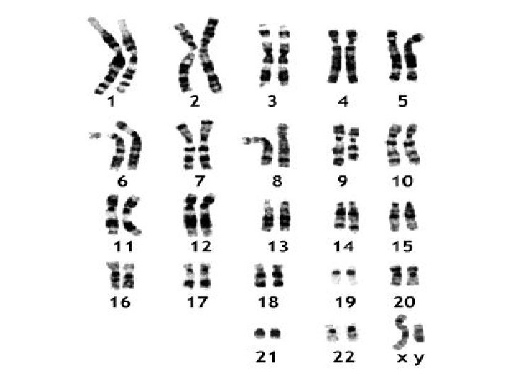
What is a Karyotype? • A display or photomicrograph of an individual’s somatic-cell metaphase chromosomes that are arranged in a standard sequence (usually based on number, size, and type)

Performing a Karyotype • The slides are scanned for metaphase spreads and usually 10 to 30 cells are analyzed under the microscope by a cytogeneticist. • When a good spread (minimum number of overlapping chromosomes) is found, a photograph is taken or the analysis is done by a computer. • The chromosomes are arranged in a standard presentation format of longest to shortest. • Actually chromosome 21 is smaller than chromosome 22.

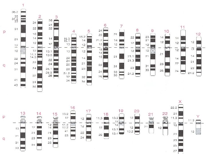
How Do Scientists Identify Chromosomes? • • Three key features to identify their similarities and differences: • Size. This is the easiest way to tell two different chromosomes apart. • Banding pattern. The size and location of Giemsa bands on chromosomes make each chromosome pair unique. • Centromere position. Centromeres are regions in chromosomes that appear as a constriction. Using these key features, scientists match up the 23 pairs

In metacentric chromosomes, the centromere lies near the center of the chromosome. Submetacentric & very Submetacentric chromosomes, have a centromere that is off-center, so that one chromosome arm is longer than the other. In acrocentric chromosomes, the centromere resides very near one end.

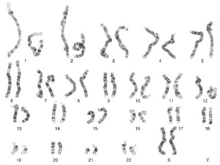
Chromosome banding • Chromosomes are stained with various dyes enabling the chromosome segments to be identified • Most methods can distinguish 550 bands/ haploid set • High resolution methods can distinguish up to 850 bands/ haploid set that can allow identification of small interstitial deletions

G-Banding • Regions that stain as dark G bands replicate late in S phase of the cell cycle and contain more condensed chromatin, • While light G bands generally replicate early in S phase, and have less condensed chromatin.

• The difference between dark- and lightstaining regions was believed to be caused by differences in the relative proportions of bases: – G-light bands being relatively GC-rich – G-dark bands AT-rich Mazen Zaharna Molecular Biology 1/2009

Chromosome Groups Group Chromosomes Description A 1– 3 Largest; 1 and 3 are metacentric but 2 is submetacentric B 4, 5 Large; submetacentric with two arms very different in size C 6– 12, X Medium size; submetacentric D 13– 15 Medium size; acrocentric with satellites E 16– 18 Small; 16 is metacentric but 17 and 18 are submetacentric F 19, 20 Small; metacentric G 21, 22, Y Small; acrocentric, with satellites on 21 and 22 but not on the Y Autosomes are numbered from largest to smallest, except that chromosome 21 is smaller than chromosome 22.

Overview of Procedure 1. 2. 3. 4. 5. 6. 7. 8. 9. Collection of blood Cell culture Stopping the cell division at Metaphase Hypotonic treatment of red & white blood cells Fixation Slide preparation Slide dehydration Treatment with enzyme Staining

Monitor the quality of chromosome spreading • Monitor the quality of chromosome spreading under phase contrast. • Chromosomes should be well spread üwithout visible cytoplasm, üshould appear dark grey under phase contrast

7 - Slide dehydration • Place fixed, dry slides on slide rack in 60 o. C oven • Bake for 3 days • Allow to cool before proceeding to the next step

8 - Treatment with enzyme • Prepare 0. 025% trypsin solution fresh, by mixing 5 ml of 0. 25% trypsin with 45 ml Hank’s solution • Immerse slide in 0. 025 % trypsin for 10120 seconds • Remove slide from trypsin and immediately immerse in phosphate buffer to stop trypsin action
 Staining Time (minutes) Lymphoblastoid 30")
Determination of Trypsin and Staining time Trypsin Time (seconds) Staining Time (minutes) Lymphoblastoid 30 4. 0 Blood Lymphocytes 15 3. 0 0 -3 days 15 3. 0 3 -20 days 30 3. 5 20+ days 45 4. 0 Previously Banded 45 4. 0 < 20 mitosis 15 3. 0 20 -50 mitosis 30 3. 5 50+ mitosis 45 4. 5 Cell Source Age of Oven Dried Slides Cell Concentration

9 - Staining • Prepare a dilution of Giemsa stain by mixing 1 part of Giemsa stain with 3 parts of Phosphate buffer • Flood slide with Giemsa stain for 2 minutes • Rinse slides thoroughly with distilled water • Allow slides to drain, then place on 60 o. C slide warming tray until completely dry

21 22 x y

• http: //www. youtube. com/watch? v=7 Sh. Pzz r. Cet. E • http: //www. youtube. com/watch? v=yox. MG HNj 3 ZU Mazen Zaharna Molecular Biology 1/2009

Thank You
- Slides: 31