Experiment 8 Separation of proteins by ion exchange
![Experiment 8 Separation of proteins by ion exchange chromatography BCH 333[practical]](https://slidetodoc.com/presentation_image_h2/5a2771d20a359116921cace8b4cdeeac/image-1.jpg "Experiment 8 Separation of proteins by ion exchange chromatography BCH 333[practical]")
Experiment 8 Separation of proteins by ion exchange chromatography BCH 333[practical]

Objectives: 1 - The objective of this experiment is for students to learn the principles of ion exchange chromatography by separating the charged molecules using a salt gradient. 2 - A practical experience on ion exchange chromatography in the laboratory 3 - Importance of ion exchange chromatography and procedures in purification.
: - One of The most commonly used chromatographic method")
4 -Ion exchange chromatography (IEC): - One of The most commonly used chromatographic method for protein purification. -Its popularity stems from: - The possibility of high resolution protein separation. - The relative ease of use, reproducibility and availability.
.")
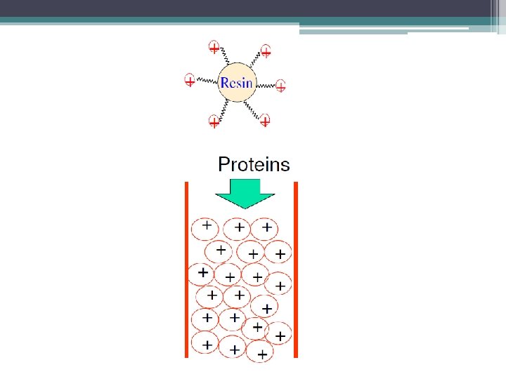
Principle: Separation of molecules based on their charge. The mobile phase: (buffered aqueous solution). -Stationary phase material: is a resin or gel matrix beads with covalently bonded charged functional groups. [charged beads].

Stationary phase

Ion-exchange chromatography

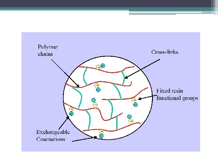
The stationary phase carries ionizable functional groups coupled to an inert matrix material. Because of the principles of electroneutrality, these immobilized charges are electrostatically associated with exchangeable counterions from the solution. So, the stationary phase surface displays ionic functional groups (R-X) that interact with analyte ions of opposite charge. This type of chromatography is further subdivided into: 1. 2. cation exchange chromatography. anion exchange chromatography.

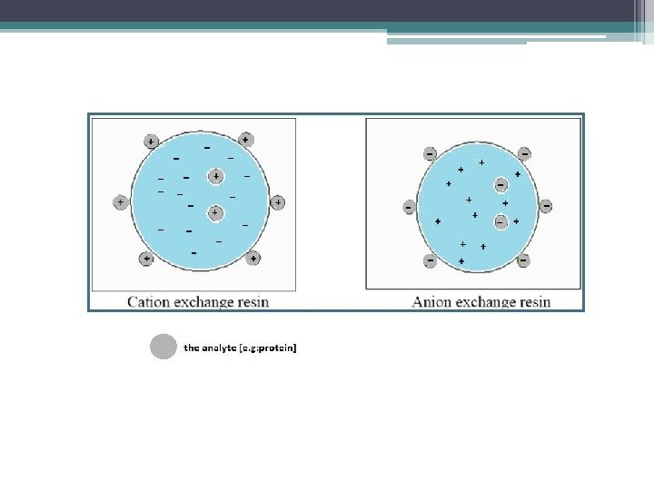
-Cation exchange chromatography: retains positively charged cations because the stationary phase displays a negatively charged functional group. [R-X ] -Anion exchange chromatography: retains negatively charged ions because the stationary phase displays positively charged functional group. [R-X+ ] Remember: -The mobile phase: (buffered aqueous solution). -Stationary phase material is a resin or gel matrix beads with covalently bonded charged functional groups. [differ from gel filtration chromatography].
 retained on the stationary phase can be eluted")
The retarded analytes (+ or -) retained on the stationary phase can be eluted by: -Using buffer that will cause the analytes having net charged same as the charge of the stationary phase, or neturant net charged. Mobile phase Increasing the ionic strength By adding salts[e. g. Na. Cl ] to compete with bound proteins. [To bind to the charged beads]

-At any given p. H, proteins may have a range of charges, since each protein has its own characteristic p. I. -The charge on biomoleucles is related to the p. H of the buffer: At p. H = p. I [net charge is zero]. At p. H higher than p. I [negative charge]. At p. H lower than p. I [positive charge].
![1 -Example for elution of the analyte by using salts [increase ionic strength]: -In](http://slidetodoc.com/presentation_image_h2/5a2771d20a359116921cace8b4cdeeac/image-13.jpg "1 -Example for elution of the analyte by using salts [increase ionic strength]: -In")
1 -Example for elution of the analyte by using salts [increase ionic strength]: -In cation exchange chromatography, to elute the positively charged analyte we could displace the positively charged analyte by the addition of the positively charged sodium ions of Nacl. -The sodium ions[+] of Nacl, will compete with the analyte[+] to bind to the charged stationary phase[-]. -This will cause the elution of the analyte from the column.

2 -Elution of the analyte by using buffers: -at a given p. H[dependent on the composition of the mobile phase. ], a protein will possess an overall net charge. -At p. H of buffer lower than p. I of the analyte , the net charge will be more positive. -At p. H of buffer higher than p. I of the analyte , the net charge will be more negative. So, we can change the net charge of the analyte in a way that it will elute from the column if it has the same charge of the stationary phase, by using the suitable p. H of the buffer and knowing the p. I of the analyte. Or by using buffer has p. H that will cause the net charge of the analyte equal to zero[no charge], leading to the analyte to be eluted.

-For IEC, a good rule to follow when separating a protein whose isoelectric point is known is to select a working p. H which is 1 unit away from the PI of the protein. At this p. H, the protein will possess a high enough net charge to bind well to the ion exchange column. -The p. H at which the positive charges equal the negative charges (in other words, the net charge of the protein is zero) defines that protein’s isoelectric point (PI).

Advantages: -The ion exchange principle permits the protein to bind even when a large buffer volume is applied, making this method especially useful for an initial purification step from a crude extract. -Ion-exchange chromatography separates molecules based on their charged groups, which cause the molecules to interact electrostatically with opposite charges on the stationary-phase matrix.

How does an ion exchanger bind protein? Assuming that the protein possesses a net charge, an ion exchanger needs an opposite charge in order to bind that protein. Ion exchangers are typically composed of a charged (ion exchange) group attached covalently to an insoluble matrix. A positively charged group, such as DEAE (diethylamino ethyl), defines the matrix as an anion exchange matrix whereas a negatively charged group, such as CM (carboxymethyl) makes a cation exchange matrix. DEAE: -CH 2 -N+H(CH 2 CH 3) 2. CM: - CH 2 – COO Thus, if a protein has a negative net charge at a given p. H, an anion exchange matrix should be used for its purification.

Practical: -You are provided with Cation exchange chromatography column, separate the sample given to you. -The sample is a mixture of two proteins, myoglobin and cytochrom C, [p. I=7. 2 and 10. 2 respectively] dissolved in phosphate buffer[p. H=8]. Separate the mixture by using the buffer and 1 M Na. Cl. 1 -carefully remove the layer of the phosphate buffer from above the resin using a pasture pipette, and let only a very thin layer of buffer. 2 - Again using the pasture pipette, very slowly layer the sample mixture solution on the top of the resin, by adding the tip of pipette on the wall of column. Care should be taken not to disturb the gel beads. 3 - Open the screw clip, and start to collect the fractions of about 3 ml each. 4 - After the sample mixture penetrates the gel so that you can see the gel beads, carefully fill up the column with the phosphate buffer [p. H=8] , and complete the collection of fractions. 5 -Collect the fraction of the first sample, and make sure that it is completely eluted.

6 -start adding the 1 M Na. Cl to elute the second sample, and collect the fraction, then make sure that it is eluted completely. 7 - Read the absorbance of each fraction at 410 nm by using spectrophotometer using phosphate buffer as blank for the fractions was collected by using the buffer to elute them, and use 1 M Na. Cl as a blank for the fractions was collected by using the salt. 8 -plot the curve[absorbance at 410 nm against fraction number].

- Slides: 21