
ExerciseInduced Protection Against Type 2 Diabetes Kristin I
Exercise-Induced Protection Against Type 2 Diabetes Kristin I. Stanford, Ph. D. Joslin Diabetes Center Harvard Medical School
Outline Ø Introduction - What is diabetes? Ø Epidemiological data on type 2 diabetes in the U. S. Ø Why is exercise important to combat type 2 diabetes? Ø Effects of acute exercise on skeletal muscle glucose uptake. Ø Effects of chronic exercise training on skeletal muscle and whole-body glucose uptake.
Outline Ø Introduction - What is diabetes? Ø Epidemiological data on type 2 diabetes in the U. S. Ø Why is exercise important to combat type 2 diabetes? Ø Effects of acute exercise on skeletal muscle glucose uptake. Ø Effects of chronic exercise training on skeletal muscle and whole-body glucose uptake.
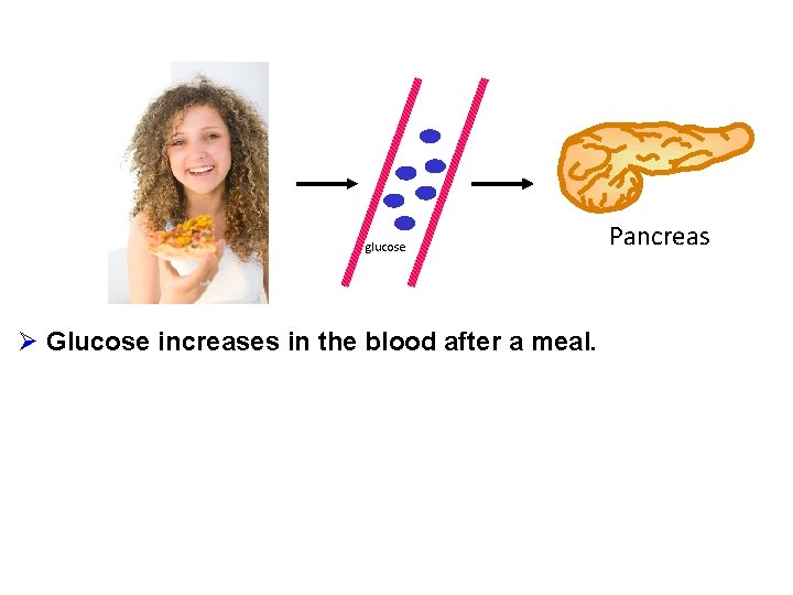
glucose Ø Glucose increases in the blood after a meal. Pancreas
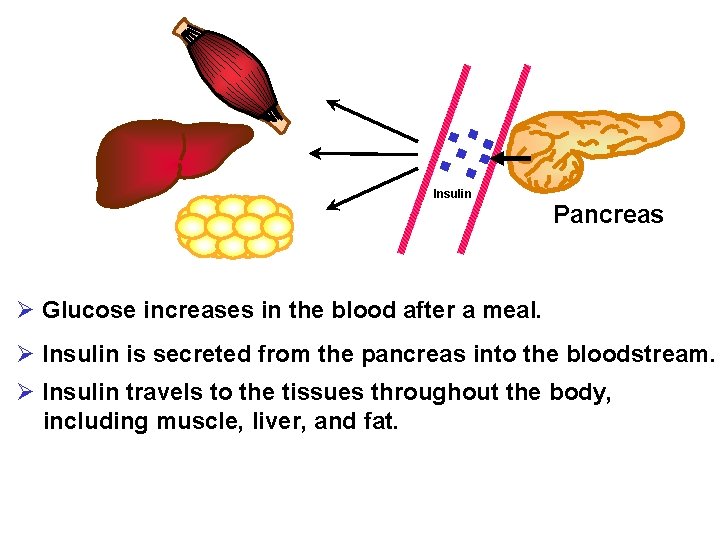
Muscle Liver Insulin Pancreas Fat Ø Glucose increases in the blood after a meal. Ø Insulin is secreted from the pancreas into the bloodstream. Ø Insulin travels to the tissues throughout the body, including muscle, liver, and fat.
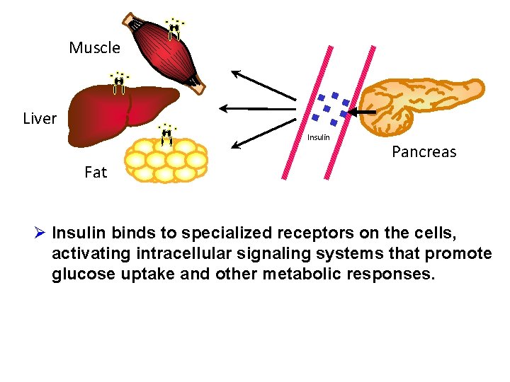
Muscle Liver Insulin Fat Pancreas Ø Insulin binds to specialized receptors on the cells, activating intracellular signaling systems that promote glucose uptake and other metabolic responses.
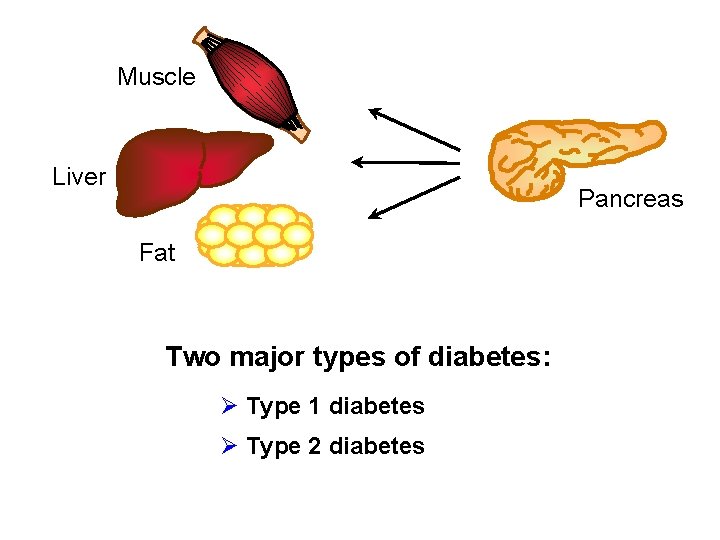
Muscle Liver Pancreas Fat Two major types of diabetes: Ø Type 1 diabetes Ø Type 2 diabetes
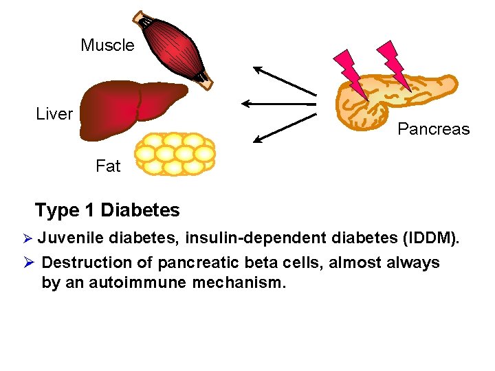
Muscle Liver Pancreas Fat Type 1 Diabetes Ø Juvenile diabetes, insulin-dependent diabetes (IDDM). Ø Destruction of pancreatic beta cells, almost always by an autoimmune mechanism.
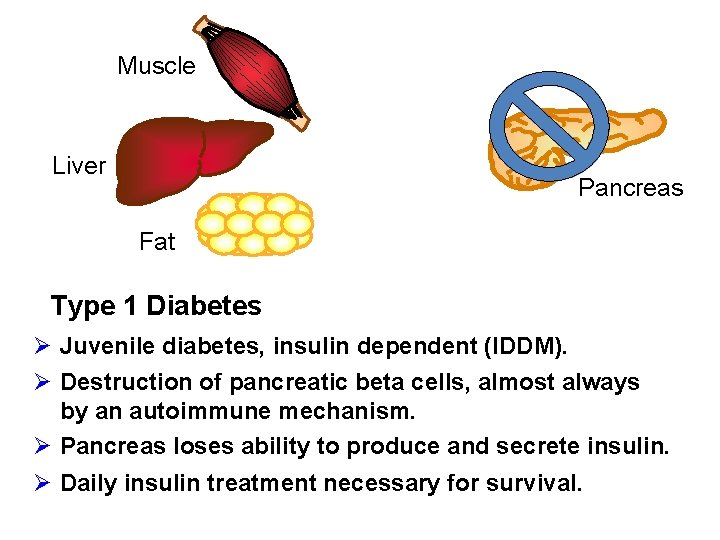
Muscle Liver Pancreas Fat Type 1 Diabetes Ø Juvenile diabetes, insulin dependent (IDDM). Ø Destruction of pancreatic beta cells, almost always by an autoimmune mechanism. Ø Pancreas loses ability to produce and secrete insulin. Ø Daily insulin treatment necessary for survival.
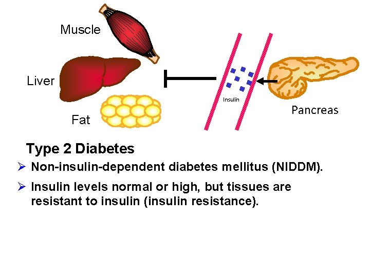
Muscle Liver Insulin Fat Pancreas Type 2 Diabetes Ø Non-insulin-dependent diabetes mellitus (NIDDM). Ø Insulin levels normal or high, but tissues are resistant to insulin (insulin resistance).
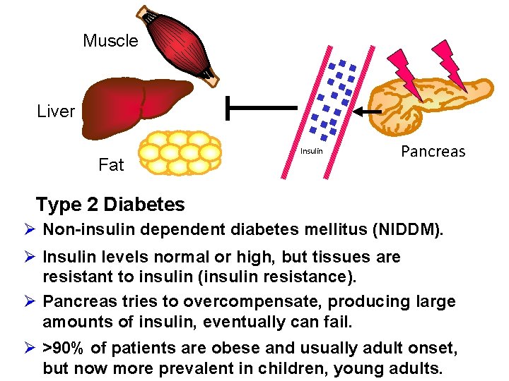
Muscle Liver Fat Insulin Pancreas Type 2 Diabetes Ø Non-insulin dependent diabetes mellitus (NIDDM). Ø Insulin levels normal or high, but tissues are resistant to insulin (insulin resistance). Ø Pancreas tries to overcompensate, producing large amounts of insulin, eventually can fail. Ø >90% of patients are obese and usually adult onset, but now more prevalent in children, young adults.
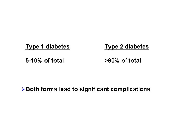
Type 1 diabetes Type 2 diabetes 5 -10% of total >90% of total ØBoth forms lead to significant complications
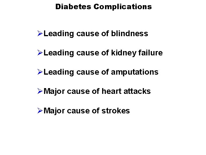
Diabetes Complications ØLeading cause of blindness ØLeading cause of kidney failure ØLeading cause of amputations ØMajor cause of heart attacks ØMajor cause of strokes
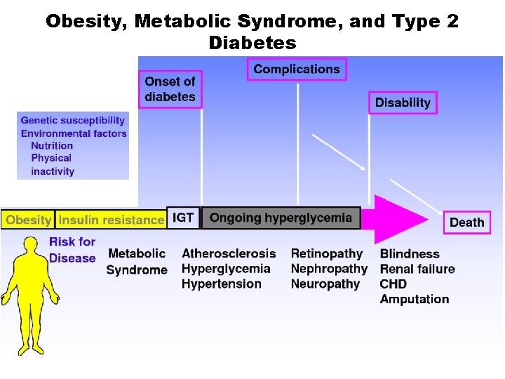
Obesity, Metabolic Syndrome, and Type 2 Diabetes
Outline Ø Introduction - What is diabetes? Ø Epidemiological data on type 2 diabetes in the U. S. Ø Why is exercise important to combat type 2 diabetes? Ø Effects of acute exercise on skeletal muscle glucose uptake. Ø Effects of chronic exercise training on skeletal muscle and whole-body glucose uptake.
Type 2 Diabetes is Increasing at Epidemic Rates Figure from The State newspaper, November 24, 2013. Freely available at thestate. com/2013/11/24/3118223_diabete s-shares-november-with. html? rh=1
Figure on “Type 2 Diabetes in the United States” from Los Angeles Times article. Freely available at latimes. com/health/latb 82485127 z. 1 -20101029223751000 gs 20101029 -graphic. html

Diabetes and Obesity are Increasing in Parallel Figure on “Diabetes and Obesity: The Continuing Epidemic. ” Freely available at http: //www. indiana. edu/~oso/Fructose/ Fructose. html
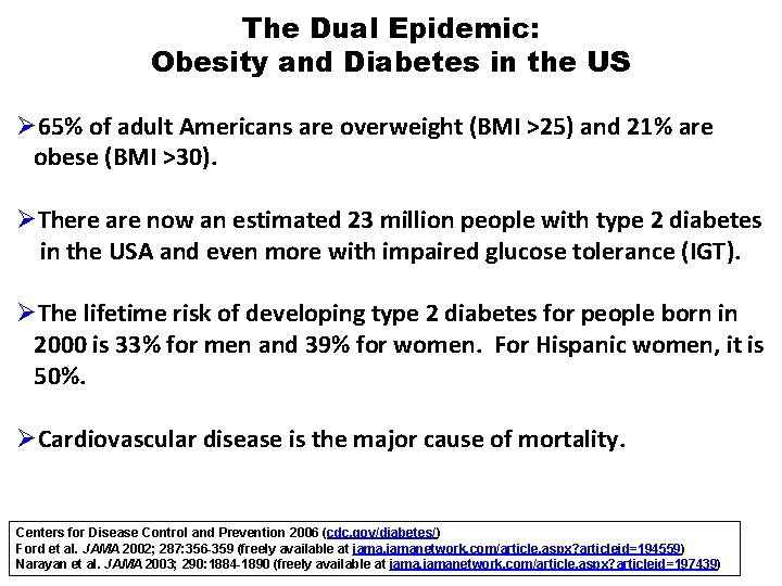
The Dual Epidemic: Obesity and Diabetes in the US Ø 65% of adult Americans are overweight (BMI >25) and 21% are obese (BMI >30). ØThere are now an estimated 23 million people with type 2 diabetes in the USA and even more with impaired glucose tolerance (IGT). ØThe lifetime risk of developing type 2 diabetes for people born in 2000 is 33% for men and 39% for women. For Hispanic women, it is 50%. ØCardiovascular disease is the major cause of mortality. Centers for Disease Control and Prevention 2006 (cdc. gov/diabetes/) Ford et al. JAMA 2002; 287: 356 -359 (freely available at jamanetwork. com/article. aspx? articleid=194559) Narayan et al. JAMA 2003; 290: 1884 -1890 (freely available at jamanetwork. com/article. aspx? articleid=197439)
Type 2 Diabetes is Increasing in Children Figure on “Percentage of 10 - to 17 -year-olds Who Are Overweight or Obese. ” Freely available at http: //rsmccain. blogspot. com/2008/11/us-solveschildhood-obesity-epidemic. html
Outline Ø Introduction - What is diabetes? Ø Epidemiological Data on type 2 diabetes in the U. S. Ø Why is exercise important to combat type 2 diabetes? Ø Effects of acute exercise on skeletal muscle glucose uptake. Ø Effects of chronic exercise training on skeletal muscle glucose uptake.
Obesity, Metabolic Syndrome, and Type 2 Diabetes Figure on “Natural History of Obesity Leading to Type 2 Diabetes” Freely available at healthsciencedegree. info/type-2 diabetes-obesity/
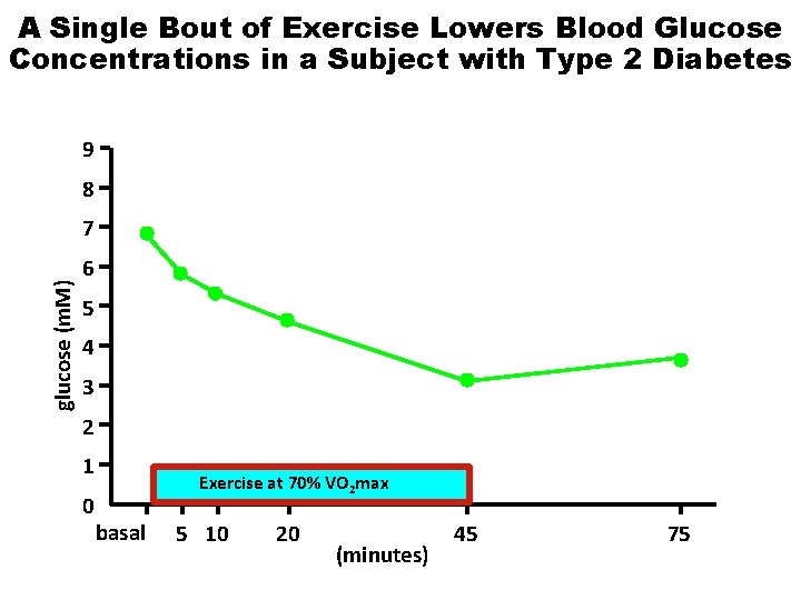
A Single Bout of Exercise Lowers Blood Glucose Concentrations in a Subject with Type 2 Diabetes 9 8 glucose (m. M) 7 6 5 4 3 2 1 0 Exercise at 70% VO 2 max basal 5 10 20 (minutes) 45 75
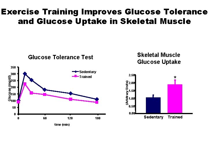
Exercise Training Improves Glucose Tolerance and Glucose Uptake in Skeletal Muscle Glucose Uptake Glucose Tolerance Test 350 Sedentary Trained Glucose (mg/dl) 300 (Arbitrary Units) 250 200 150 100 1. 50 1. 00 50 0 0. 00 0 60 120 time (min) 180 * 2. 00 Sedentary Trained
Outline Ø Introduction - What is diabetes? Ø Epidemiological data on type 2 diabetes in the U. S. Ø Why is exercise important to combat type 2 diabetes? Ø Effects of acute exercise on skeletal muscle glucose uptake. Ø Effects of chronic exercise training on skeletal muscle whole-body glucose uptake.
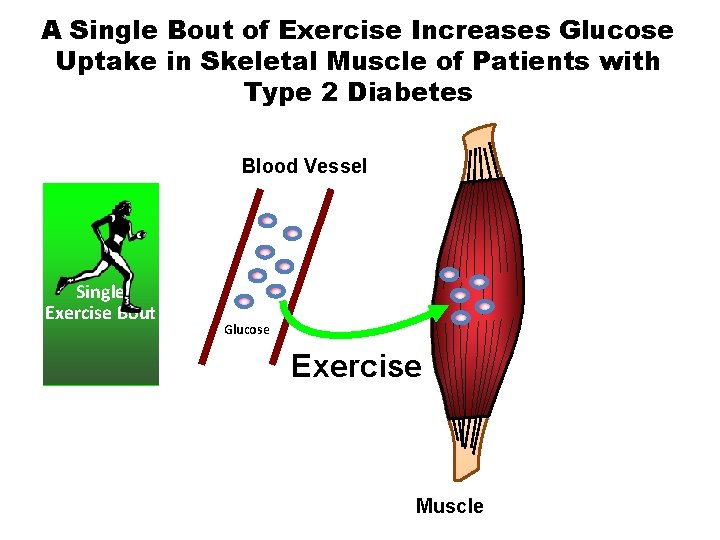
A Single Bout of Exercise Increases Glucose Uptake in Skeletal Muscle of Patients with Type 2 Diabetes Blood Vessel Single Exercise Bout Glucose Exercise Muscle
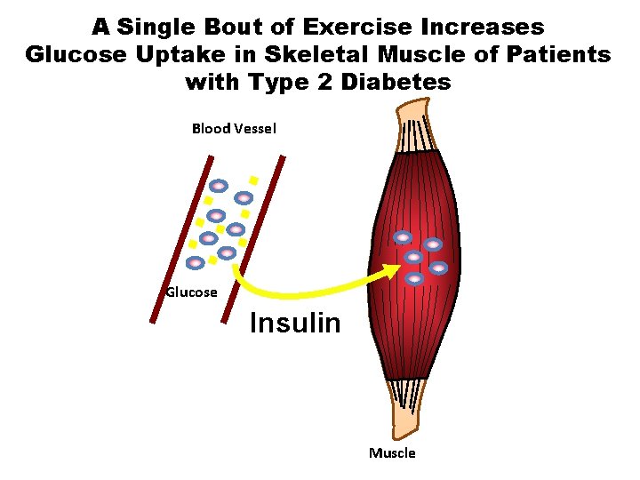
A Single Bout of Exercise Increases Glucose Uptake in Skeletal Muscle of Patients with Type 2 Diabetes Blood Vessel Glucose Insulin Muscle
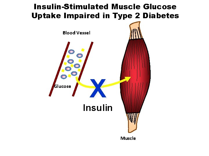
Insulin-Stimulated Muscle Glucose Uptake Impaired in Type 2 Diabetes Blood Vessel Glucose X Insulin Muscle
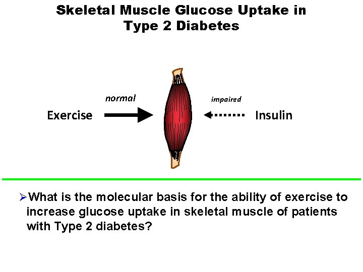
Skeletal Muscle Glucose Uptake in Type 2 Diabetes normal Exercise impaired Insulin ØWhat is the molecular basis for the ability of exercise to increase glucose uptake in skeletal muscle of patients with Type 2 diabetes?
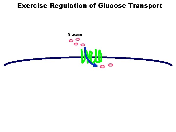
Exercise Regulation of Glucose Transport Glucose
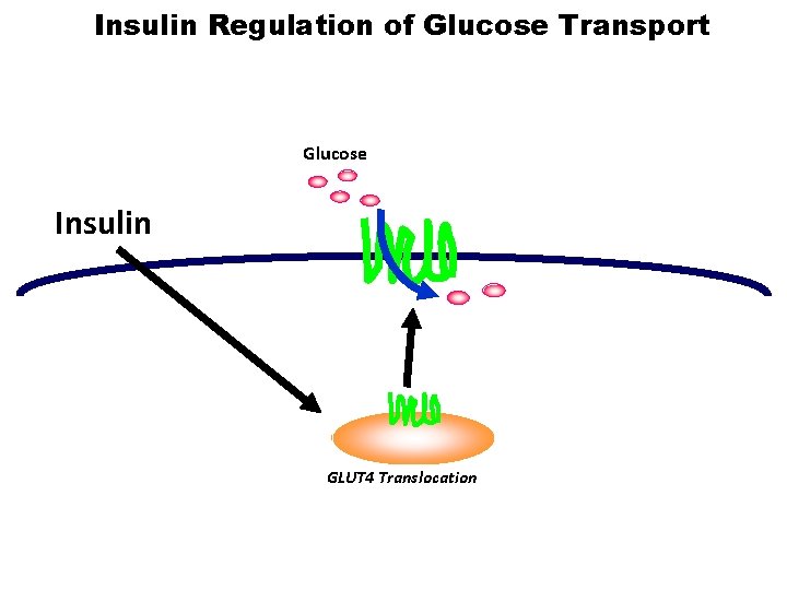
Insulin Regulation of Glucose Transport Glucose Insulin GLUT 4 Translocation

Insulin and Exercise Increase GLUT 4 Translocation in Skeletal Muscle Glucose Insulin Exercise GLUT 4 Translocation
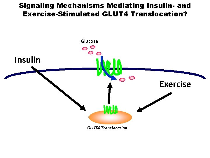
Signaling Mechanisms Mediating Insulin- and Exercise-Stimulated GLUT 4 Translocation? Glucose Insulin Exercise GLUT 4 Translocation
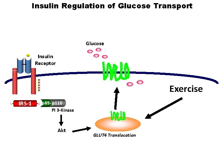
Insulin Regulation of Glucose Transport Glucose Insulin Receptor P P P IRS-1 P Exercise p 85 - p 110 PI 3 -Kinase Akt GLUT 4 Translocation
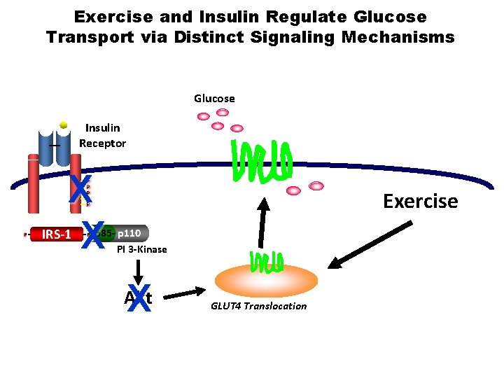
Exercise and Insulin Regulate Glucose Transport via Distinct Signaling Mechanisms Glucose Insulin Receptor X X P P P IRS-1 P Exercise p 85 - p 110 PI 3 -Kinase X Akt GLUT 4 Translocation
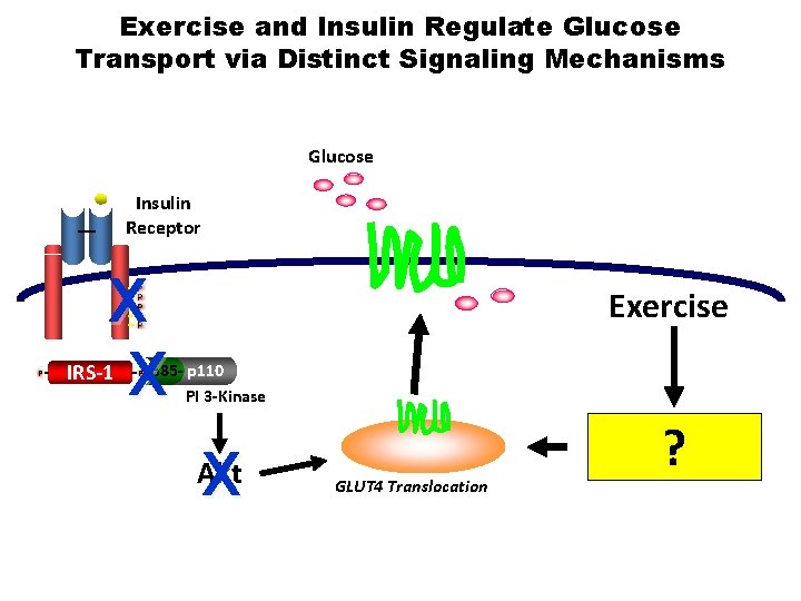
Exercise and Insulin Regulate Glucose Transport via Distinct Signaling Mechanisms Glucose Insulin Receptor X X P P P IRS-1 P Exercise p 85 - p 110 PI 3 -Kinase X Akt GLUT 4 Translocation ?
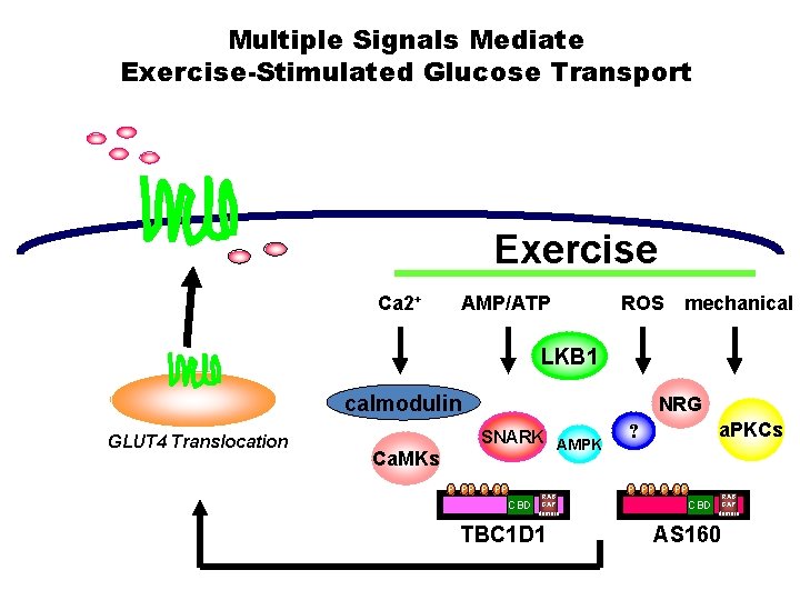
Multiple Signals Mediate Exercise-Stimulated Glucose Transport Exercise Ca 2+ AMP/ATP ROS mechanical LKB 1 calmodulin GLUT 4 Translocation Ca. MKs NRG SNARK AMPK P PP P CBD RAB GAP domain TBC 1 D 1 a. PKCs ? P PP P CBD RAB GAP domain AS 160
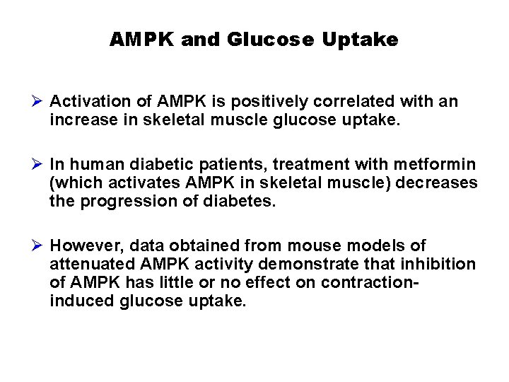
AMPK and Glucose Uptake Ø Activation of AMPK is positively correlated with an increase in skeletal muscle glucose uptake. Ø In human diabetic patients, treatment with metformin (which activates AMPK in skeletal muscle) decreases the progression of diabetes. Ø However, data obtained from mouse models of attenuated AMPK activity demonstrate that inhibition of AMPK has little or no effect on contractioninduced glucose uptake.
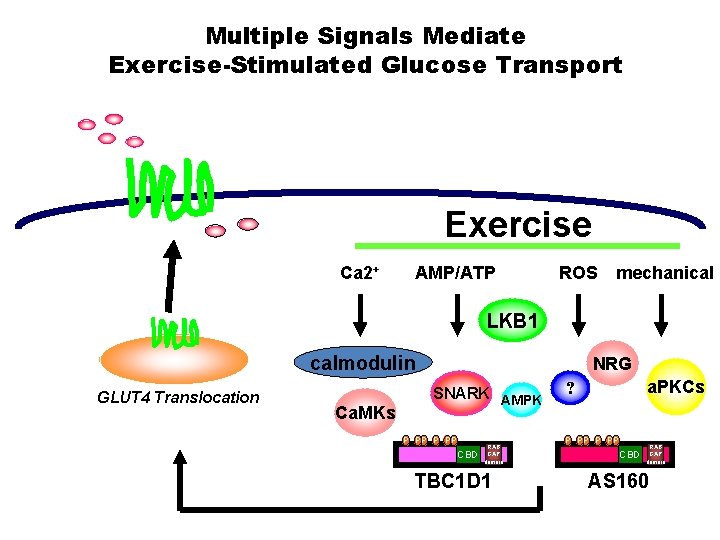
Multiple Signals Mediate Exercise-Stimulated Glucose Transport Exercise Ca 2+ AMP/ATP ROS mechanical LKB 1 calmodulin GLUT 4 Translocation Ca. MKs NRG SNARK AMPK P PP P CBD RAB GAP domain TBC 1 D 1 a. PKCs ? P PP P CBD RAB GAP domain AS 160
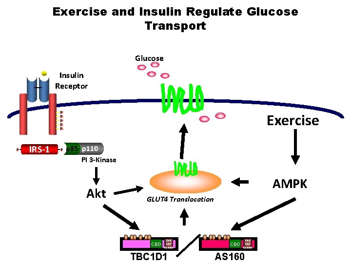
Exercise and Insulin Regulate Glucose Transport Glucose Insulin Receptor P P P IRS-1 P Exercise p 85 - p 110 PI 3 -Kinase Akt AMPK GLUT 4 Translocation P PP P CBD RAB GAP domain TBC 1 D 1 P PP P CBD RAB GAP domain AS 160
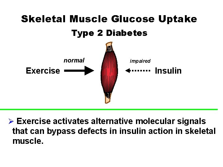
Skeletal Muscle Glucose Uptake Type 2 Diabetes normal Exercise impaired Insulin Ø Exercise activates alternative molecular signals that can bypass defects in insulin action in skeletal muscle.
Outline Ø Introduction - What is diabetes? Ø Epidemiological data on type 2 diabetes in the U. S. Ø Why is exercise important to combat type 2 diabetes? Ø Effects of acute exercise on skeletal muscle glucose uptake. Ø Effects of chronic exercise training on skeletal muscle and whole-body glucose uptake.
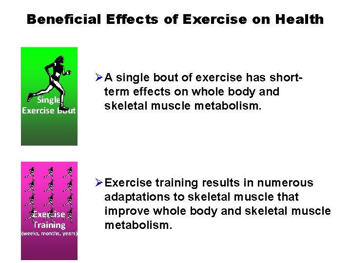
Beneficial Effects of Exercise on Health Single Exercise Bout Exercise Training (weeks, months, years) ØA single bout of exercise has shortterm effects on whole body and skeletal muscle metabolism. ØExercise training results in numerous adaptations to skeletal muscle that improve whole body and skeletal muscle metabolism.
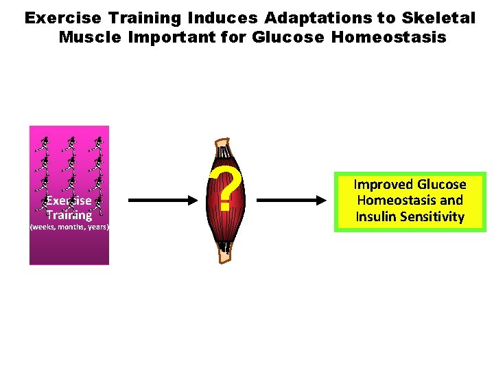
Exercise Training Induces Adaptations to Skeletal Muscle Important for Glucose Homeostasis Exercise Training (weeks, months, years) ? Improved Glucose Homeostasis and Insulin Sensitivity
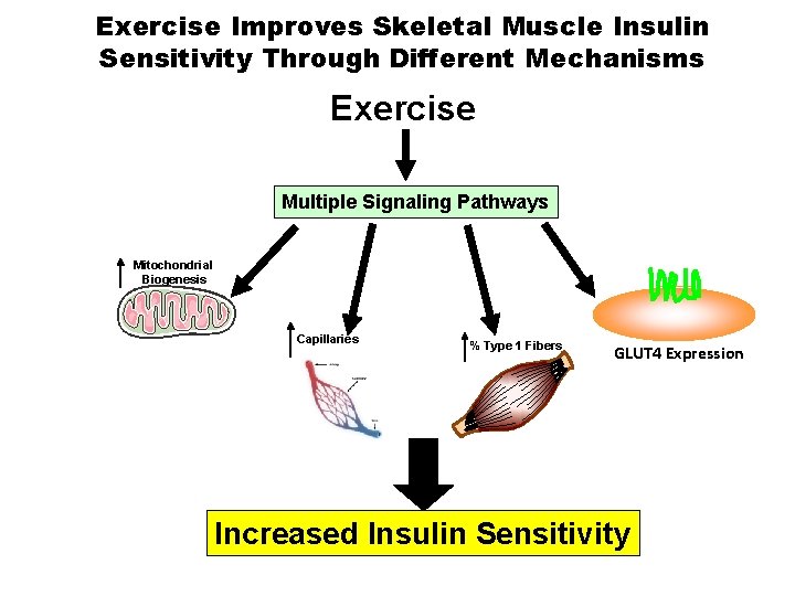
Exercise Improves Skeletal Muscle Insulin Sensitivity Through Different Mechanisms Exercise Multiple Signaling Pathways Mitochondrial Biogenesis Capillaries % Type 1 Fibers GLUT 4 Expression Increased Insulin Sensitivity

Exercise Improves Skeletal Muscle Insulin Sensitivity Through Different Mechanisms Exercise Multiple Signaling Pathways Mitochondrial Biogenesis Capillaries % Type 1 Fibers GLUT 4 Expression Increased Insulin Sensitivity
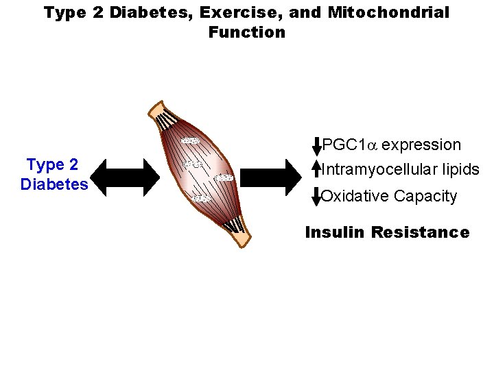
Type 2 Diabetes, Exercise, and Mitochondrial Function Type 2 Diabetes PGC 1 a expression Intramyocellular lipids Oxidative Capacity Insulin Resistance
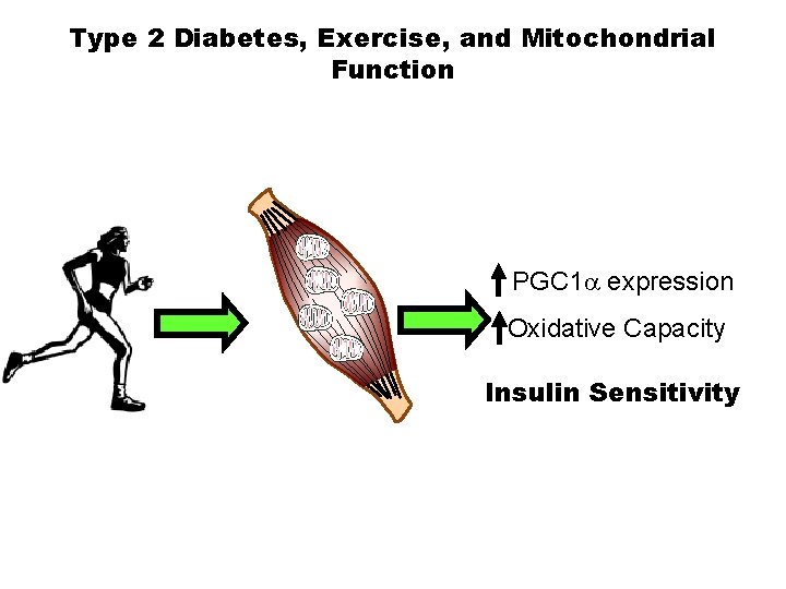
Type 2 Diabetes, Exercise, and Mitochondrial Function PGC 1 a expression Oxidative Capacity Insulin Sensitivity
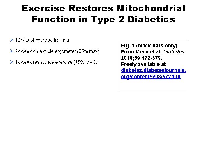
Exercise Restores Mitochondrial Function in Type 2 Diabetics Ø 12 wks of exercise training Ø 2 x week on a cycle ergometer (55% max) Ø 1 x week resistance exercise (75% MVC) Fig. 1 (black bars only). From Meex et al. Diabetes 2010; 59: 572 -579. Freely available at diabetesjournals. org/content/59/3/572. full
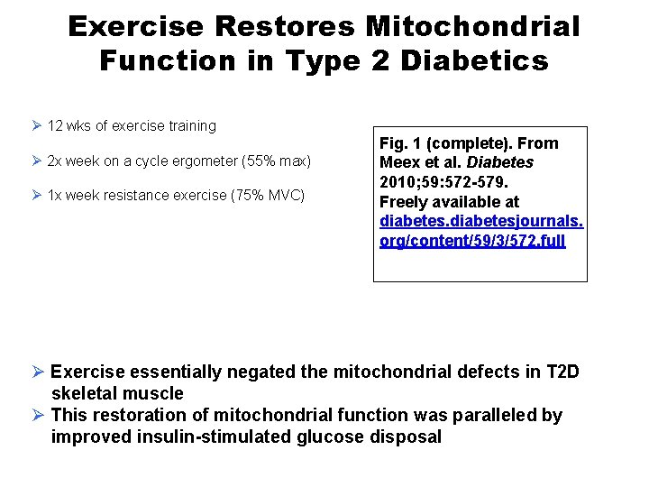
Exercise Restores Mitochondrial Function in Type 2 Diabetics Ø 12 wks of exercise training Ø 2 x week on a cycle ergometer (55% max) Ø 1 x week resistance exercise (75% MVC) Fig. 1 (complete). From Meex et al. Diabetes 2010; 59: 572 -579. Freely available at diabetesjournals. org/content/59/3/572. full Ø Exercise essentially negated the mitochondrial defects in T 2 D skeletal muscle Ø This restoration of mitochondrial function was paralleled by improved insulin-stimulated glucose disposal
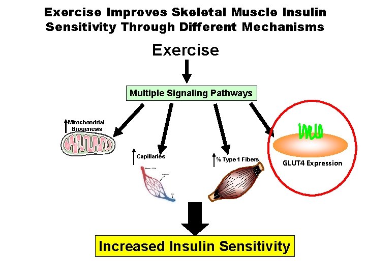
Exercise Improves Skeletal Muscle Insulin Sensitivity Through Different Mechanisms Exercise Multiple Signaling Pathways Mitochondrial Biogenesis Capillaries % Type 1 Fibers GLUT 4 Expression Increased Insulin Sensitivity
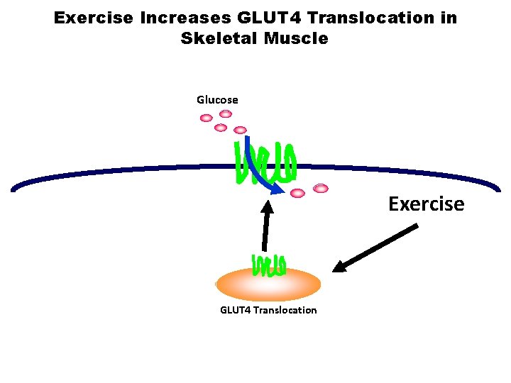
Exercise Increases GLUT 4 Translocation in Skeletal Muscle Glucose Exercise GLUT 4 Translocation

Chronic Exercise Increases GLUT 4 Expression in Skeletal Muscle Chronic Exercise GLUT 4 Expression
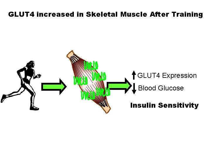
GLUT 4 increased in Skeletal Muscle After Training GLUT 4 Expression Blood Glucose Insulin Sensitivity
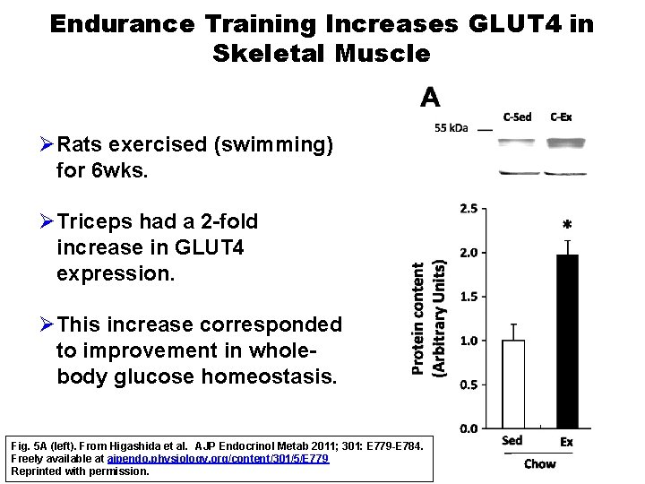
Endurance Training Increases GLUT 4 in Skeletal Muscle ØRats exercised (swimming) for 6 wks. ØTriceps had a 2 -fold increase in GLUT 4 expression. ØThis increase corresponded to improvement in wholebody glucose homeostasis. Fig. 5 A (left). From Higashida et al. AJP Endocrinol Metab 2011; 301: E 779 -E 784. Freely available at ajpendo. physiology. org/content/301/5/E 779 Reprinted with permission.
GLUT 4 increased in Skeletal Muscle After Training in Type 2 Diabetics ØLow-intensity exercise training has been established as an effective strategy to improve the metabolic health of type 2 diabetic patients. ØHowever, the amount of exercise required to see improvements in whole-body and skeletal muscle metabolic health is debatable. ØProposed that low-volume high-intensity exercise interval training, a time-efficient strategy to improve health and fitness, could be applied to a type 2 diabetic patient and improve metabolic health. Little et al. J Appl Physiol 2011; 111: 1554 -1560. Freely available at ap. physiology. org/content/111/6/1554
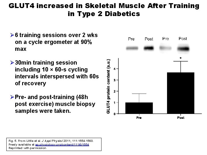
GLUT 4 increased in Skeletal Muscle After Training in Type 2 Diabetics Ø 6 training sessions over 2 wks on a cycle ergometer at 90% max Ø 30 min training session including 10 × 60 -s cycling intervals interspersed with 60 s of recovery ØPre- and post-training (48 h post exercise) muscle biopsy samples were taken. Fig. 5. From Little et al. J Appl Physiol 2011; 111: 1554 -1560. Freely available at ap. physiology. org/content/111/6/1554 Reprinted with permission
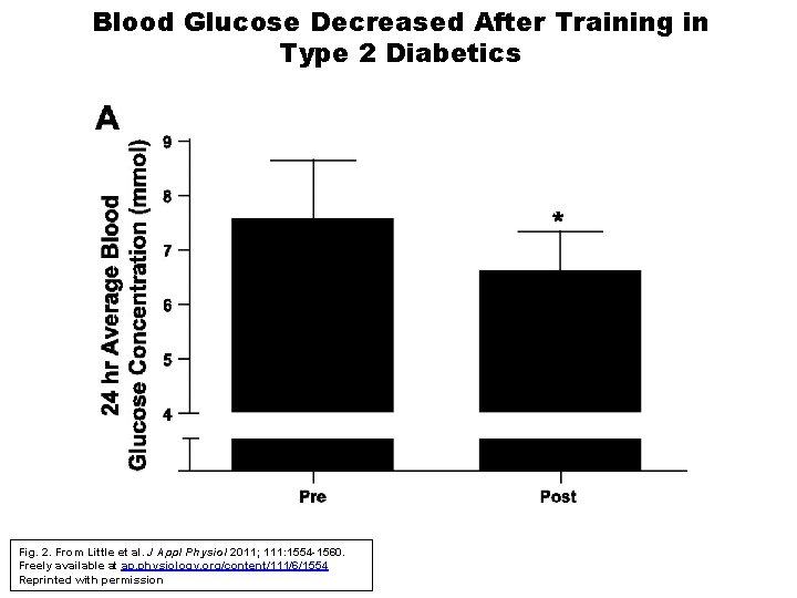
Blood Glucose Decreased After Training in Type 2 Diabetics Fig. 2. From Little et al. J Appl Physiol 2011; 111: 1554 -1560. Freely available at ap. physiology. org/content/111/6/1554 Reprinted with permission
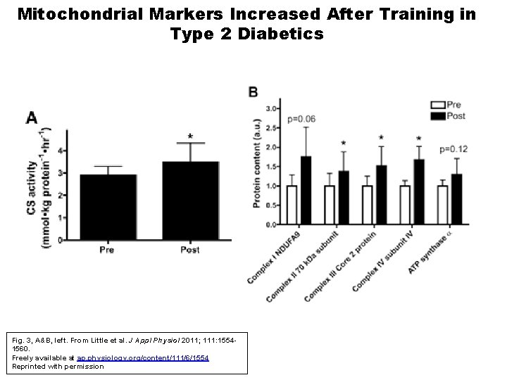
Mitochondrial Markers Increased After Training in Type 2 Diabetics Fig. 3, A&B, left. From Little et al. J Appl Physiol 2011; 111: 15541560. Freely available at ap. physiology. org/content/111/6/1554 Reprinted with permission

Summary ØExercise training increases GLUT 4 expression and increases and improves mitochondrial function in skeletal muscle. ØThese increases correspond to improved glucose metabolism and whole-body insulin sensitivity.
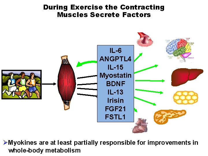
During Exercise the Contracting Muscles Secrete Factors IL-6 ANGPTL 4 IL-15 Myostatin BDNF IL-13 Irisin FGF 21 FSTL 1 ØMyokines are at least partially responsible for improvements in whole-body metabolism
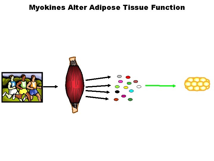
Myokines Alter Adipose Tissue Function
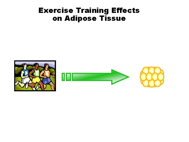
Exercise Training Effects on Adipose Tissue
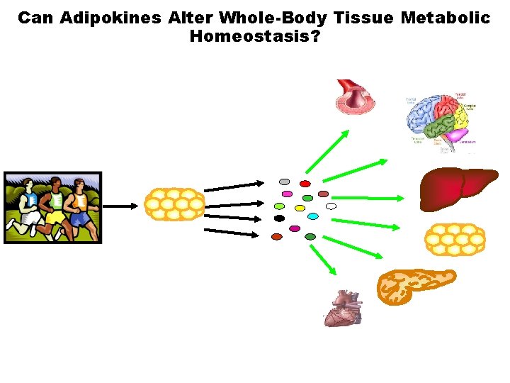
Can Adipokines Alter Whole-Body Tissue Metabolic Homeostasis?
White Adipose Tissue White adipose tissue exists in two major forms in the body Ø Visceral adipose tissue (v. WAT) § The adipose tissue surrounding internal organs § Excess v. WAT is linked to insulin resistance, cardiovascular disease, and stroke Ø Subcutaneous adipose tissue (sc. WAT) § The adipose tissue found underneath the skin § Predisposition to accumulate sc. WAT is linked to insulin sensitivity
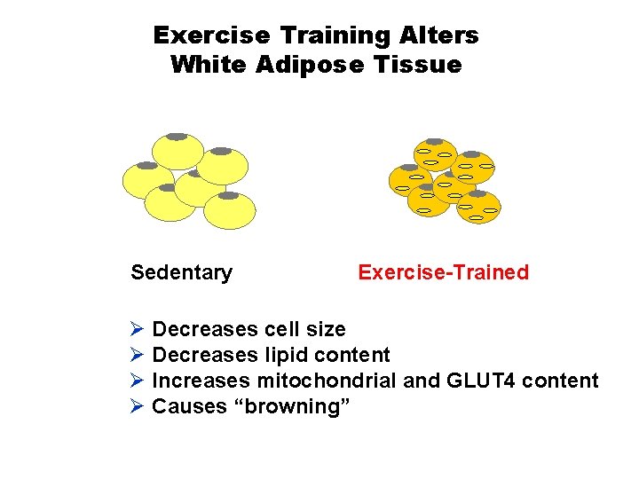
Exercise Training Alters White Adipose Tissue Sedentary Exercise-Trained Ø Decreases cell size Ø Decreases lipid content Ø Increases mitochondrial and GLUT 4 content Ø Causes “browning”

Sedentary Housed in static cages 11 days Exercise Trained Housed in wheelcages 11 days, ~8 km/day subcutaneous (sc. WAT) visceral (v. WAT) Ø gene expression (microarray and q. PCR) Ø histology
Exercise Training Increases Ucp 1 m. RNA in Subcutaneous (sc. WAT), but not Visceral (v. WAT) Supplementary Fig. 2 D. In Stanford et al. Diabetes published ahead of print January 20, 2015, doi: 10. 2337/db 14 -0704. Available at diabetesjournals. org/content/early/2015/ 01/15/db 14 -0704. full
Exercise Training Increases Metabolic Genes in sc. WAT Figure showing m. RNA expression of metabolic genes Prdm 16, Pgc 1α, Adrb 3, dio 2, and Vegfa in sedentary vs. exercise trained animals. Stanford, unpublished data.
Exercise Training Increases Multilocular Droplets and Decreases Cell Size and Lipid Content in sc. WAT Fig. 4 G. In Stanford et al. Diabetes published ahead of print January 20, 2015, doi: 10. 2337/db 14 -0704. Available at diabetesjournals. org/content/early/2015/ 01/15/db 14 -0704. full
Exercise Training Significantly Increases Expression of >1800 Genes in sc. WAT Figure showing gene expression in sedentary vs. exercise trained animals. Stanford, unpublished data. Ø metabolism Ø mitochondrial biogenesis Ø oxidant stress and signaling Ø membrane transport

Exercise Training Protocol Ø Subjects § males (n=10) § 18 -40 yr § BMI <30 Ø Training § 12 weeks § 5 days/wk § 65 -85% VO 2 max Figure showing VO 2 max pre- and post-exercise training. Stanford, unpublished data.
Training Increased UCP 1, VEGFA in Human sc. WAT Figure showing m. RNA expression of metabolic genes UCP 1, Prdm 16, Pgc 1α, ADRB 3, VEGFA in sedentary vs. exercise trained humans. Stanford, unpublished data.
Effects of Exercise Training on Gene Expression Profile in Human sc. WAT Figure showing gene expression pre- and post-training in humans. Stanford, unpublished data. Ø metabolism Ø mitochondrial biogenesis Ø oxidant stress and signaling Ø membrane transport
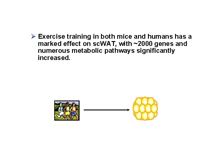
Ø Exercise training in both mice and humans has a marked effect on sc. WAT, with ~2000 genes and numerous metabolic pathways significantly increased.
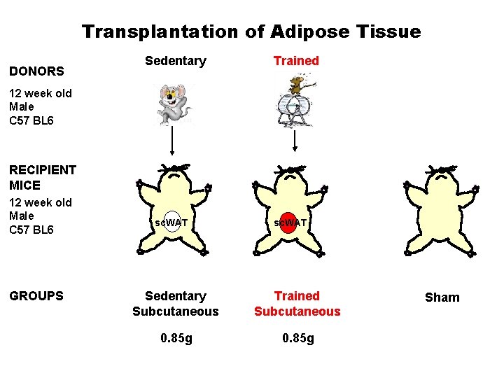
Transplantation of Adipose Tissue DONORS Sedentary Trained 12 week old Male C 57 BL 6 RECIPIENT MICE 12 week old Male C 57 BL 6 GROUPS sc. WAT Sedentary Subcutaneous Trained Subcutaneous 0. 85 g Sham
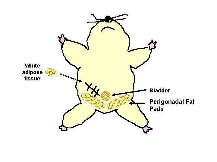
White adipose tissue Bladder Perigonadal Fat Pads
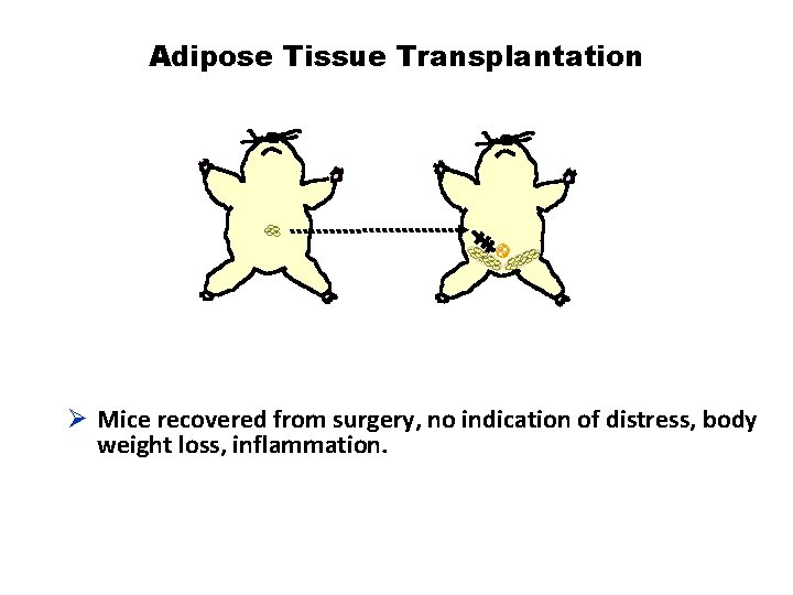
Adipose Tissue Transplantation Ø Mice recovered from surgery, no indication of distress, body weight loss, inflammation.
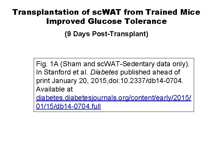
Transplantation of sc. WAT from Trained Mice Improved Glucose Tolerance (9 Days Post-Transplant) Fig. 1 A (Sham and sc. WAT-Sedentary data only). In Stanford et al. Diabetes published ahead of print January 20, 2015, doi: 10. 2337/db 14 -0704. Available at diabetesjournals. org/content/early/2015/ 01/15/db 14 -0704. full
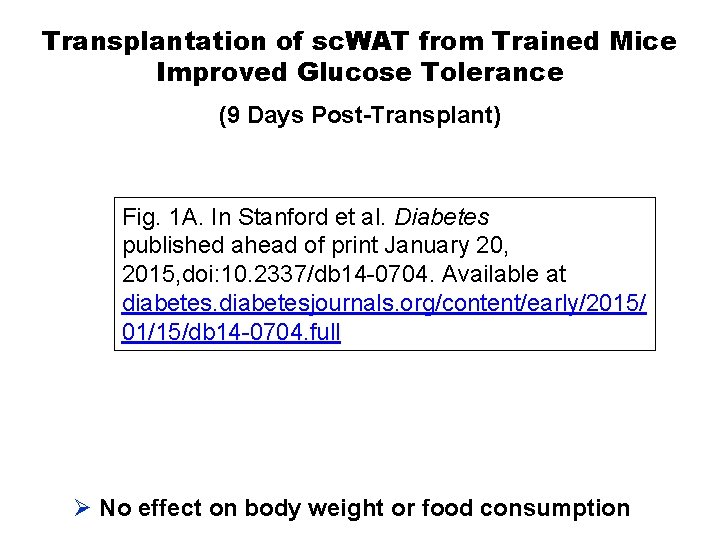
Transplantation of sc. WAT from Trained Mice Improved Glucose Tolerance (9 Days Post-Transplant) Fig. 1 A. In Stanford et al. Diabetes published ahead of print January 20, 2015, doi: 10. 2337/db 14 -0704. Available at diabetesjournals. org/content/early/2015/ 01/15/db 14 -0704. full Ø No effect on body weight or food consumption
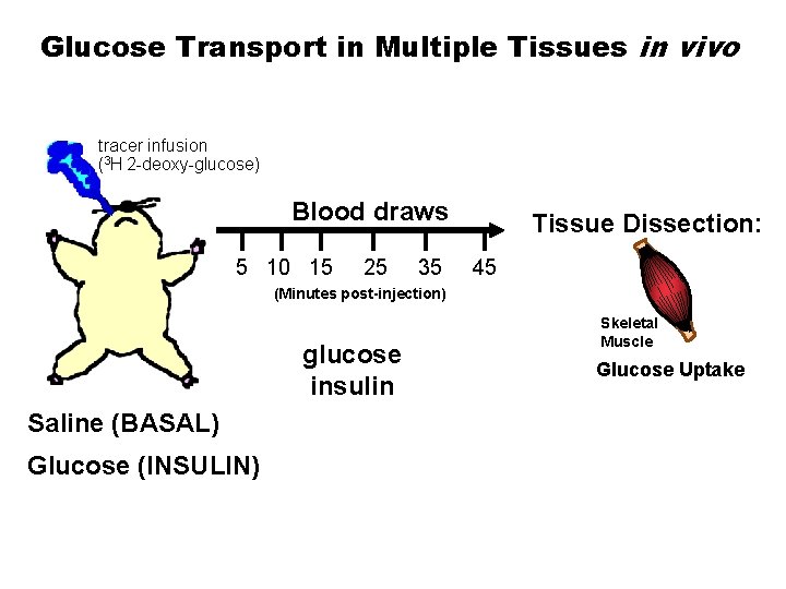
Glucose Transport in Multiple Tissues in vivo tracer infusion (3 H 2 -deoxy-glucose) Blood draws Tissue Dissection: 5 10 15 25 35 45 (Minutes post-injection) glucose insulin Saline (BASAL) Glucose (INSULIN) Skeletal Muscle Glucose Uptake
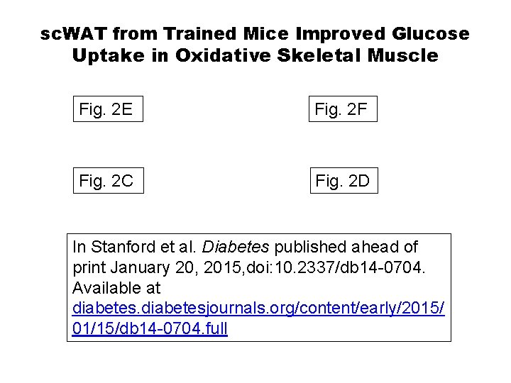
sc. WAT from Trained Mice Improved Glucose Uptake in Oxidative Skeletal Muscle Fig. 2 E Fig. 2 F Fig. 2 C Fig. 2 D In Stanford et al. Diabetes published ahead of print January 20, 2015, doi: 10. 2337/db 14 -0704. Available at diabetesjournals. org/content/early/2015/ 01/15/db 14 -0704. full
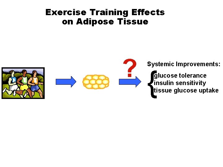
Exercise Training Effects on Adipose Tissue ? Systemic Improvements: { glucose tolerance insulin sensitivity tissue glucose uptake
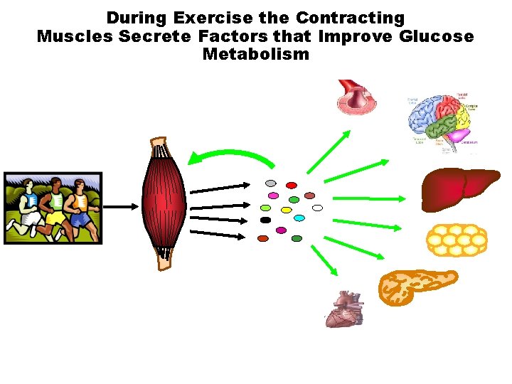
During Exercise the Contracting Muscles Secrete Factors that Improve Glucose Metabolism
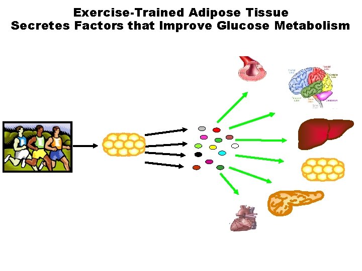
Exercise-Trained Adipose Tissue Secretes Factors that Improve Glucose Metabolism
Summary Ø Transplantation of sc. WAT from trained mice into the visceral cavity of sedentary mice resulted in: § Improved glucose tolerance § Increased glucose uptake in tibialis anterior and soleus ØNovel adipokines from sc. WAT are a potential mechanism for the improved whole-body and tissue glucose homeostasis observed after transplantation.
Conclusions ØType 2 diabetes is increasing at epidemic rates. ØExercise activates alternative molecular signals that can bypass defects in insulin action in skeletal muscle and restore skeletal muscle glucose uptake. ØExercise alters metabolic function of other tissues, in a similar manner as skeletal muscle. ØExercise is essential to combat and treat the epidemic of type 2 diabetes.

Thank You! Laurie Goodyear Sarah Lessard
- Slides: 88