EXCRETORY SYSTEM Intended Learning Outcomes Learning Outcomes Text


")


: most of the important")

: the blood dumps things it wants to get")


![Recall that osmosis is the movement of water (from [High] to [Low])across a selectively](https://slidetodoc.com/presentation_image/f2f4d7a7d61ced4ce4f8b0f0a5646b4a/image-66.jpg "Recall that osmosis is the movement of water (from [High] to [Low])across a selectively")


. Plasma Filtrate Final")


 This hormone controls water reabsorption with a negative feedback")
, the osmoreceptor cells in the hypothalamus")


- Slides: 103
EXCRETORY SYSTEM
Intended Learning Outcomes # Learning Outcomes Text 1 Identify and give functions for each of the following: kidney, urethra, urinary, bladder, renal cortex, renal medulla, renal pelvis Page 302 2 Identify and give functions for each of the following: Page 306 nephron, glomerulus, Bowman's capsule, afferent and efferent arterioles, -309 peritubular capillary network, proximal and distal convoluted tubules, collecting duct, loop of henle 3 Contrast the blood in the renal artery and the renal vein with respect to urea and glucose content 4 Identify the source glands for ADH and aldosterone and explain how these P. 310, hormones are regulated) 394, 399400 5 Relate ADH, aldosterone, and the nephron to the regulation of water and sodium levels in the blood P. 310, 394, 399400 6 Describe how the kidney maintains blood p. H Page 313 Not in text
VOCABULARY _____ Afferent arteriole _____ Aldosterone _____ Antidiuretic hormone _____ Blood pressure _____ Bowman’s capsule _____ Collecting duct _____ Constriction _____ Cortex _____ Dehydration _____ Distal convoluted tubule _____ Efferent arteriole _____ Excretion _____ Filtrate _____ Glomerulus _____ Hypertonic _____ Hypothalamus _____ Juxtoglomerular apparatus _____ Kidney _____ Loop of Henle _____ Medulla _____ Nephron _____ Osmotic pressure _____ Peristalsis _____ Peritubular capillaries _____ Permeable _____ Posterior pituitary _____ Pressure filtration _____ Proximal convoluted tubule _____ Renal arteries _____ Renal pelvis _____ Renal vein _____ Renin _____ Selective re-absorption _____ Tubular secretion _____ Tubule _____ Urea _____ Urinary bladder _____ Urination _____ Water re-absorption
• If you knew there was poison hidden in your house, you would surely do everything possible to find and remove that poison. • If you didn't, you and your family would slowly die. • How would you find and then remove it? • You would probably figure out a system of searching and removing. • That is what the excretory system does!!
1. The Human bladder can stretch to hold about 1. 5 cups (400 ml) of urine. 2. All the blood in our body passes through each kidney 400 times every day. 3. When the bladder is ½ full, our brain tells the bladder to relax and we get the urge to urinate. We can’t control or stop urination until we are 2 years old. 4. Even if 75% of the nephrons are lost, the kidney will still function. It is possible to live a healthy life with only one kidney. 5. Normal urine is sterile (germ-free). It is composed of water, salts, and waste products.
6. The kidneys have 1 million little filters which filter 1. 3 litres of blood every minute. That is 1, 620 litres every day. 7. 99. 9% of the materials passing through the kidneys are reabsorbed back into the blood. 8. We made approximately 1. 5 litres of urine per day. 9. A traditional medical practice in India is called urine therapy, and they apply urine to the skin or drink it to cure various health problems. 10. Urine has a lot of urea (waste), and it is a good source of nitrogen for plants. Gardeners often recommend you add 1 cup of urine to 15 cups of water and apply it to pot plants and flower beds to help them grow.
11. The ancient Romans used to bleach their clothes with urine. 12. People lost at sea or in the desert for a long time often resort to drinking their urine when no rainwater is available. However, this won't prevent you from dying of dehydration, especially if it causes vomiting. 13. Urine has also been used as an antiseptic. In times of war, when other antiseptics were unavailable, urine, the darker the better, was used on open wounds to kill bacteria. 14. The yellow colour of urine was once thought to come from gold. Alchemists in the middle ages spent a lot of time trying to extract gold from urine. They were, of course, unsuccessful.
1. Filters the blood 2. Removes wastes from the blood wastes 3. Keeps the amount of water and salts in the blood constant 4. Helps to regulate Blood Pressure
Humans will die if they lose about 12% of their body water. 12 The threat of dessication is the most dessication important problem confronting terrestrial life. How do we control our loss of water? 1. Multiple layers of dead, keratinized skin cells 2. Drink and eat moist foods 3. Nervous and hormonal mechanisms control thirst 4. Behaviour: (ie: stay out of intense heat) : 5. Kidneys and other excretory organs help conserve water
Water loss can be considerable: a person in a 38 o. C temperature will lose 1 Litre of H 2 O/hour. We acquire most of our water in our food and drink, and obtain a smaller amount by dehydration synthesis and as a by-product of cellular respiration We lose water by urinating, defecating, sweating, and breathing. defecating breathing
The Excretory System is made up of the kidneys, liver, lungs, and skin. Each plays a role in excretion. Skin: sweat with water, salts, heat and some urea Lungs: excretes carbon dioxide, water, and heat Liver: gluconeogenesis – makes urea as a byproduct Intestines: excretes certain heavy metals Kidneys: the main excretory organ. Excrete toxins, drugs, wastes (urea…), and water.
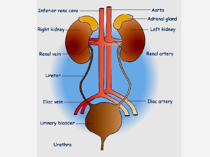
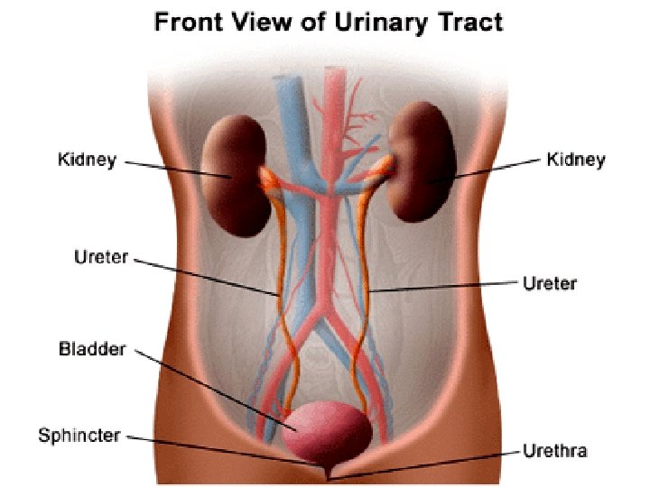
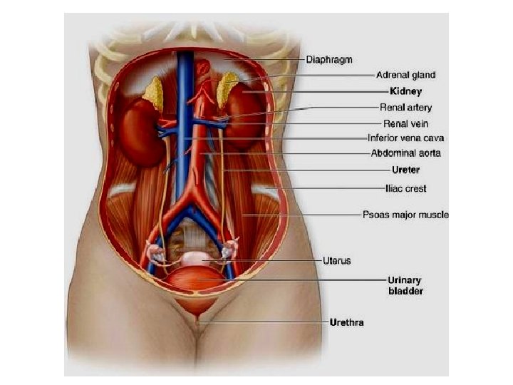
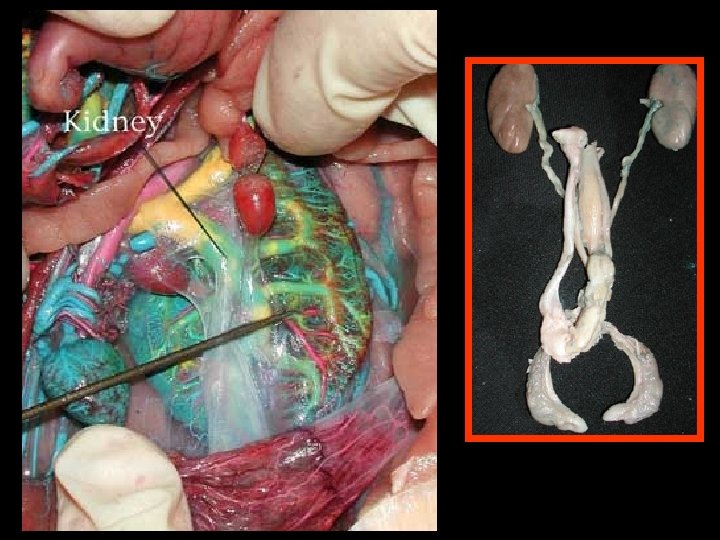
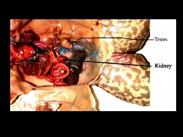
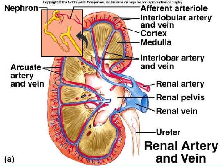
KIDNEY Kidney: • Bean-shaped • 10 cm long • Lower, dorsal part dorsal of the abdomen • Blood enters the kidney via the renal artery, is cleaned, artery and leaves the kidney in the renal vein
The kidneys account for less than 1% of the weight of the human body, but they receive about 20% 20 of the blood pumped with each heartbeat
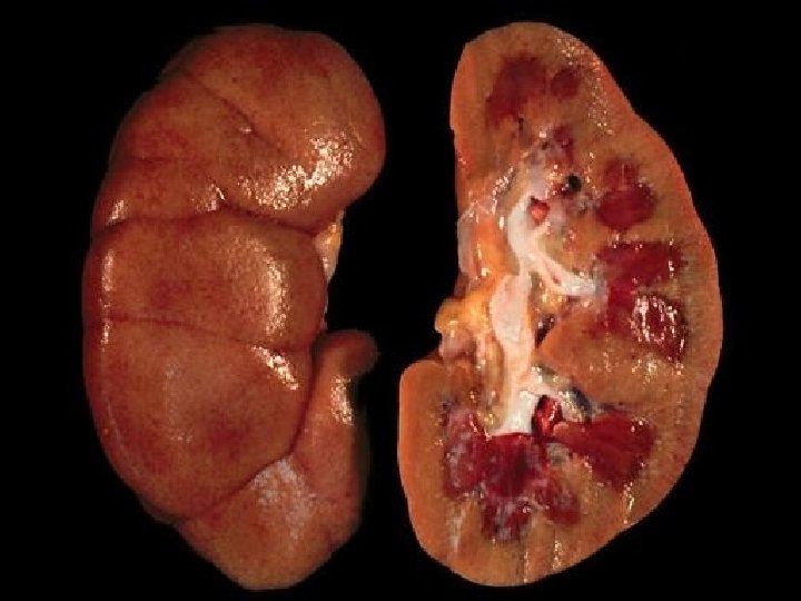
Renal cortex
Renal cortex renal medulla
Renal cortex renal medulla renal pelvis
Renal cortex renal medulla renal pelvis Renal pyramid
Renal cortex renal medulla renal pelvis Renal pyramid Ureter
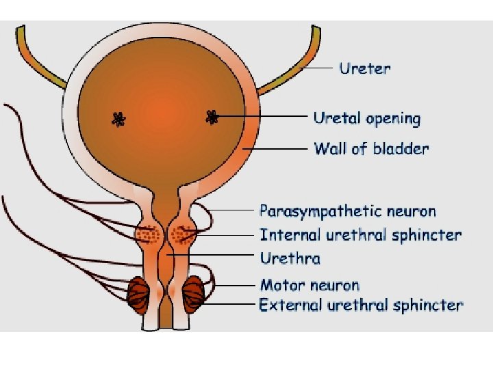
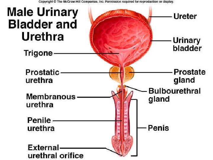
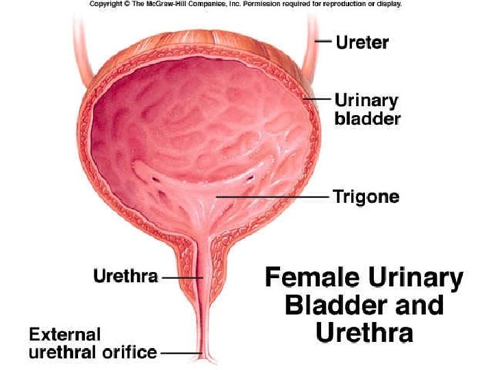
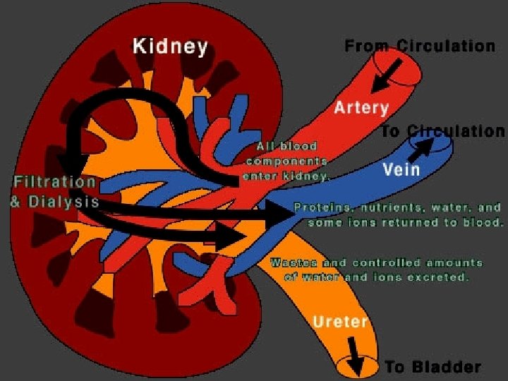
KIDNEY Ureter: Waste fluid made in the kidney exits through a duct called a ureter Moves the urine to the bladder via peristalsis URETER
KIDNEY Bladder: stores the urine An average bladder can hold bladder a maximum of 1. 5 to 2 cups of urine. When full, the BLADDER sphincter muscles sphincter control the release of urine from the bladder URETER
KIDNEY Urethra: when the sphincter relaxes, the urethra carries the urine outside the body. 20 cm in males URETER 4 cm in females BLADDER Females are more prone to infections of the urinary system. URETHRA
The kidneys perform a number of homeostatic functions, as they are the chief regulators of our internal environment: 1. Regulates blood volume and osmotic balance by excreting or conserving water as the situation demands. water 2. Regulates the ionic balance of the blood by controlling the excretion of inorganic salts (especially sodium). 3. Regulates Blood p. H by excreting excess acids or base 4. Excretes toxic metabolic by-products such as urea, ammonia, urea uric acid, and creatine (a product of muscle activity). creatine
Nitrogen wastes are a by-product of protein metabolism When proteins are turned into glucose during gluconeogenesis, gluconeogenesis ammonia is made. Because ammonia is very toxic and must be diluted as it travels around the body, however, terrestrial animals usually need to conserve water What do we do? The liver converts the ammonia to urea, which is then transported to the kidneys, urea where it is concentrated and excreted out of the excreted body as urine. This takes a lot of energy. urine energy
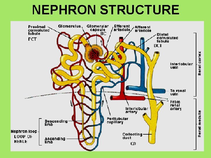
The functional unit of the kidney is the NEPHRON, which consists of a NEPHRON renal tubule and its associated blood vessels Each kidney contains approximately 1 million nephrons, which represents approximately 80 km of tubules. 80 km
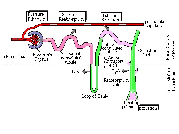
Water, urea, salts, and other small molecules in the blood flow from the capillaries into the renal tubules, where the fluid is now called FILTRATE The epithelial cells that line the renal tubule adds and removes things from the filtrate to eventually form URINE From the 1100 to 2000 L of blood that flows through the human kidneys each day, the nephron processes about 180 L of filtrate, but filtrate excretes only ~1. 5 L of urine The rest of the filtrate, including ~99% of the water, is water reabsorbed into the blood. reabsorbed
1 2 3 4 5
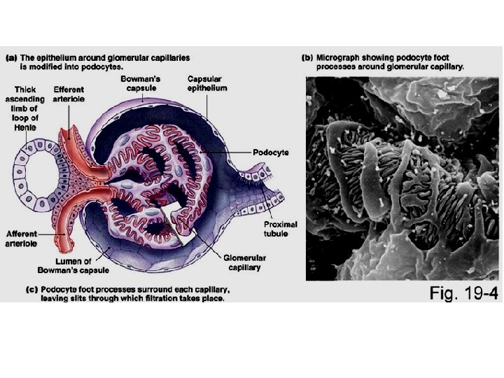
filtrate blood 1. Bowman’s Capsule: Capsule this cup shaped receptacle is the blind end of the renal tubule, which receives filtrate from the blood It encloses a ball of capillaries called the glomerulus
salt r wate nutrients 2. The proximal convoluted tubule (PCT): most of the important things (nutrients, water, salt…) are reabsorbed from the PCT.
3. The ascending and descending loop of Henle: Henle a lot of salt and water are reabsorbed from Loop of Henle. water salt
4. The distal convoluted tubule (DCT): the blood dumps things it wants to get rid of into the DCT so it can be removed in the urine acids drugs
5. The collecting duct collects filtrate from many tubules and pass the urine into the renal pelvis Water and urea are also reabsorbed here. water urea
2 3 1 4 5
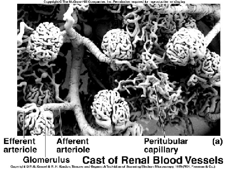
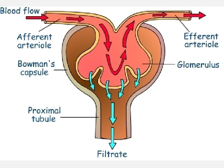
1. Afferent Arteriole: this arteriole enters the bowman’s capsule Arteriole from the renal artery. Renal artery Renal vein
2. Glomerulus: the capillaries of the glomerulus are porous and Glomerulus porous very twisted up. They sit within the Bowman’s capsule Renal artery Renal vein
Bowman’s Capsule
3. Efferent Arteriole: this arteriole leaves the bowman’s capsule. Arteriole capsule The blood is very hypertonic as most of the plasma has left the blood. The blood carries RBC, WBC, platelets and blood proteins. Renal artery Renal vein
4. Peritubular Capillaires: these are the capillaries that surround Capillaires the Proximal and Distal convoluted tubules Renal artery Renal vein
5. Vasa Recta: these are the capillaries that surround the Loop of Recta Henle. Renal artery Renal vein
1 2 The Nephron has 4 main functions: 3 4
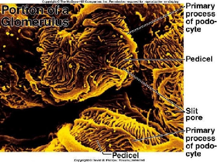
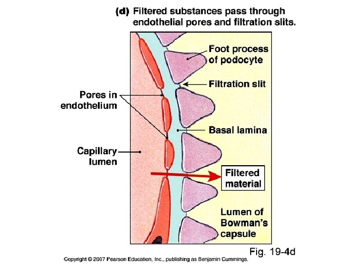
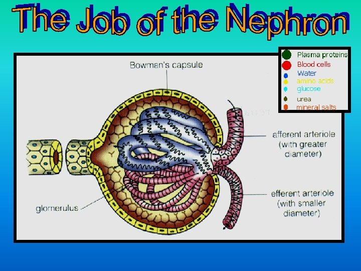
1. Pressure Filtration: the blood pressure forces water and Filtration solutes through the pores of the glomerulus into the Bowman’s solutes capsule. The holes in the glomerulus are small, so they are permeable to small water and small solutes, but not to blood cells or blood proteins The filtrate contains a mixture of solutes such as salts, nutrients, water and other small molecules At this point, the concentration of substances in the blood plasma is equal to that of the filtrate
Blood plasma in glomerulus Filtrate in Bowman’s Capsule salts, nutrients (glucose, fatty acids, glycerol, nucleotides, amino acids), water, hormones, antibodies, water soluble vitamins, penicillin and other drugs, histamines, nitrogenous wastes (urea, uric acid…) and other small molecules.
Composition of Plasma, and Glomerular Filtrate Urea Glucose Amino acids Water Plasma 0. 03 0. 10 0. 05 52. 0 Filtrate 0. 03 0. 10 0. 05 52. 0 Salts (ie: Na+, Cl-) 0. 9 Protein RBC, WBC, Platelets 8. 0 Lots None
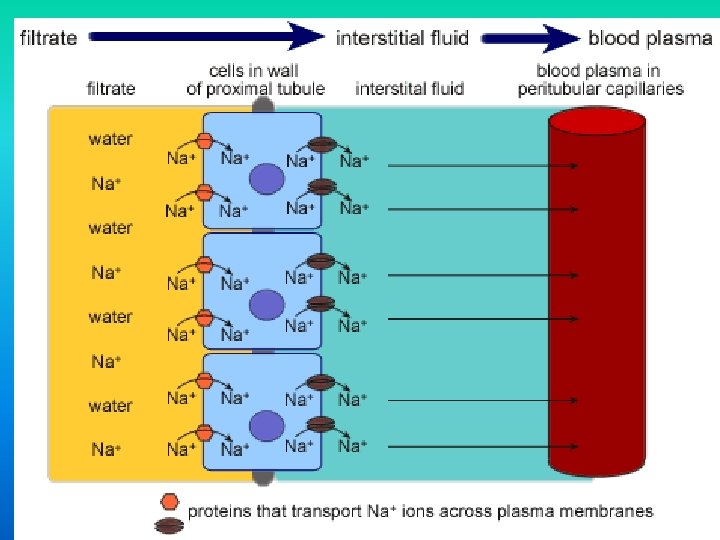
2. Selective Reabsorption: since filtration is nonselective, the Reabsorption nonselective body MUST make sure that the important molecules are returned to the blood plasma as soon as possible. returned Nearly all of the nutrients (esp. sugar), salts and salts water are reabsorbed. water This happens at the PCT and it is a very selective process. There a lot process of mitochondria in the mitochondria cells of the PCT. Why? This costs ENERGY!! ENERGY
WHAT IS REABSORBED? : 70% of the water, and 75% of the 70% 75% salts, glucose, amino acids, and other nutrients move back into the blood. The positive sodium ions are actively transported into the interstitial fluid and the negative chlorine ions follow passively, and finally, water follows by osmosis The glucose, amino acids, and other nutrients are actively transported into the blood. Lots of ATP required!! ATP
Recall that osmosis is the movement of water (from [High] to [Low])across a selectively permeable membrane. This movement occurs whenever two solutions separated by a membrane differ in total solute concentration or osmolarity The osmolarity of human blood is ~300 mosm/L.
When two solutions differ: Hyperosmotic The one with the greater concentration of solutes (salt, urea) is HYPEROSMOTIC Hypo-osmotic The one with the more dilute (watery) solution is HYPOOSMOTIC Hyperosmotic Remember: Water always moves towards a hyperosmotic environment. SALT SUCKS!! SUCKS Changes in the body fluids are controlled by a variety of regulatory mechanisms, usually involving negative feedback The control of water gain/loss is called OSMOREGULATION
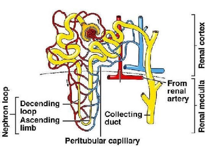
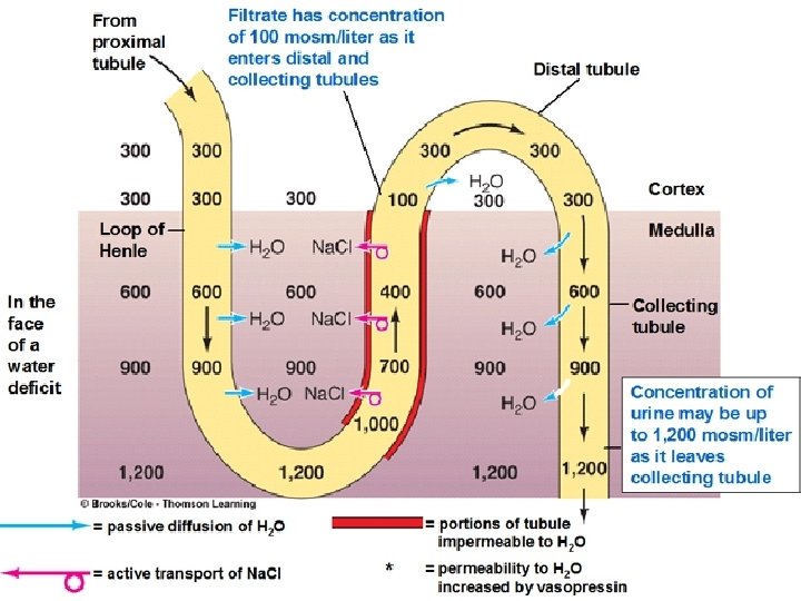
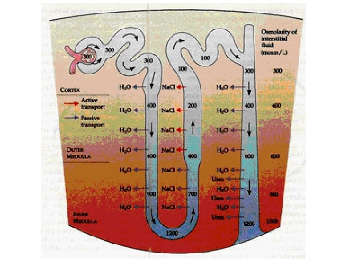
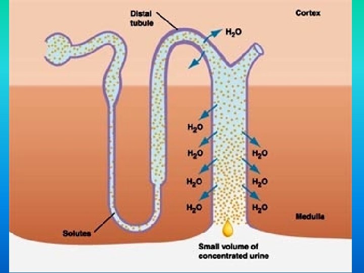
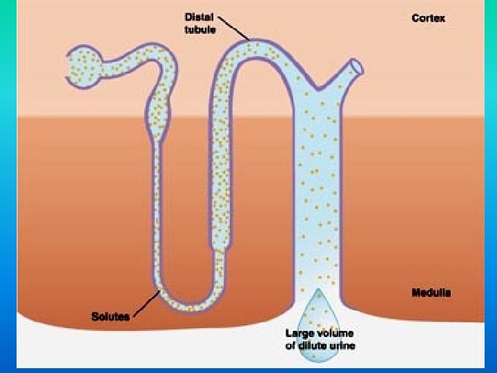
3. Water Reabsorption: water reabsorption occurs mainly at the Reabsorption loop of henle and in the collecting duct. The nephron pumps out salt and urea in these areas to make the blood and medulla extremely hyperosmotic Water then moves back in to the blood by osmosis
It is important to note that the blood in the vasa recta blood moves in the OPPOSITE DIRECTION that the filtrate is moving. This allows the nephron to create a hyperosmotic environment around the descending loop of henle So we’ll talk about the ascending Loop of Henle 1 st.
The ASCENDING LOOP OF HENLE: This part of the loop of henle is NOT PERMEABLE to water, but the salt can move. water move So, the Na. Cl passively and actively moves out Na. Cl of the tubule and into the blood and the medulla of the kidney. This creates a very HYPERTONIC solution in the blood. As the filtrate moves towards the DCT, most of the salt has left and the filtrate becomes more and more dilute
The DESCENDING LOOP OF HENLE: HENLE The loop of henle continues the reabsorption of water as the filtrate moves from the cortex and down into the concentrated medulla. The epithelium of this portion of the renal tubule is permeable to water, but not very permeable water to salt and other solutes. Lots of WATER moves into the HYPERTONIC blood (created at the ascending loop) by osmosis as it follows the salt. As a result of this great loss of water, the filtrate water becomes more and more concentrated
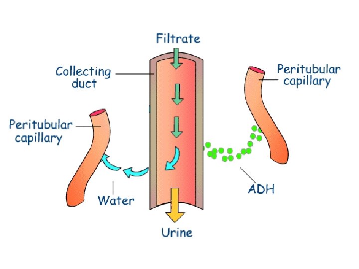
The COLLECTING DUCT: DUCT Water is also reabsorbed here. The epithelium of this duct is permeable to water and urea As the duct moves down into the VERY HYPERTONIC medulla, the filtrate loses more and more water by osmosis as it moves back into the blood. This loss of water concentrates the urea in the filtrate, and because there is so much UREA in the collecting duct, some of it duct diffuses down its concentration gradient diffuses and back into the blood and medulla.
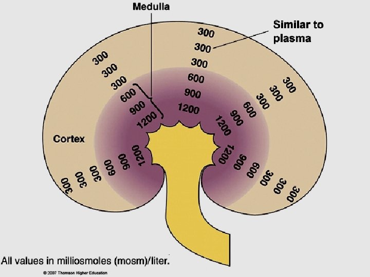
The kidney is made up of two sections: the cortex and the medulla The medulla is HYPEROSMOTIC as it has high concentrations of the solutes Na. Cl and urea renal medulla HYPERTONIC renal cortex
4. Secretion: if the blood still needs to rid itself of other ions and Secretion waste products (hydrogen ions, ammonia, potassium ions), it will secrete them into the Distal Convoluted Tubule (DCT). This is a very selective process that involves both active and passive transport. Lots of mitochondria in the DCT too! ATP required!!! mitochondria
REVIEW OF NEPHRON: http: //www. youtube. com/watch? v=c 05 m. Jae. IQu. Y
Composition of Plasma, Glomerular Filtrate, and Urine (g/100 ml of fluid). Plasma Filtrate Final Urine % Reclaimed Urea Glucose Amino acids Water 0. 03 0. 10 0. 05 52. 0 Salts (ie: Na+, Cl-) 0. 9 Protein RBC, WBC, Platelets 8. 0 Lots None 50% 100% 99% < 0. 9 -3. 6 99. 5% 1. 8 None 1. 0 None
If the blood is ACIDIC, 2 things will happen: HCO 3 H+ 1. The bicarbonate ions are actively transported back into the blood The HCO 3 - is an important buffer which will join with H+ to remove it from the solution. 2. The blood will also secrete hydrogen ions into the filtrate so we will pee it out
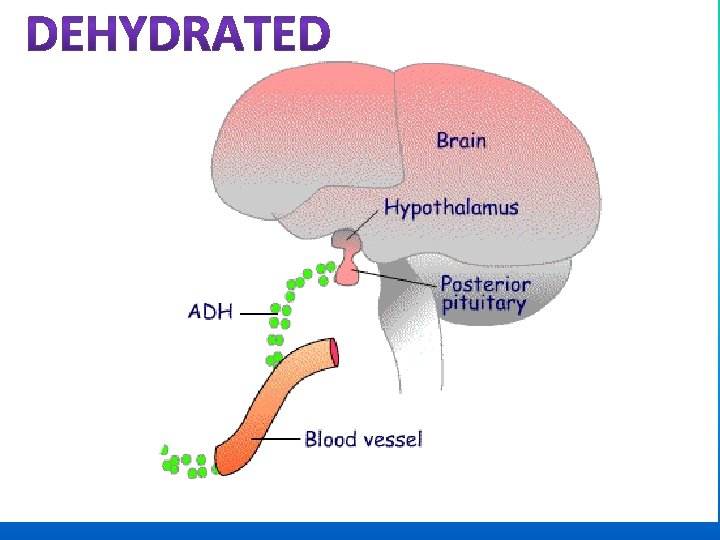
If you are dehydrated, the kidneys can excrete a small, dehydrated concentrated volume of urine and reabsorb the majority volume reabsorb of water from the filtrate. You will have a very small water amount of dark yellow urine. If you have consumed an excessive amount of fluid, the excessive fluid kidneys can excrete a large, dilute volume of urine, with very little water being reabsorbed from the filtrate. You will have lots of pale yellow urine. How do we maintain the proper amount of water in our bodies? It is controlled homeostatically with 2 hormones
FIRST HORMONE: ANTIDIURETIC HORMONE (ADH) This hormone controls water reabsorption with a negative feedback cycle. The hypothalamus makes ADH and it is stored and released from hypothalamus the posterior pituitary gland It is released in response to an increase in the osmolarity of the blood (very concentrated blood) when you are dehydrated (inadequate intake of dehydrated water).
When you have low blood volume (high osmolarity), the osmoreceptor cells in the hypothalamus triggers the posterior pituitary to release ADH increases the permeability of the DCT and collecting ducts to water. This causes more water to be reabsorbed and ultimately increases blood volume (amount of blood). volume At the same time, the removal of water from the filtrate increases the concentration of the urine. You urinate less and it will be dark yellow.
When you have high blood volume and low osmolarity, sensors in the heart signal the hypothalamus to cause a reduction of the amounts of ADH in the blood. ADH This decreases the amount of water that is reabsorbed into the blood, and large quantities of a more dilute urine are produced. This is a negative feedback cycle, because as the osmolarity of the cycle blood is reduced (blood volume increases) less ADH will be secreted.
Alcohol can perturb water balance by inhibiting the release of Alcohol ADH, causing excessive loss of water in the urine and dehydrating ADH the body. This leads to the symptoms of a hangover. Which is why some hangover people drink a lot of water when drinking alcohol. This keeps them hydrated.
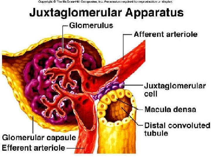
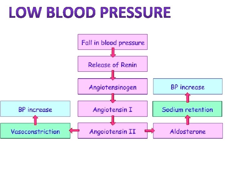
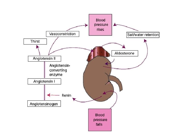
Second Hormone: ALDOSTERONE There is tissue surrounding the afferent arteriole called the juxtaglomerular apparatus (JGA) which measures Blood Pressure + ] in • When the BP drops, or if the [Na drops the blood is too low (due to diarrhea, a severe injury…), the JGA releases an enzyme called RENIN • The liver makes a protein called ANGIOTENSIN which is always circulating around the blood. • RENIN activates angiotensin
The active form of ANGIOTENSIN does two things: 1. It stimulates the adrenal gland to release the hormone ALDOSTERONE Aldosterone tells the DCT to reabsorb lots of Na+, and water follows the sodium by osmosis. This increases the amount of water in the blood, which increases the BP. 2. It also causes VASOCONSTRICTION of the VASOCONSTRICTION arterioles, which also raises blood pressure
Why are the kidneys so concerned with the BLOOD PRESSURE? Low blood pressure is bad because there is not enough pressure to filter the blood properly. This makes the kidneys less efficient at ridding the body of drugs and toxins. High blood pressure is bad because the glomerulus can burst and then we would see blood and blood proteins in the urine.
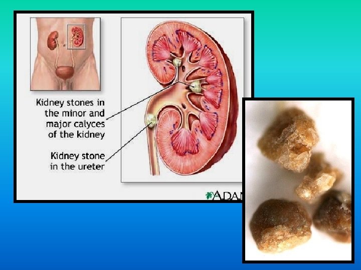
Bladder infections - usually caused by bacteria. More common in women due to their short urethra. 33% of women and 10% of men will have one or more in their lifetime. Enlarged prostate - in men, this can make it difficult to empty the bladder. Incontinence - when urine leaks out of the urethra. Common after natural childbirth. Kidney infections - when a bladder infection ‘backs up’ the ureters and makes it difficult to urinate. Kidney stones - caused by infection and high blood levels of calcium.
Symptoms of kidney disease Kidney disease is called a ‘silent disease’ as there are disease often few symptoms. Some signs and symptoms include: • Pee more and more often especially at night • Blood in the urine • Foaming urine • Puffiness around the eyes and ankles • Pain in the back (under the lower ribs, where the kidneys are located) • Pain or burning when passing urine. KIDNEY TRANSPLANT ANIMATION http: //www. kidneypatientguide. org. uk/site/TRAanim. php
You are more ‘at risk’ of chronic kidney disease if you: • Have diabetes • Have high blood pressure • Are obese • Are over 50 years of age • Have a family history of kidney disease • Smoke • Are of Aboriginal descent.