Excitable tissue Muscle physiology Dr A K Dwivedi

Excitable tissue : Muscle physiology Dr. A. K. Dwivedi

Muscle physiology Introduction: Nature in its supreme wisdom has therefore given muscles about half a man’s body weight. Locomotion is achieved by skeletal muscle so called they are attached to bones and bring about movement at joints. Muscle cells like neurons can excited chemically, electrically and mechanically to produce an action potential that is transmitted along there cell membrane, unlike neurons they have a contractile mechanism that is activated by the action potential. The contractile proteins actins and myosin are abundant in muscles where they bring about

muscles may contract voluntarily or involuntarily. All muscles movement results from contraction since movement is possible in opposite direction when a group of muscle contract, the opposing group of muscle must relax.

Classification of muscles : Basis of classification There are three methods to classify the muscles. The basis of this classification is as follows : 1. Depending upon the presence or absence of striations 2. Depending upon the control and 3. Depending upon the functions Depending upon striations : depending upon the presence or absence of the cross striations, the muscles are derived into two groups namely

a. Striated muscle and b. Non striated muscle Depending upon the control, the muscles are classified into two types namely : a. Voluntary muscle and b. Involuntary muscle Depending upon the function : The muscles are classified into three types depending upon the function : a. Skeletal muscle. b. Cardiac muscle. c. Smooth muscle.

Functions of muscle tissue 1. Body movements : total body movements like walking, running or fine movement like writing, holding the pen or holding light or heavy objects is integrated functions of bones, joints and skeletal muscles. 2. Stabilizing body position : skeletal muscle contraction stabilize joints and help to maintain body positions such as sitting. Postural muscles contract continuously when a person is awake, for eg : sustained contraction in neck muscle hold the head upright. 3. Regulating organ volume : storage of food, :

4. Moving substance within the body : cardiac muscle contract and pump blood throughout the body through the vessels contraction and relaxation of smooth muscles in body help to regulate the rate of flow. smooth muscles contract and also move by and other enzymes through gastro-intestinal tract. 5. Generating heat : as the muscle tissue contract they produce heat, this is used to maintain body temperature. Involuntary contraction of skeletal muscle is called as shivering can increase the late of production of heat many lines. It is a type of metabolic activities.

Properties : Muscle tissue have four special properties : 1. Electrical excitability : property of both muscle and nerve cells, is the ability to respond to certain stimuli by producing electrical signals such as action potential. Action potential can propagate along a cells plasma membrane due to the presence of specify voltage gated channels for muscles cells. Two main types of stimuli trigger action potential one is the auto systemic electrical signals arising in the muscle tissue itself such as occurs in the heart pacemaker. The other is chemical stimuli, such as neurotransmitter released by neurons, hormones distributed by the

2. Contractibility : it is ability of muscle tissue to contract forcefully when stimulated by action potential. When muscle contract, generates tension ( force of concentration) while pulling on its attached points. In an isometric contraction (iso-equal, metricmeasure or length ). The muscle developed tension but does not shorten. An e. g. : holding a book is an out stretched hand. If the tension generated is great enough to overcome the resistance of the object to being moved. The muscle shortens and movement occurs. In an isotonic contraction ( tonic – tension ), the tension developed by the muscle remain almost constant while the muscle shortens. An e. g. : lifting a book from a table.

3. Extensibility : it is the ability of muscle stretch without being damaged. Extensibility allows a muscle to contract forcefully even if it is already stretched. Normally, smooth muscle is subject to the greatest amount of stretching. For e. g. : each time the stomach fills with food, the muscle in its wall is stretched. Cardiac muscles also is stretched is each time the heart fills with blood. During normal activities, the stretched on skeletal muscle remains almost constant 4. Elasticity ; it is the ability of muscle tissue to return to its original length and shape after contraction or extension.

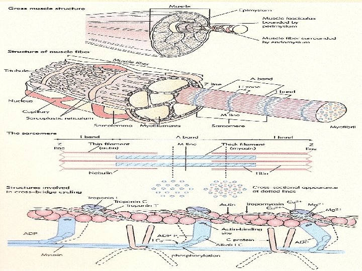
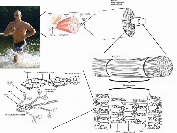
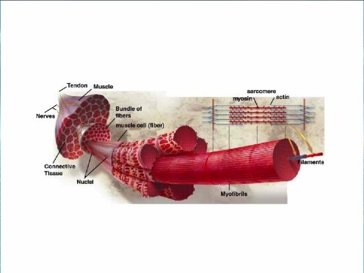
Description of muscle 1. Skeletal muscle Introduction : skeletal muscle is made up of individual muscle fibers that are the ‘building block’ of the muscular system in the same sense that the neurons are the building block of the nervous system. The skeletal muscle fibers are multinucleated cylindrical structures having a clear display of longitudinal and cross striations. Most skeletal muscle began and end in tendons, and the muscle fibers are arranged in parallel between the tedious ends, so that the force of contraction of the units is additive.

Distribution : These muscles mostly in all instances are attached to osseous tissue, innervated with somatic nerves through which voluntary control is performed. In the fresh state the human skeletal muscle is pink in colour due to the presence of muscle pigments and high vascularity. Due to variations in colour there are red and white muscles. In most skeletal muscles, each fiber extend the entire length of muscle, except for about two percent of the fibers. Each fiber is innervated by only one nerve ending, located near the middle of the fiber.

Types of skeletal muscle There are two types of muscle fibers – red and white with intermediate varieties between the extremes red, white and intermediate fibers are present within a single muscle. However, some muscles have predominantly red fibers and some predominantly white. 1. Red fibers : red fibers are small in diameter and rich in mitochondria and myoglobine. Their red colour to the higher content of myoglobin which is reddish brown in colour. Their speed of contraction is low and they contract less forcefully but do not get

2. White muscle fibers : they are larger in diameter then red fibers, have fewer mitochondria, less myoglobine and a higher velocity of contraction. They contract more forcefully but fatigue quickly. There is evidence to suggest that the muscle fiber type is determined by the motor nerve fibers.

surrounds and protect muscle tissue. Three layers of connective tissue extend from the deep fascia to further protect and strengthen skeletal muscle. The outer most layer including the whole muscle is the “epimysium” , “perimysium” surrounds groups of 10 -100 or more including muscle fibers, separating them into bundles called fascicles many fascicles are large enough to be seen with the naked eye, they give a cut of meat its characteristics “grain” and if you tear a piece of a meat , reps a part along the fascicles. Both epimysium and perimysium are dense irregular connective tissue. Penetrating the interior of each fascicle and separating individual muscle fiber from one another

Is “endomysium”, a then sheath of areolar connective tissue Histology The skeletal muscle fiber are cylindrical elongated cells with multiple nuclei. The extend of muscle fiber in the bulk may be form – a. One end to the other b. One end somewhere at midway or c. Both the ands within the muscle having no attachment with either side. The length and breadth of the muscle fiber vary from 1. 0 -40 mm and 0. 01 mm to 0. 1 mm respectively.

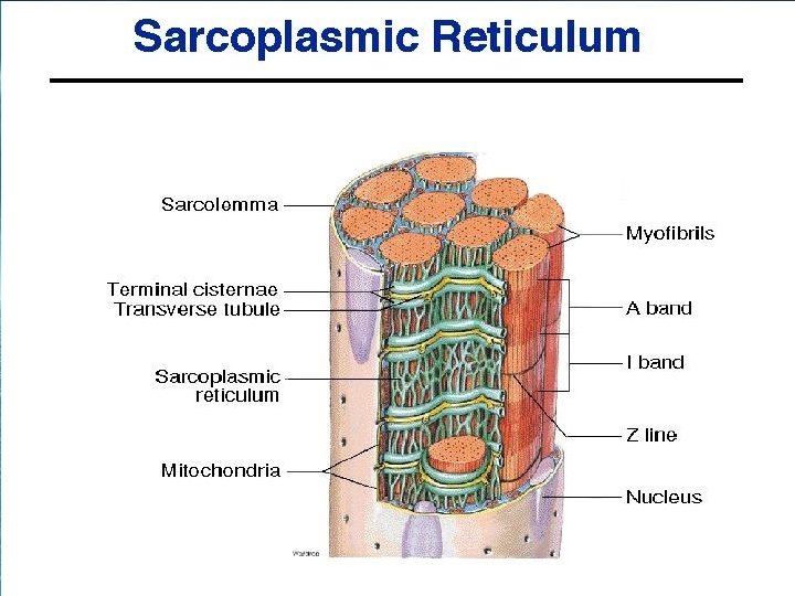
The transparent cell wall of the muscle fiber is named as “sarcolemma” under light microscope it is visible when fresh muscle fiber are teased electron micrographs shows that it is made up not only of the plasma lemma but also of an extrinsic coat of amorphous layer is and by reticular fibers. The plasma lemma is of same structure as that of reticular fiber. The sarcoplasm contains other constituent as that of any other cell e. g. . Numerous mitochondria, a small gorge apparatus near each nucleus, myoglobin, lipid, glycogen, sarcoplasmic reticulum etc. fiber are riches in sarcoplasma.

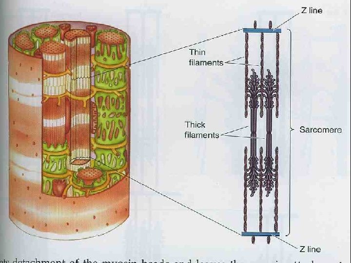
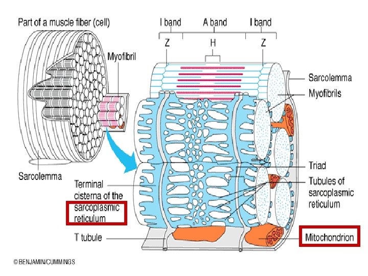
Myofibrils : The characteristics feature of skeletal muscle, the alternate legit and dark shades and thick longitudinal strands can be studied with light microscope. Electron microscope reveals that the longitudinal striation is due to the presence of myofibrils of different thickness where case the transverse striation is due to presence of alternate light and dark segments of longitudinally arranged. In cross section myofibrils appear as fine dots either in a group poly gonad areas and are separated from adjacent bundle by clear sarcoplasm. The separated myofibrils by the sarcoplasmic known as fields of
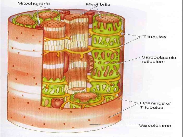

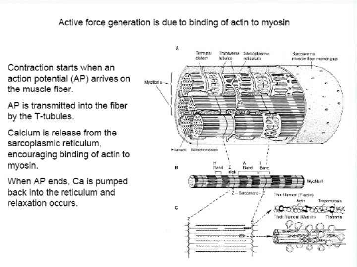
Sarcotubular system : under electron microscope, the myofibrils are seen to have surrounded by a canalicular network of membrane limited tubules – “Sarcoplasmic reticulum” is identical with the enoplasmic reticulum of other cell type but with the difference that its membrane does not possess ribosome. The sarcoplasmic reticulum is extended longitudinally along the A- band with frequent anastomosis in the region of the H- band also in the I- band. The sarcoplasmic reticulum is connected at its both longitudinal and terminal ends by another set of the transvere cisterns – terminal cisterns. The terminal cistern have got larger calibre and are thus continsous and confluent with the longitudinal

H Band

each other by a slender transverse tubule which is known as T- tubule. This T- tubule is not confluent the terminal cisternae and is a tubular invagination of the sarcolemma but not a part of sarcoplasmic reticulum. It is continuous with the extra cellular space. These tubules are generally called as T- system. The pair of terminal cisternae and the central T-tubules are collectively called as “triads”. In amphibian muscle the triads encircle the I- band at the region of the Z- line but in mammalian muscle, the same is present at the junction of each A- band with the adjacent I- band so in mammals are there two sets of triads in each sarcomere. The T- system plays an important role in quick transmission of impulse from the cell surface to

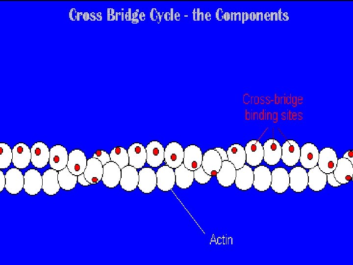
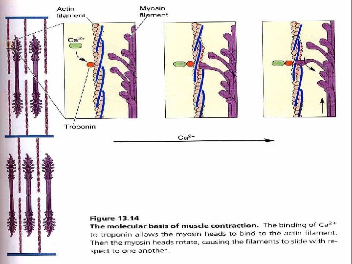
Muscle protein : Myofibrils are built from three kinds of proteins : 1. Contractile proteins, which genrate force during contraction. 2. Regulatory protein which help switch the contraction process on and off and 3. Structural proteins, which keep the thick and thin ligaments in the proper ligament, give the myofibril elasticity and extensibility and link the myofibrils to the sarcolemma and extracellular matrix. the two contractile proteins in muscle are “myosin and actin” which are the main components of thick and thin filaments, respectively myosin function as a motor protein in all three types of muscle tissue.
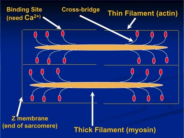

Motor proteins push or pull their cargo to achieve movement by converting the chemical energy in ATP to the mechanical energy of motion of force production. In skeletal muscle, about 300 molecules of myosin from a single thick filament each myosin molecules of is shape like two golf clubs handle points towards the M – line in the center of the sarcomere. The two projections of each myosin molecules are called myosin head or cross bridge. The head project outward from the shaft in a spilling fashion, each extending towards one of the six then filament that surround each thick filament.

Z discs. Their main component is protein actin. Individual actin molecules four to form an actin filament that is twisted into one each actin molecules is a myosin binding site, where myosin head can attach smaller amount of two regulatory protein – “Tropomyosin and Troponin” are also part of their filament. In relaxed muscle myosin blocked from binding to actin because strands of tropomysin could the myosin binding sites on actin. The tropomysin strands, in turn are held in place by Troponin molecules. Beside contractile and regulatory proteins, muscle contains about a does not structural proteins which contractile to the alignment, stability, elasticity and extensibility of myofibrils.

Binding Site Troponin Tropomyosin

Myosin

Several key structural proteins are titan myosin and dystrophic. Titan ( titan – gigantic ) is the third most plentiful protein in skeletal muscle. This molecules name reflects its huge size. with a molecular weight of about 3 million Daltons , tint is 50 times larger then an average size protein.

Titin anchors a thick filament to both a Z disc and the M- line. There by helping stabilize the position of the thick filament. The part of the titin molecule that extends from the Z disc to the beginning of the thick filament is very elastic. Because it can stretch to at least four times its resting length and then spring back unharmed, titin accounts fore much of the elasticity and extensibility of myofibrils. Titin probably help the sarcomere return to its resting length after a muscle has contraction or been stretched. Molecules of the protein myosin form the M-

And connect adjust thick filament s to one another. Dystrophin is a cytoskeletal protein that links thin filament of the sarcomere to integral membrane proteins of the sarcolemma. In turn the membrane proteins attach to protein in the connective tissue matrix that surrounds muscle fiber. Hence, Dystrophin and its associated protein are thought reinforce the sarcolemma and help transmit the tension generated by the sarcomeres to the tendons.

General mechanism of contraction Introduction : Muscle fibers cells are connected with connective tissue. When the muscle fibers shortens, the pull is , there fore transmitted through the connective tissue to the bone and there in flexion / extension etc movement of the joint. Finer details : at rest is when the muscle is in a state of relaxation : 1. H- zone is wide 2. I- bands are wide ( because good deal of thin filament is not overlapped by the thick filament in the

Contracted state of muscle 1. The H- zone greatly narrowed or disappear 2. The width of the band is reduced but the width of the A- band remain we changed. Thus in each sarcomere the I- bands becomes narrows -> the muscle fiber as a whole short ends -> the muscle belly as a whole shortens. Explanation : as the muscle begins to contact , the thin filaments starts to move -> the thin filaments move towards the H- zone of a sarcomere move towards the H- zone.

1. H- zone becomes obliterated , 2. I- band is narrowed 3. The sarcomere as a whole is shortened. Types of contraction 1. Isometric contraction : means contraction on which there is no change in length of muscle, but there is increase in tension. 2. Isotonic contraction : means contraction in which there is change of length at constant tension. The tension is equal to the weight lifted during contraction of the muscle.

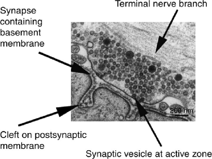
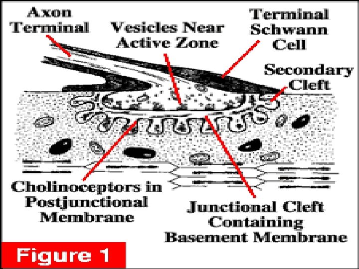
Neuromuscular junction The skeletal muscle fibers are innervated by large, myelinated nerve fibers that originated in the large motoneuron of the anterior horns of the spinal cord. As pointed out in nerve fiber, after interning the muscle belly , normally because and stimulates from three to several hundred skeletal muscle fiber. Each nerve ending makes a junction called the “Neuromuscular junction” , with the muscle fiber near its midpoint, and the resulting action potential in the muscle fiber travels in both direction towards the muscle fiber ends with excention of about 2% of the muscle fiber there is only one such function per muscle fiber.

Neuromuscular Junction

Spread of excitation : electrical excitation spreads to the interior of the muscle fiber by means of the transversely oriented T- tubules which are formed by invagination of the sarcolemma since T- tubules aig. Deep into the muscle fiber encircle every myofibril and are repeated at every A-I junction. They conduct the action potential throughout the length and breadth of the muscle fiber within a very short time.

Role of calcium in contraction and relaxation Calcium mediated the effect of excitation to the contractile proteins of muscle that is why inerea in the contraction of sarcoplasmic calcium leads to contraction. Relaxation is due to removal of calcium ions into the cisternae by active transport. An active calcium pump resides in the cisternal membrane which concentrates calcium ions in the cisternae. The pump is overpowered only movementasily during excitation by the opening up of a large membrane of calcium channels.

In order due to understand how calcium ions bring about contraction it is essential to learn more about muscle proteins.

Characteristics of muscle contraction One have so fore discussed muscle contraction at the level of a single sarcomere now we shall study some characteristics of the contraction of a whole muscle and examine how they can be explained in tern of function at molecular and microscopic level. This excuse will also lead to the discovery of some defects in the cross bridge and sliding filament theories of muscle contraction.

Z line

Mechanism of skeletal muscle Many mechanical characteristics of skeletal muscle were explained by means of to very similar models proposed by Hill in 1951 and Albert in 1956. This models are no longer very popular because of there short comings and because of over better understanding today of the real structure in muscle at molecular level but it is still quiet convenient to make these models becomes of their simplicity.

These are basically three components models in which skeletal muscle is believed to have contractile elements. Contractile elements are the filaments which being about contraction through their rearrangement , series elastic elements are mainly the tendons, and parallel elastic elements are the sarcolemmal and connective tissue element disposed parallel to muscle fiber.
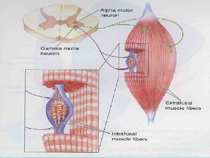

Motor unit : The unit of contraction Skeletal muscle are supplied by motor nerves. A motor nerve consist of several nerve fiber. Each motor nerve fiber is the axon of a motor neuron. The cell body of leis in the anterior column of the spinal cord. After entering the muscle belly, a motor nerve fiber supplies several muscle fiber. A motor neuron together with the all muscle fiber supplied by it is called “motor unit. ” the muscle fibers of a motor unit are scattered throughout the muscle belly. Therefore even when only the muscle fiber of one motor unit contract, the whole muscle

Motor Unit All the muscle cells controlled by one nerve cell

A muscle may be activated by activation of a variable number of nerve fibers since the unit of activation is a nerve fibers, the unit of contraction is a motor unit. The number of muscle fiber in a motor unit varies from to several hundred the precision with which the contraction of a muscle may be geaded depends on the size of the motor unit. For e. g. if a motor unit has 5 muscle fibers, number of muscle fibers activated at a time may 5, 10, 15, 20 and so on the other hand.

If a motor unit has 100 muscle fibers , the number of muscle fiber activated at a time may be 100, 200, 300, 400 and so on. The other hand muscle of lag have large motor units and there fore only poorly graded contractions. The size of motor units in a muscle seen to be related to the functional necessity for precision in grading of the contractive strength. Further all the motor units in a given muscle are not exactly equal. During weak contraction of a muscle, only small motor units are activated. Stronger contraction is achieved by recruitment of progressively larger motor units.

Length- tension relationship : The forcefulness of muscle contraction depends on the length of the sarcomere within the muscle before contraction begins at a sarcomere length of about 2. 0 – 2. 4 um. The zone of overlap in each sarcomere is optimal and the muscle fiber can develop maximum tension. As the sarcomeres of a muscle fiber are stretched to a longer length the zone of overlap shortens , and few of myosin heads can make contact with thin filaments.

So the tension, the fiber can produce decreases. when a skeletal muscle fiber is stretched to 170% of its optimal length, there is no overlap between the thick and thin filaments. Because none of the myosin heads can bind to thin filaments. The muscle fibers can not contract, and tension is zero. As sarcomere length become increasingly shorter than the optimum. The tension that can develop again decreases. this is because thick filaments. Crumple as they are compressed by the z discs, resulting in fewer myosin heads making contact with thin filaments.

Normally resulting muscle fiber length is held very close to the optimum by firm attachments of skeletal muscle to bones and to other inelastic tissue, so that our stretching does not occur.

Energetic of muscle contraction : Work output during muscle contraction : when a muscle contracts against a load, it perform work. This means that energy is transferred from the muscle to the external load, for eg, to lift an object to a greater height or to overcome resistance to movement. In mathematical terms work is defined by the following equation : W=l *d In which w is the work output, l is the load , and d is the distance of movement against the load , the energy required to perform the work derived from the chemical reactions in the muscle cells during contraction.

Skeletal muscle tone : Even when muscles are at rest, a certain amount of tautness usually remains. This is called muscle tone. Because skeletal muscle fibers do not contract without an action potential to stimulate the fibers , skeletal muscle tone results entirely from a low rate of nerve impulse coming from the spinal cord. This in turns are controlled partially by impulse transmitted from the brain to the appropriate anterior motor neurons and partly by impulses that originated in muscle spindles located in the muscle itself.

Motor unit recruitment : The process in which the number of active motor units increases is called motor unit recruitment. typically. , the different motor units in a whole muscle are not stimulated to contract in unison. While some motor units are contracting, others are relaxed. This pattern of motor units actively delays muscle fatigue by allowing alternately contracting motor units to relieve one another. In this way , contraction of a whole muscle can be sustained for a long period. The weakest motor units are recruited first, with progressively stronger motor units being added if the task requires more force.

Recruitment is one factor responsible for producing smooth movements rather than a series of jerks. As mentioned, the number of muscle fibers innervated by one motor neuron varies greatly, precise movements are brought about small changes in muscle contraction. Therefore, the muscles that produce precise movement are made up of small motor units for this reason when a motor unit is recruited of turn of only slide changes occur in muscle tension. By contract, large motor unit are active where large tension is needed and precision is less important.

Energy supply for muscle contraction : We have seen that the immediate source of energy for muscle contraction is ATP. But the ATP used must be replenished promptly if the contraction has to continue for any length of time because all the ATP in a muscle can provide energy for muscle contraction for only LS. The immediate source of replenishment of ATP is creative phosphate.

Creatine phosphate + ADP --> creatine + ATP. Creatine phosphate is stored in muscle set depleted completely in just 3 s. If the muscle is contracting maximally for replenishment of creatine phosphate and APP the next fuel, which can last much longer is glucose. A muscle can get glucose from two source : from the blood flowing through the muscle and from the breakdown of glycogen stored in muscle can also last only a few minutes. Therefore the only steady source of glucose is blood glucose which in turn is replenished is the post absorptive state by the breakdown of liver glycogen.
 may be divided into two")
The breakdown of glucose into lower energy compounds (glycolysis) may be divided into two stapes. One stage up to the formation of the 3 – carbon compound pyruvic acid yields only two molecules of ATP for each molecule of glucose broken down. But this stapes does not need oxygen and is therefore also known as aerobic glycolysis. Aerobic glycolysis yields much more energy than anaerobic glycolysis another 34 molecules of ATP. Thus a total of 36 molecules of ATP is obtained from the oxidative breakdown of each molecule of glucose. The body stored of glucose and glycogen also cannot sustain heavy exercise beyond 100 min.

The reason why we can continue with exercise much longer is because the body has yet another fuel in reserve ie fat. The fat stored in muscles and else where can be broken down to yield free fatty acids which are a concentrated source of energy. Theoretically, amino acids can also be broken down to live energy they are spared for protein synthesis. It is only in starvation conditions that protein reserves of the body are drawn upon for providing energy.

To summarize there is a chain of fuels to replenish the ATP which is broken down for muscle contraction there is considerable overlap in the stapes at which different links in the chain become operative. For example even brief bursts of contraction for which ATP and creatine phosphate. Longer exercise for which enough glucose is available in the body are also partly energized by breakdown of fat. As the duration of exercise increases, the proportion of energy provided by fat increases and that provided by carbohydrates decreases.

fat are a more concentrated source of energy is that they provide more calories per gram but they are less efficient in that they provide less energy per liter of oxygen consumed. Also the maximum rate of energy production in case of fats than carbohydrates. That is perhaps the reason why the body show slight preference for using carbohydrates as compared to fats. In general the rate of energy production by a fuel smaller is the amount stored in the body with high rate of energy production serve as immediate source of fuel where as those of lower rates of energy production serve as energy stores.

Heat production in muscle : From a biochemical point of view, heat production by a tissue is a by product of energy expenditure. Energy is spent primarily for staying alive and for staying alive for specific function of the tissue concerned. But since the utilization of energy is not perfect part of the energy released in the tissue is dissipated as heat. Muscle is a tissue the energy expenditure of which differs markedly at rest as compared to that during activity. Therefore although an un stimulated muscle produces heat, the heat production increases during and immediately

The amount of heat produced is small during any phase but is of great theoretical and historical interest. The heat production was first measured very accurately by A. V hill in 1939, long before the molecular basis of contraction has been worked out. On the basis of heat production hill had deduced many aspects of muscle function which have been borne out by subsequent research. Heat produced during different stapes of rest and activity is designated by specific terms, some of which are confusing or ambiguous.

Resting heat : Resting heat is the heat produced in stimulated muscle before shortening. It is likely to be a by product of energy spent on released of calcium from the terminal cisternae binding of calcium by the sarcoplasmic reticulum. The last named process begins as soon as the calcium released and accounts for about half the activation heat. There is also a term, initial heat, which is best avoided because some author used it as a synonym for activation heat while others use it to imply the sum of activation heat and shortening heat.

Shortening heat : As the name indicates this is the heat associated with shortening. The amount of shortening heat depends on the degree of shortening and the velocity of shortening. Since there is no shortening in isometric contraction, there is no shortening heat associated with it. shortening heat seems to be a by product of the energy spent on ualchet mechanism involving myosin cross bridges and the achieve sites on action filaments.

Maintain heat : Maintain heat is the heat produced during tetanus. It is partly made of activation heat associated with each stimulus and partly of the heat produced due to actin myosin interaction.

Relaxation heat : As the name indicates, relaxation heat is associated with relaxation. It is due to the energy expenditure associated with the uptake of calcium by the terminal cisternae.

Recovery heat or delayed heat : This is due to the over and above the resting heat – after contraction and relaxation are over. This is due to the restoration of the resting state. Replenishment of the energy stored of the muscle and correction of the slight imbalance of in sodium and potassium concentration brought about by the action potential need energy expenditure. Consequently this process also lead to dissipation of heat. Although these process soon side by side with contraction, they also continue for some time afterwards,

Neuromuscular Junction. : The functional region between the motor nerve fiber and the corresponding skeletal nerve fiber is called neuromuscular junction. A typical neuromuscular is seen only in the skeletal muscle, smooth muscle or cardiac muscle do not have such typical structure.
 it generated a")
Load and Tension When a muscle contract ( Isotonic or Isometrically) it generated a force. This force is called tension. This tension is applied to an object which is moved (lifted). The resistance offered by the object is called the load and is expressed in engineering units. E. g. kg wt is mass in kg x gravity ) To take a concrete example assume that the biceps ( flexor of elbow joint ) is contracting. When it contracts, the weight of the forearm ( i. e. the mass of forearm x gravity)

opposes the shortening of the biceps, therefore the wt of the forearm is the load against which the tension generated by the contraction of the biceps is working. Muscle Twitch : - A single action potential causes a brief contraction followed by relaxation. This response is called a muscle twitch. The action potential and the twitch are plotted on the same time scale. The twitch starts about 2 m after the start of depolarization of the membrane before repolarization is complete.

the duration of the twitch varies with the type of muscle being tasted “fast” muscle fibers, primarily those concerned with fine, rapid. precise movement have twitch duration as short as muscle “slow” muscle fibers principally. Those involved in strong, gross, sustained Movements, have twitch duration up to 100 ms. The twitch contraction is the brief contraction of all the muscle fibers in a motor unit in response a single. Action potential in its motor neuron of its muscle fibers.

The record of muscle contraction called a myogram is shown in twitches of skeletal muscle fibers last anywhere from 20 to 200 on sec. this duration is very long compared to the brief 1 to 2 m sec duration of an action potential. Note that a brief occurs b/w application of the stimulation (time zero on the graph) and the beginning of contraction. The delay, which lasts, about two milliseconds, is termed the latent period. During the latent period, calcium ions are being related from the sarcoplasmic retinaculum, the filament start to exert tension.

Others, such as those that move the legs are slowtwitch fibers, with contraction and relaxation period of about m sec each. If two stimuli are applied one immediately after the other, the muscle well respond to the first stimulus but not to the second, when a muscle fibers receives enough stimulation to contract. It temporarily loses its excitability, called the refractory period, is a of all muscle and nerve cells. The duration of the refractory period varies with the muscle involved.

skeletal muscle has a short refractory period of about five milliseconds, whereas cardiac muscle has a long refractory period of about 300 milliseconds. Types of contraction : - Isotonic contraction are used for body movements and for moving objects. The two types of isotonic contraction are concentric and eccentric. In a concentric isotonic contraction a muscle shortens and pulls on another structure such as a tendon, to produce movement and to reduce the angle at a joint.

picking a book up off a table involves concentric isotonic contraction of the biceps brachia muscle in the arm. By contract, as you lowers the book to place it back. On the table the previously shortness biceps lengthens in a controlled manner while it continues to contract. when the length of a muscle arises during a contraction the contraction is an eccentric isotonic contraction. During an eccentric contraction, the tension exerted by a myosin cross bridges resists movements of a load (the book in this case) and slows the lengthening process.

for isotonic contraction produce more muscle damage and more delayed – onset muscle soreness than to concentric isotonic contraction. In isometric contractions, the myosin cross bridges generate considerable tension but the muscle doesn’t shorten because the force of the load equals the muscle tension. An example would be holding a book steady using an outstretched arm. These contraction are important for maintaining posture and for supporting objects as a fixed position.

although isometric contractions. Do not result in body movement, energy is still expanded. The book pulls the arm downward stretching the shoulders and arm muscles. The isometric contraction of the shoulder and arm muscles counteracts the stretch. Isometric contraction are important because they stabilize. Some joints as others are moved. Most activities include both isotonic and isometric contraction.

Benefits of stretching : The overall goal of stretching is to achieve normal range of motion of joints and mobility of soft tissues surrounding the joints. For most individuals, the best stretching routine involves static stretching, that is, slow sustained the muscle should be stretched to the point of slight discomfort ( not pain ) and held for about 15 to 30 seconds. Stretching should be done after warming up as this will increase range of motion, among the benefits of stretching are

1. Improved physical performance : a flexible joint has the ability to move through a greater range of motion, which improves physical performance. 2. Decreased risk of injury : stretching decreases : resistance in various soft tissues and thus there is less likelihood of exceeding maximum tissues extensibility during all activity. This decreases the risk of injury.

3. Reduced muscle sources : stretching can reduce some of the muscle soreness that results from delayed on set muscles soreness (DOMS) after exercise. 4. improved posture : poor posture results from improper posture and gravity over a number of years, stretching can help realign soft tissues to improve and maintain good posture.

Applied physiology : Rigor mortis : After death cellular membrane start to become leaky. Ca++ ion leak out of the sarcoplasmic reticulum into the cytosol and allow myosin heads to bind to actin. ATP synthesis has ceased. However , so the cross bridge can not detach from actin. The resulting condition, in which muscles are in a state of rigidity (can not contract or stretch) is called rigor mortis (rigidity of death).

Rigor mortis begins 3 – 4 hours after death and lasts about 24 hours, then it disappears as proteolytic enzymes from lysosomes digest the cross bridge. Aerobic training versus strength training : regular the supply of oxygen reach blood available to skeletal muscle for aerobic cellular respiration by contract, activities such as weight lifting rely more on anaerobic production of ATP through glycolysis. Such anaerobic activities stimulate synthesis of muscle proteins and result, over time, in increased muscle size.

As a result aerobic training builds endurance for prolong ate activities, where as aerobic training builds muscle strength for short term feat. Internal training is a work out regimen that incorporates both types of training for example alerting sprints with jogging. Anabolic steroids : the illegal use of anabolic steroids by athletes has received wide spread attention. This steroid hormones, similar to testosterone, are taken to increase muscle size and thus strength during athletic contests.

The large doses needed to produce an effect. Hormones have damaging , some times even demonstrating side effects, including lever cancer , kidney damage increased rest of heart disease, stunted growth and increased irritability, females who take anabolic steroid may experience atrophy of the breast and uterus, menstrual regulates, sterility, facial hair growth, and deepening of the voice. Males may experience diminished testosterone secretion atrophy of the testis, and baldness.

Tenosynovitis : Tenosynovitis commonly known as tendonitis, is a pain full inflammation of the tendon. Tendon sheaths and synovical membranes of joint the tendon most often affected are at the wrist shoulders, elbows resulting finger joints, ankles, and feet. The affected sheaths some times become visibly swollen due to fluid accumulation. The joint is tender and movement of the body part often causes pain. Trauma, strain, or excessive exercise may cause Tenosynovitis. For instance, tying shoelaces too tightly may cause Tenosynovitis of the dorsum of the foot. Also gymnasts are prone to developing maximum hypertension of the wrists.

Smooth muscle : Introduction : smooth muscle is distinguished anatomically from skeletal and cardiac muscle because cross striation. Actin and myosin-2 present and they slide on each other to produce contraction. Smooth muscle also contain tropomyosin, but it is poorly developed.
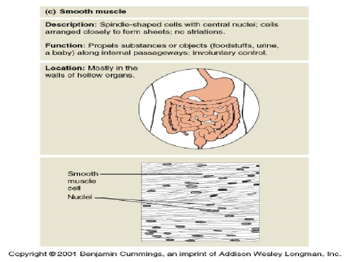

Types : Smooth muscle like cardiac tissue, smooth muscle is usually activated involuntarily of the two types at smooth muscle tissue. The more common type is visceral smooth muscle tissue. It is found in up round sheets that from part of the walls of small arteries and veins and hollow organs such as the stomach, intestine, uterus and urinary bladder like cardiac muscle, visceral smooth muscle is autothymic because the fiber connect to one another by gap junctions, muscle action potential spread throughout the network when a neurotransmitter, hormones, or autothymic signals stimulates one fiber.

Distribution : This muscle are present in all hollow viscera eg. Gastro intestinal tract, ducts of the glands, blood vessels, respiratory, and lymphatic systems at the body. This are also present in the dermis, ciliary body and iris of the eye. The automatic contraction at the smooth muscle fibers facilitates the movement at the contents that are passing though the above mentioned viscera.

Origin and Development The smooth muscles are mesenchymal in origin. The mesenchymal cells first start to stretch out. The nucleus becomes elongated and myofilament appear in the wall of the tube at regular intervals and developed in the same fashion as already mentioned and ultimately forming the continuous circular and longitudinal muscular layers of blood vessels. There is also evidence that is smooth muscle cells themselves can divide by mitosis.

Physiology of smooth muscle Although the principles of contraction are similar in all three types of muscle tissue, smooth muscle tissue exhibits some important physiological differences compared with contraction in skeletal muscle fiber. Contraction in smooth muscle fiber starts more slowly and lots much longer, moreover , smooth muscle can both shorten and stretch to a greater extent than other muscle types.

An increase in the concentration and of calcium in smooth muscle cytosol initiates contraction, just as in striated muscle. Calcium ions flow into smooth muscle cytosol from both the interstitial fluid and sarcoplasmic reticulum. Several mechanisms regulate contraction and relaxation of smooth muscle cells. In one a regulatory protein called calmodulin binds to calcium in the cytosol. Myosin light chain kinase work rather slowly, also contribution to the slowness of smooth muscle contraction. The prolonged presence of calcium in the cytosol provides for smooth muscle tone, a state of continuous partial contraction.

smooth muscle tissue can thus sustained long term tone, which is important in the gastrointestinal tract, where the walls maintain a steady pressure on the contents of the tract, and in the walls of blood vessels called arterioles, which maintain a steady pressure on blood. Most of smooth muscle fibers contract or relax in response to action potential from the autonomic nervous system. In addition, many smooth muscle fibers contract or relax in response to stretching, hormones, or local factors such as changes in ph, oxygen and carbon dioxide levels, temperature and ion concentration.

Histology : The smooth muscle fibers are elongated, fusiform with a wider central portion where the single nucleus is situated. Both the ends of the fibers are tapering towards the periphery. The average length is about 0. 2 mm which varies much and the width is about 6 u at the central widest portion. The cells of the smooth muscles are so arranged that the thick middle portion of one is juxtaposed by the thin end portion of the other.

So in the transverse section there have round or irregularly polygonal profile of various size only the largest profile will demonstrate the centrally placed nucleus. Delicate uniform chromatin network is present in the nucleoplasm. There are two or more nucleoli in ordinary preparation, the sarcoplasm is quite homogeneous, but in special preparation. “fine longitudinal striations” may be demonstrate running full length of the fiber.

This are the myofibrils and are interpreted as the myofibrils and are of the smooth muscles. A small golgi apparatus is also present near the nucleus. Sarcoplasm contents considerable amount of glycogen. The surface of the smooth muscles has a thick basal lamina.

Fine structure : In electron micrograph, the nucleus appears as elongated, smooth surfaced and rounded at the ends. The mitochondria are present at the poles of the nucleus. Myofilaments are intepressed by the mitochondria which are also arranged along the long axis.

Cell to cell relation : the adjacent smooth muscle cells are separated by the thick basilar lamina. These are the suggested site for cell to cell cohesion. At some areas the basal lamina are absent where the unit membranes are coming in close contact to each other having a similar fusion pattern as that at the tight junctions at the cardiac intercalated disc. The fascia occludents or nexuses. The nexuses is probably a low resistance area through which rapid propagation of excitation impulse is possible from one cell to other in case at the smooth muscle.

Mechanism of contraction, excitation, contraction, coupling : Smooth muscle contain actin and myosin. At the time of contraction, myosin heads bind with the actin filaments as in cardiac or skeletal muscle leading to sliding of the actin filaments shortening. However, mechanism of excitation contraction coupling is different smooth muscle, is as follows : When the smooth muscle is activated, say by an action potential or a chemical like ACH, Ca ions from ECF, via Ca++ channels of cell membrane

Here, the ca ions combine with a protein calmodulin to a ca calmodulin complex. Calcium calmodulin complex now binds with an enzyme MLCK myosin light chain kinase now MLCK is activated myosin is now phosphorylated by the active MLCK enzyme. ATP acting as phosphate donor. This phosphorylated myosin can attach with actin so that the sliding of actin becomes possible.

General properties of smooth muscle The properties can be described under the following headings: 1. Autorythmicity 2. Factors influencing the performance of the smooth muscle 3. Tone 1. Autorythmicity : in the organs where the single unit are present (e. g. : intestine, uterus, ureter. ), there are “pace makers”. The pace maker generate impulses which are propagated and stimulate the whole bundle of the smooth muscle.

Sympathetic and parasympathetic stimulation can alter the activity of the pace maker, and hence of the Autorythmicity, but enervation cannot abolish the spontaneous contraction totally.

Factors influencing the performance of smooth muscle a. Stretch : stretch causes excitation of smooth muscle, when spontaneous rhythmic contractions are already present. Stretch causes sustained contractions. b. Effects of catecholamine : effect of catecholamine depends upon the organ in which the smooth muscle is present, the nature of receptors as well as on the species. c. Effect of acetyl choline : acetyl choline stimulates the smooth muscle and increases the force of contraction.

d. Effects of autonomic stimulations Like the catecholamines , stimulations of the autonomic nerves produces effects which depend on the organ, the receptors and the species.

e. Gastro intestinal hormones : Gastrointestinal hormones have profound effects on the contractions of the smooth muscles of the gastrointestinal tract.

f. Effects of the classical hormones: On uterus, estrogen causes excitation where as, progesterone causes depression of the excitability, oxytocin causes contraction of both, uterine muscles and myoepithelial cells of the mammary gland thus helps in parturition and milk ejection.

Tone : Tone of skeletal muscles is completely last when its motor nerves are destroyed. On the other hand the isolated intestine, in the tissue organ both, maintains a tone.

Nerve supply : Smooth muscles are supply by sympathetic and parasympathetic nerves. This control activities of smooth muscle but are not responsible for initiation actively.

Neuromuscular junctions of smooth muscle : The physiologic anatomy of smooth muscle neuromuscular junction : neuromuscular junctions of the highly structured type found on skeletal muscle fibers do not occur in smooth muscle. Instead, the autonomic nerves fibers that innervate smooth muscle generally branch diffusely on top of a sheet of muscle fibers.

In most instances , this fibers, do not make direct contact with the smooth muscle fibers but instead from so called “diffuse junction”. That secrete their transmitter substance into the matrix coating of the smooth muscle a few nanometers to a few micrometer away from the muscle cells, the transmitter substance than diffuses to the cells, the nerve fibers often innervate only the outer layers , and the muscle excitation than travels from this outer layer to the inner layer by action.

Potential conduction in the muscle mass by additional diffusion of the transmitter substance. Excitatory and inhibitory transmitter substances secreted at the smooth muscle neuromuscular junction. The most important transmitter substances secreted by the autonomic nerves innervating smooth muscle are acetylcholine and nor epinephrine but, they are never secreted by the same nerve fibers.

Acetylcholine is a excitatory transmitter substance for smooth muscle fibers in some organs but an inhibitory transmitter for smooth muscle fiber, nor epinephrine ordinarily inhibits it. Conversely, when acetylcholine inhibits a fiber nor epinephrine usually excites it some of the receptors proteins are excitatory receptors, whereas others are inhibitory receptors.

Thus, the types of receptors determine whether the smooth muscle is inhibited or excited and also determines which of the two transmitters, acetylcholine or nor epinephrine is effective in causing the excitation or inhibition.

Effects of hormones on smooth muscle contraction : Most of the circulatory hormones in the body affect smooth muscle contraction to some degree in some have profound effects. Among the more important blood born hormones that affect contraction are nor epinephrine, acetylcholine angiotensin vasopressin, oxytocin, serotonin and histamine.

A hormone causes contraction of a smooth muscle when the muscle cell membrane contains hormonesgated excitatory receptors for the respective hormones.

Type : Smooth muscle at not two sites is the same. But conventionally it is divided into two broad categories, single unit and multi unit.

Single unit smooth muscle : This type of smooth muscle behaves as it the entire muscle mass is a single unit. The gap junction between muscle fiber are so generous that the impulse of activation can spread rapidly from one cell to another. Such an arrangement is also called as functional syncytium. Although single unit smooth muscle has a nerve supply, the nerve supply only reverse to modulate its activity, which can still be modulated to a considerable extent by hormones intrinsic factors.

Multiunit smooth muscle : Multiunit smooth muscle resembles skeletal muscle in that each muscle fiber has independent nerve supply, and activation of a muscle fiber does not lead to activation of its neighboring fibers. Autonomy and automicity of multiunit smooth muscle is much less than that of single unit smooth muscle. Classical examples of this type of smooth muscle are found in the ciliary muscles of the eye, iris and vas deferens.
 contracts rhythmically and automatically, which")
Cardiac muscle : The cardiac muscle (involuntary striated ) contracts rhythmically and automatically, which is particularly maintained the life process of the living system by assisting the supply of nutrients oxygen and removal of metabolic waste products.
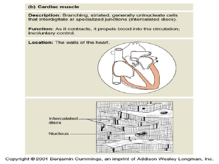

Distribution : The cardiac muscle are actually forming the muscular body of the heart. This muscles are also present in small amounts in the great vessels ending in or opening from the heart.

Histology : The cardiac muscle fibers are separated from each other by the connective tissue endomysium along with blood vessel and lymphatic. The cardiac muscle fiber are not made at one straight simple cylinder but they have gat short cylindrical branches in all directions. This branches are coming in contact with that of the adjacent fibers, ultimately forming a three dimensional network.

Under like microscope these network appears is syncytium which was also supported as the property of the cardiac muscle that if contracts it will contact as a whole. But electron micrograph reveals these branches are not acting as cytoplasmic bridge but they are separated from each other by special surface specialization, the intercalated disc. The sarcolemma of the cardiac muscle is more or less similar to skeletal muscle. The mitochondria are more numbers and the cytoplasm is more abundant. The mitochondria are arranged longitudinally and are present in between the myofibrils in rows.

A small gorge apparatus is present at one pole with a few lipid drop lets. As age increase the lipofuchsin pigments are deposited near the nucleus and may be much extensive to give the brownish appearance of the heart the brown atrophy of the heart. Fine structure under electron microscope the myofibrils made up of Myofilaments, are more are less similar to that of the skeletal muscle. This myofibrils are delineated by the sarcoplasmic reticulum and sarcoplasm. Sarcoplasm contends a large amount of mitochondria along the long axis.

Mitochondria often remain completely surrounded by Myofilaments. so the Myofilaments of the cardiac muscle from a continuum which looks like a large cylindrical mass made up of a parallel myofilaments subdivided by fusiform clefts of the sarcoplasm occupied by mitochondria.

Sarcotubular system : 1. T –system : this T-system is the tubular imagination of the sarcolemma of the cardiac muscle and is larger in diameter than that of the skeletal muscle. The T- tubules are present at the z line in the cardiac muscle fiber, but the same are at the A-I junction in case of the mammalian skeletal muscle. The functional significance of the locational difference is not yet understood. These tubules also increase the surface for metabolic exchange in between the interior of the cardiac muscle fiber and the intercellular space, over and above they

2. Sarcoplasmic reticulum : this reticulum of the cardiac muscle fiber is ill developed. They are consisted of longitudinal interconnected tubules which are expanded into small terminal sacs at the Zline. There is not well developed transverse cisternae in the cardiac muscle.

Physiology of cardiac muscle : The heart is composed of three major types of cardiac muscle. Atrial muscle, ventricular muscle, and specialized excitatory and conductive muscle fiber. The atrial and ventricular types of muscle contract in much the same way skeletal muscle. The except that the duration of contraction is much longer conversely. The specialized excitatory and conductive fibers contract only feebly because they contain few contractile fibrils instead, they exhibit rhythmicity and varying rates of conduction, providing an excitatory system that

Bibliography : 1. 2. 3. 4. 5. 6. 7. C. C. CHATTERJEE R. L. BIJLANI TORTORA WILLIAM F. GANONG GUYTON AND HALL S. K. CHAUDHARY K. SEMBULINGAM

- Slides: 142