Examination techniques in ophthalmology V Matukov Basic examination

Examination techniques in ophthalmology V. Matušková
 • Measurement of intraocular pressure")
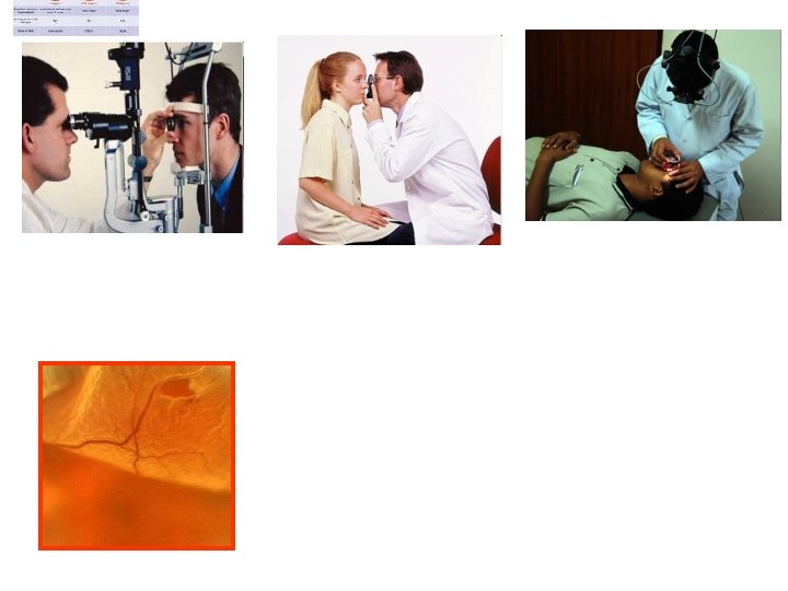
Basic examination techniques • Visual acuity (natural, best corrected) • Measurement of intraocular pressure • Slit lamp examination • Fundus examination
 • Distance vision • Near vision")
Visual acuity • Visual acuity (natural, best corrected) • Distance vision • Near vision • Charts • • Snellen Landolt ring Pictures ETDRS

Charts

Near vision Jaeger chart
 • Hand movement • Light perception")
Visual acuity • Counting finger (metres ) • Hand movement • Light perception
 • 0, 1 • Minimum separabile –")
Visual Acuity • 5/50 (metres, feet 20/200) • 0, 1 • Minimum separabile – minimum angle of resolution

Refraction • Refractive error is an optical abnormality in which the shape of the eye fails to bring light into sharp focus on the retina, resulting in blurred or distorted vision. • In optometry, a "refraction" procedure is the measurement of refractive error • Autorefractor

Visual acuity • Frame • Lenses

Tonometry Contact techniques • Schiötz tonometry • Goldmann applanation tonometry • Non contact techniques • Jet air (air puff)

Schiötz tonometry • 7/7, 5 • Topical anestesia

Schiötz tonometry

Goldmann tonometry • The normal range is 10 -21 mm. Hg • Topical anestesia, fluorescein • Force required to flatten the cornae

Goldmann tonometry

Non contact tonometry

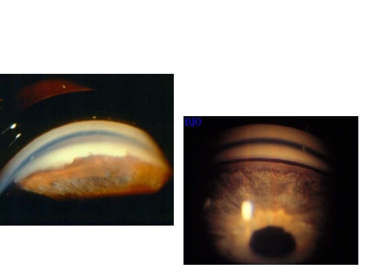
Examination of anterior segment • Aspection

Slit lamp biomicroscopy • table mounted microscope with a special adjustable illumination source attached

Slit lamp biomicroscopy nduhovka

Slit lamp biomicroscopy ,

Synechiae posterior

Sunconjunctival haemorrhage

Corneal abrasion

Hyphema

Hypopyon

Examination of fundus • Ophthalmoscopy - direct - indirect • Slit lamp biomicrocsopy (high power convex lens, the image is vertically inverted and laterally reversed)

Ocular fundus

Special examination techniques
 • Angle structures • Fundus examination")
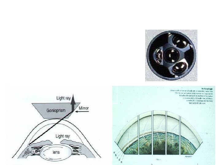
Gonioscopy • Gonio lens ( mirrors) • Angle structures • Fundus examination

Optical coherence tomography

Fluorescein angiography
 diagnosis of functional defects of retina • Record of")
Electrophysical tests • ERG (electroretinography) diagnosis of functional defects of retina • Record of an action potential produced by the retina when it is stimulated by light • VEP (visual evoked potential) -diagnosis of functional defects of visual pathways • Record of electrical activity of the visual cortex created by stimulation of the retina

Electrophysical tests

Electrophysical tests

Ultrasound

Ultrasound • Non transparent optical media • UBM- high frequency ultrasound – imaging of anterior segment
 • B scan")
• A scan ( biometry) • B scan

Ultrasound

Color vision – HUE test

Hertl exoftalmometry

Děkuji za pozornost!
- Slides: 43