Examination Procedure A B C D E F
 B) C) D) E) F) G) H) Acuity of vision")
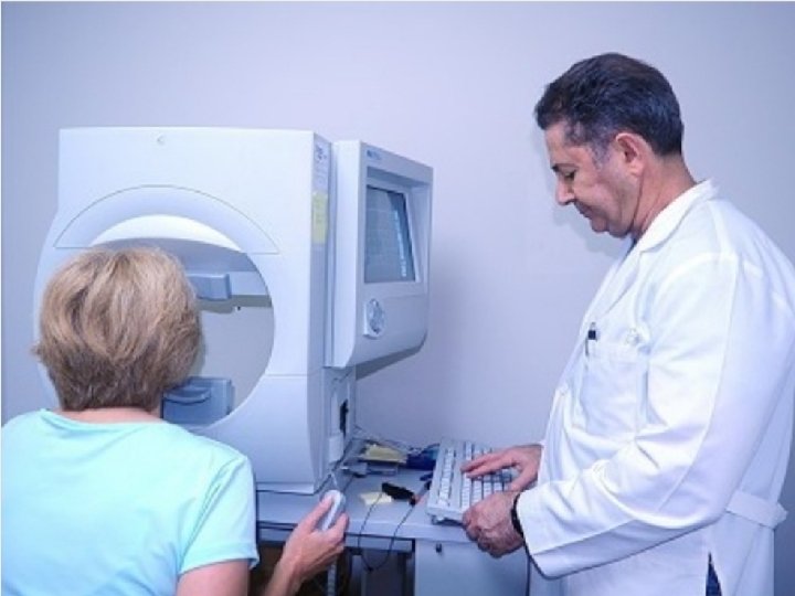
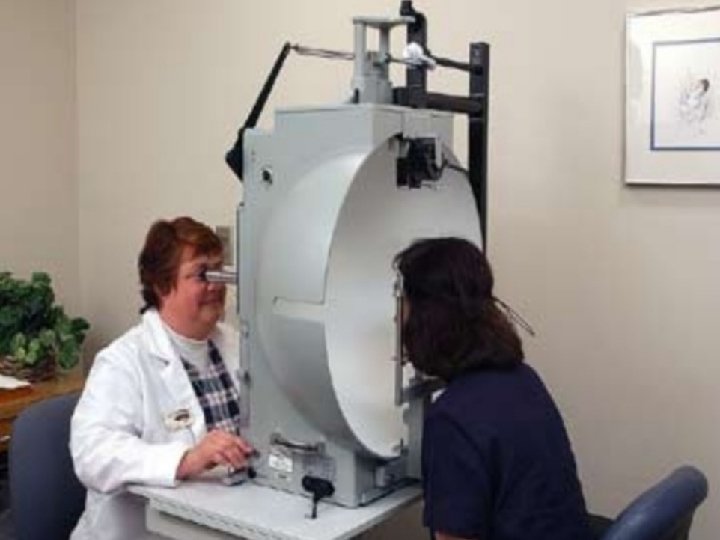
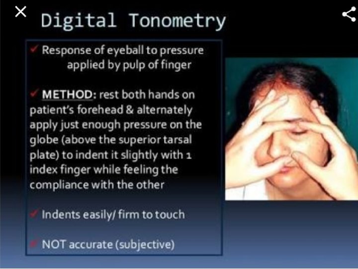
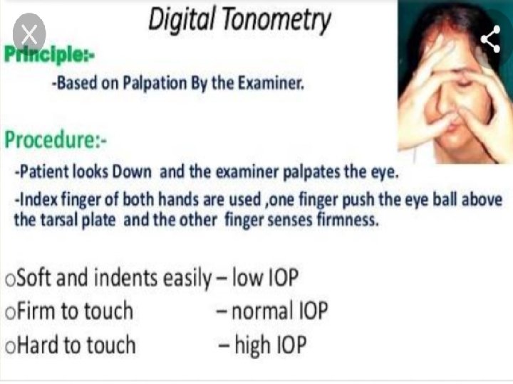
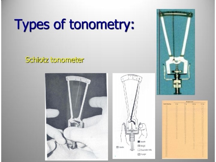
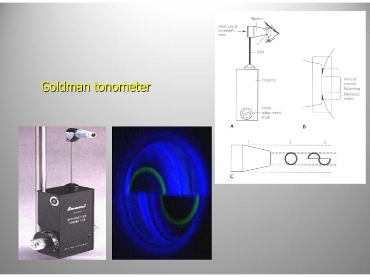
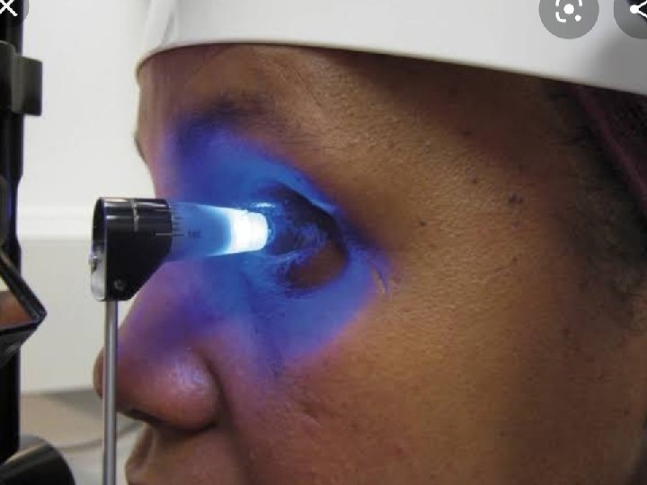
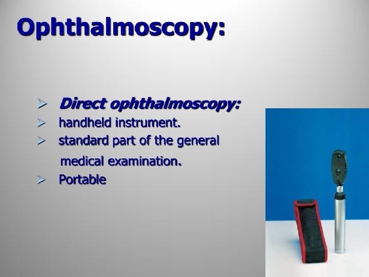
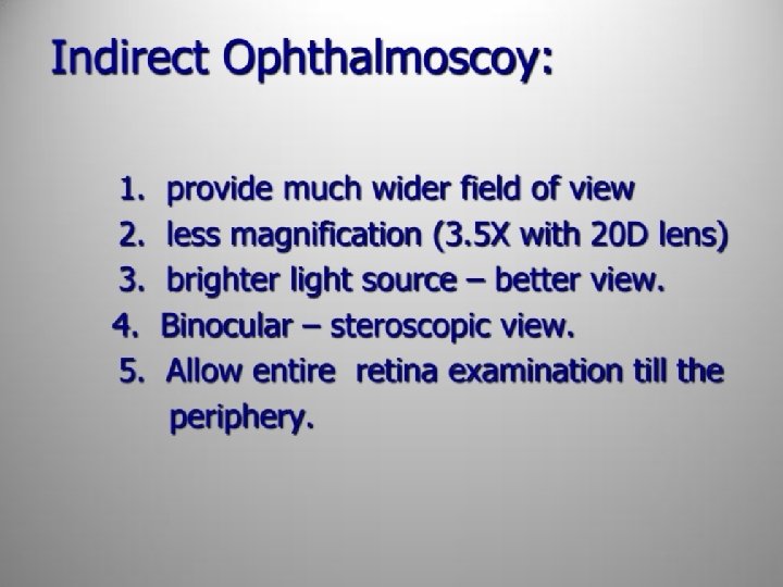
Examination Procedure : A) B) C) D) E) F) G) H) Acuity of vision Colour vision Field of vision Ocular motility and allignment Pupillary reaction Slit lamp examination Intra ocular pressure Ophthalmoscopy

 – 20 ft")
Visual Acuity Standard Test Disances : • Distance visual acuity (DVA) – 20 ft or 6 M is equivalent to optical infinity • Near visual acuity (NVA) - 40 cm • Why 6 M / 20 ft – because rays beyond 6 M are parallel and we should consider the parallel rays come from infinity • Why 40 cm – because near point of eye is 40 cm

Requirement : • • • Illuminated vision chart 6 M/ 20 FT distance Individual eye should be tested separately First unaided, then with pinhole If patient wear spectacle then unaided, then with spectacle, then spectacle and pinhole.

Snellen Chart

Tumbling E Chart

Landolt C Chart

Bailey-Lovie Chart

Pinhole Effect
 •")
Near Visual Acuity • Testing the VA at close range (usually 40 cm) • Near visual acuities are taken through the habitual correction • The purpose is to detect people with near vision difficulties (e. g. , uncorrected high hyperopia, accommodative dysfunction) • In patients over 40 years old, the reduced near visual acuity is one of the symptoms of presbyopia

Near Vision Charts • Types of notation – Reduced Snellen Acuity card • Test distance at 16 in (or 40 cm) – Jaeger Acuity Card • 20 letter sizes classified J 1 to J 20 • Test distance at 14 in – Point system • Each point is 0. 35 mm – M notation • Based on meter unit

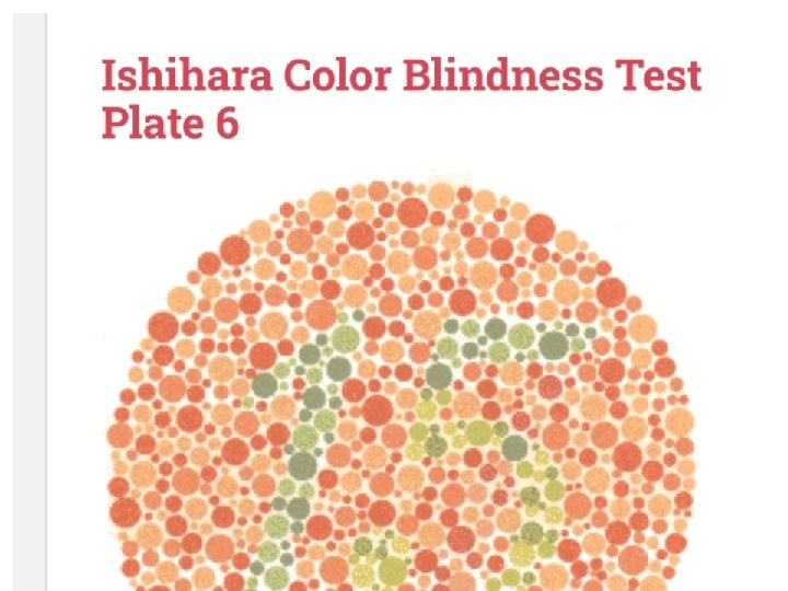
Colour vision test • Different charts • Common one is Ishihara chart • Three basic colour red/ green/ blue for bedside test.


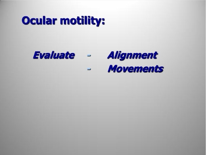
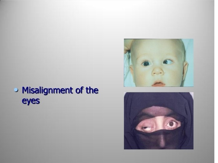

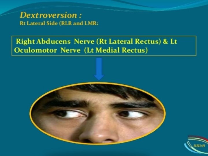
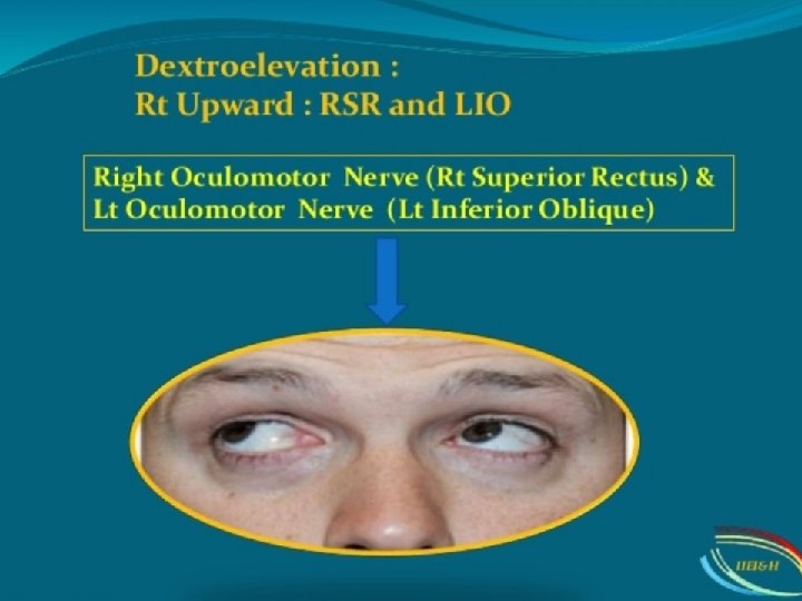
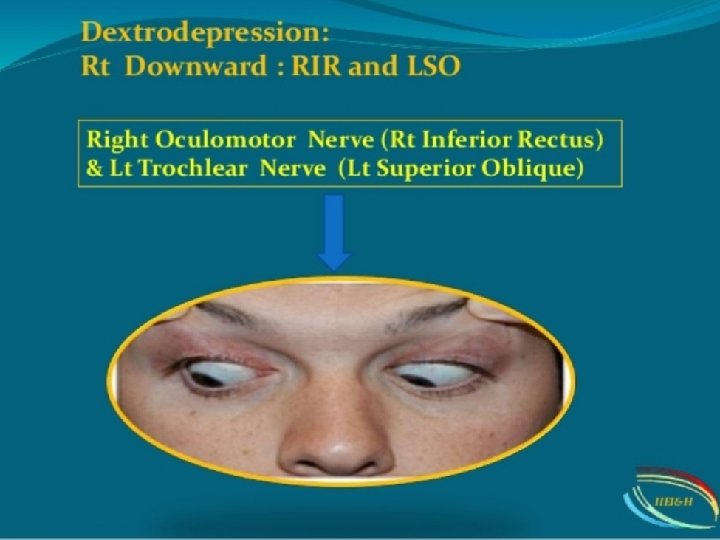
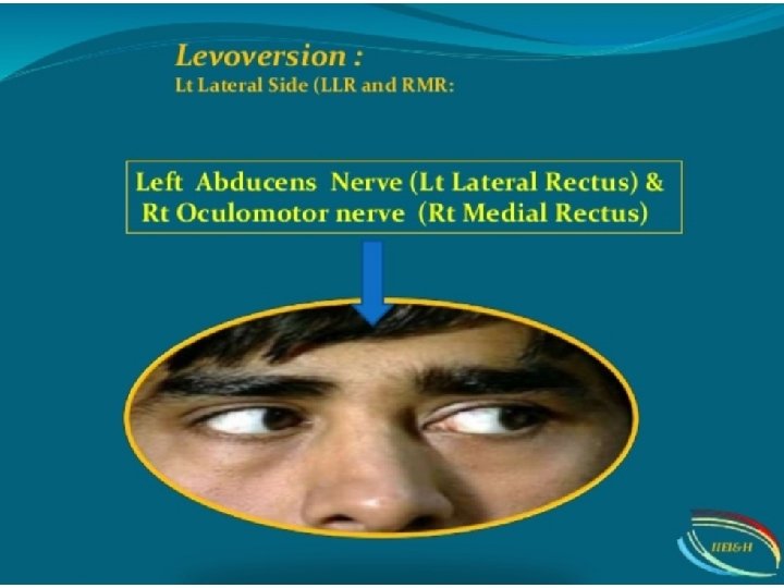
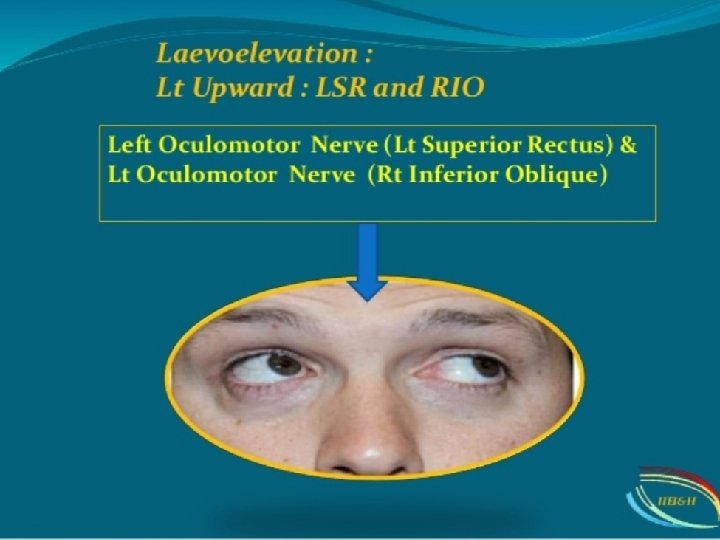
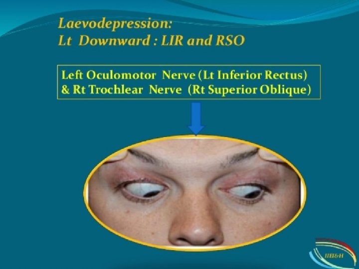
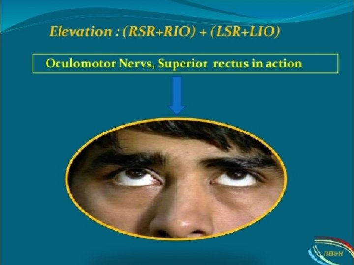
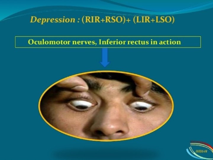
Ocular movements : Ø Duction – monocular movements, are abduction, depression, elevation, depression. Ø Version – binocular movements, are dextroversion, levoversion dextrodepression, dextroelevation, , levodepression, levoelevation. These are the cardinal positions of gaze

Pupillary reflex
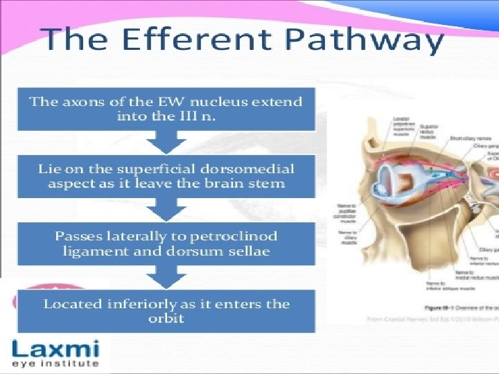

When light fall in one eye constriction of pupil in that eye is called direct pupillary reflex. Constriction of pupil in other eye is called consensual pupillary reflex.


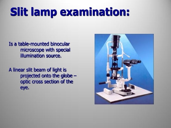

Structures evaluated by slit lamp: • • Eyelid and lashes Conjunctiva Cornea Anterior chamber Irish Pupil Lens

Slit lamp examination • • Lid and lashes Conjunctiva Cornea Anterior chamber Pupil Irish lens

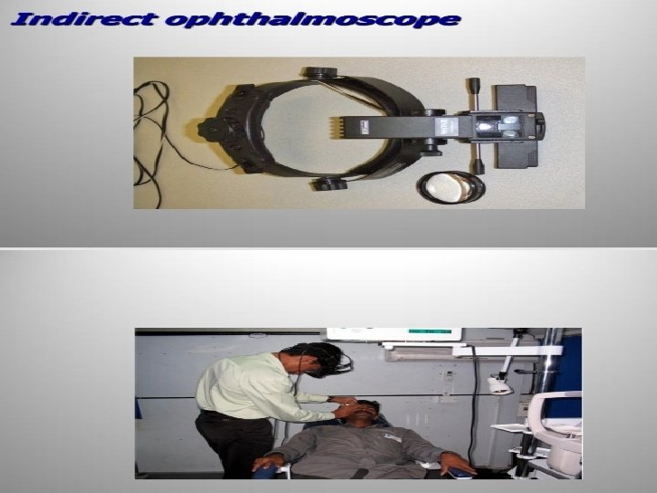
 Binocular view 2) Larger area can be seen Disadvantage : 1)")
Advantage : 1) Binocular view 2) Larger area can be seen Disadvantage : 1) Inverted and laterally reversed image 2) Bright light is uncomfortable to the patient 3) Need more expert person
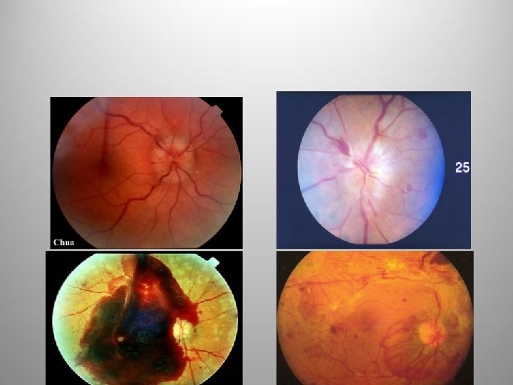
- Slides: 62