Examination of the Ears Nose Throat and Neck


 Introduction 7) Otoscopic examination 2) Position the patient 8)")
 Introduce yourself to the patient Any deafness? Communication")
 Position the patient Away from the wall In chair")
 Start with the better side Start with the dry")
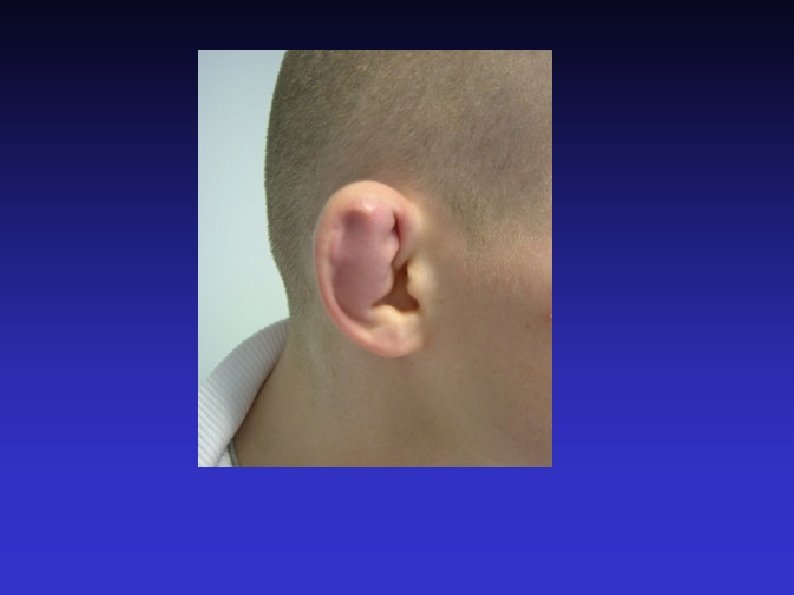
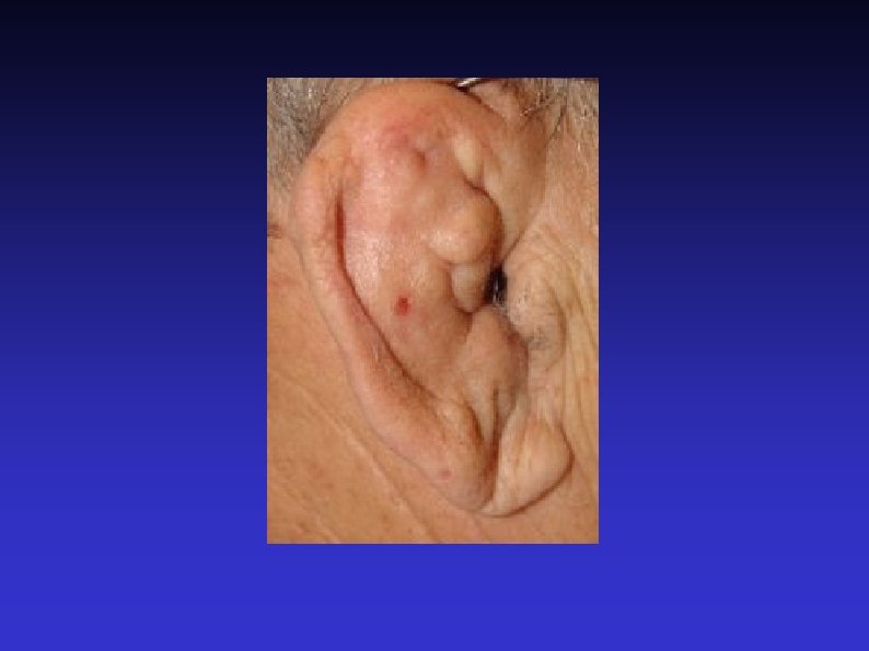
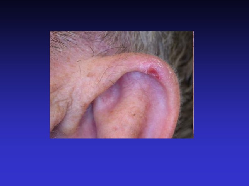
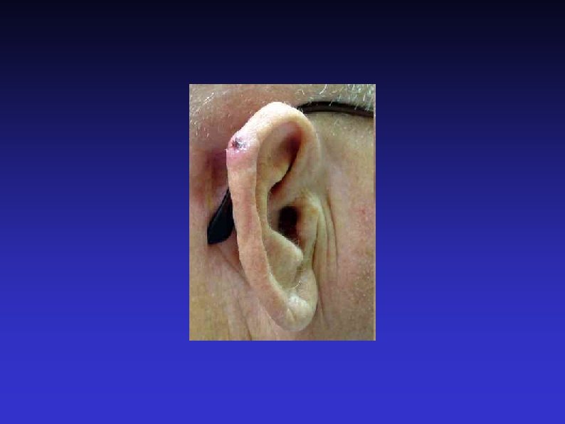
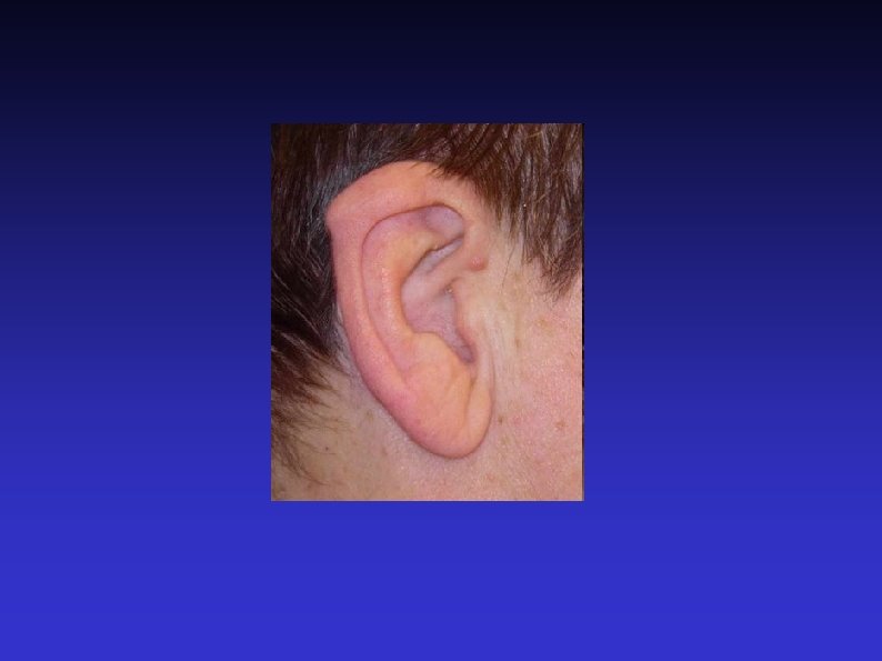
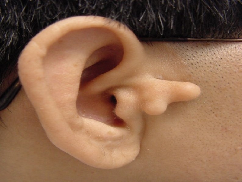
 Inspect the pinna Front and behind Skin condition Lesions")
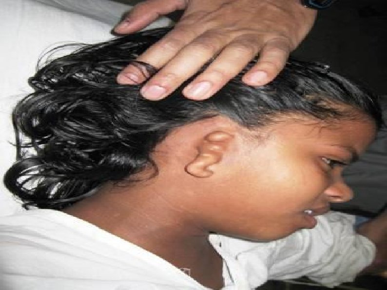
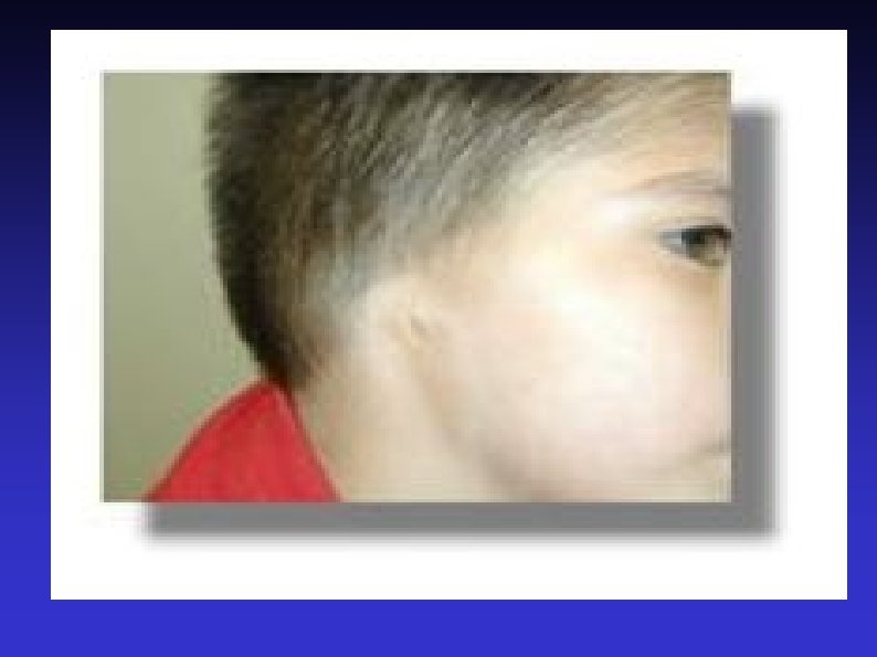
 Inspect the mastoid Mastoiditis is very, very rare Post")
 Inspect the external auditory meatus Pull pinna upwards, outwards")
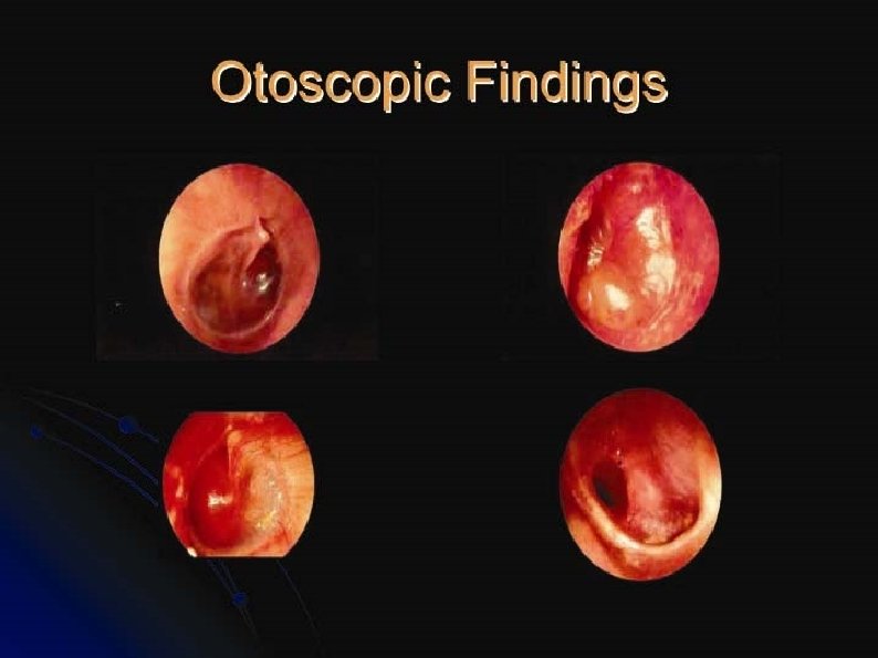
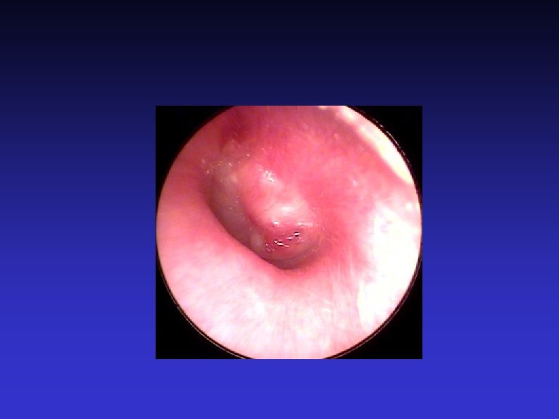
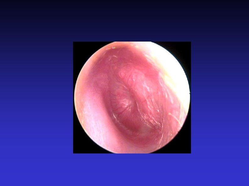
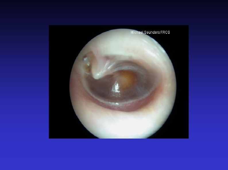
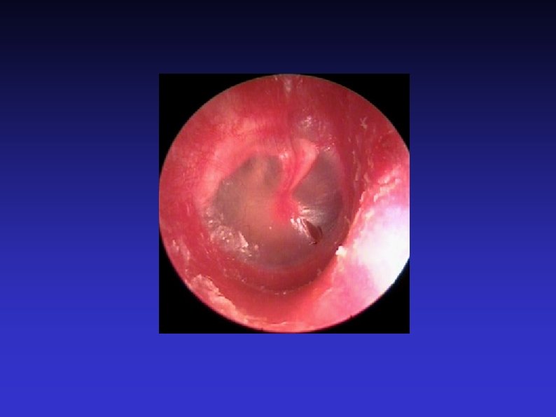
 Otoscopic examination The lateral process and handle of the")
 Fistula test A test for ENT doctors! Warn the")
 Free field testing Start with the better ear Use")
 Tuning fork tests Traditionally 512 Hz (256 Hz may")
 Facial nerve Otoneurological conditions affect the cranial nerves closest")
 Post nasal space Conditions affecting the middle ear can")
 Introduce yourself 5) Anterior rhinoscopy 6) Oral examination 7)")

 Introduce yourself Any hyponasal speech?")
 Position the patient Head-mirror or headlight?")
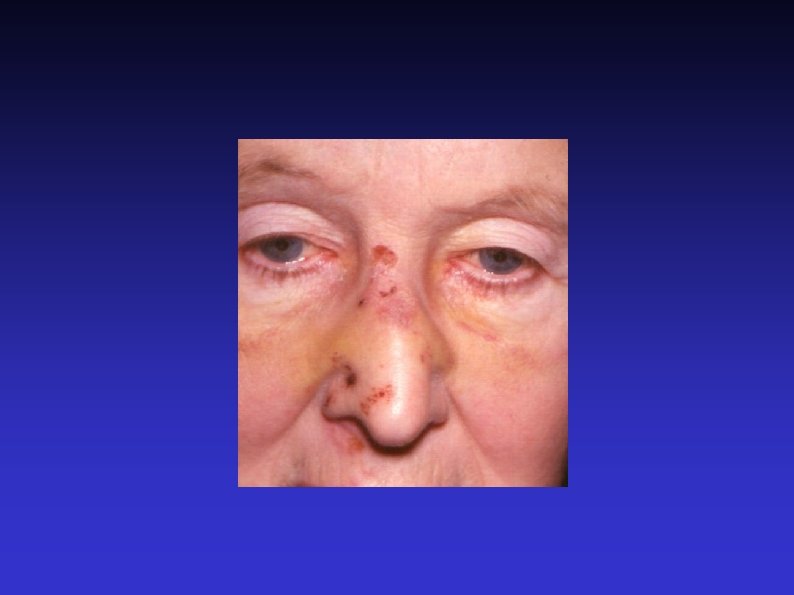
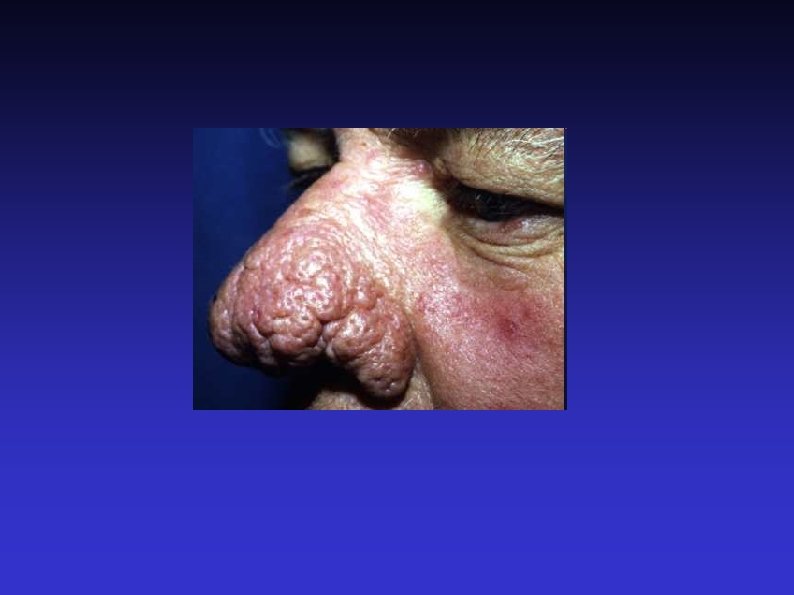
 Inspect the external nose Compare nose to rest of")
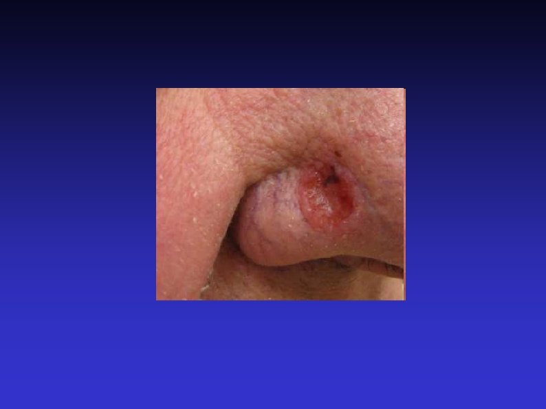
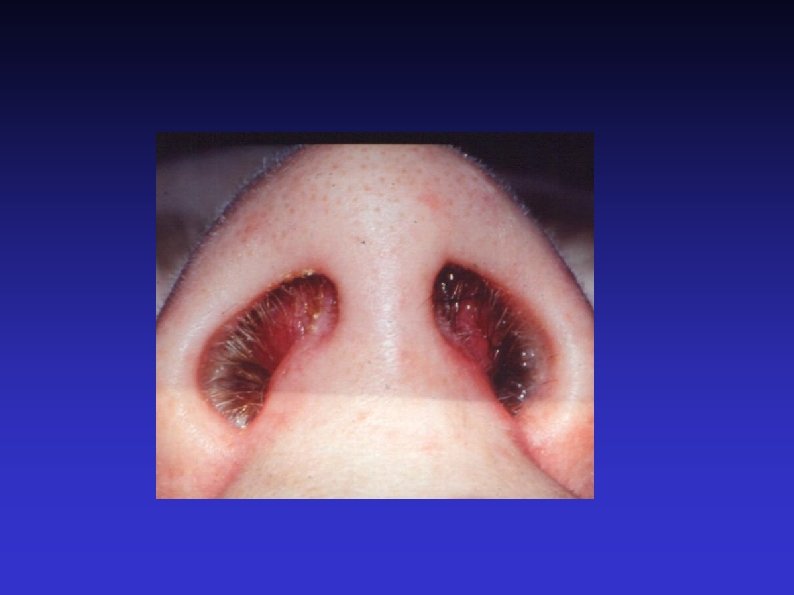
 Examine the nasal tip, vestibule, and assess the nasal")
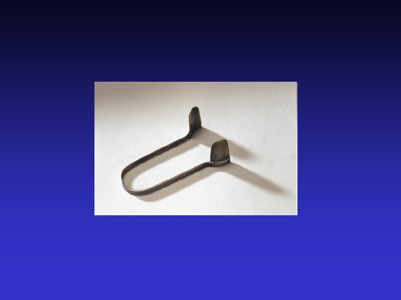
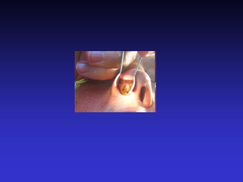
 Anterior rhinoscopy Thudichum’s speculum vs otoscope Obvious lesions Mucosa")
 Oral examination Rotten teeth Alveolar process of the maxilla")
 Post nasal space examination With mirror, Hopkins rod, or")
 Neck examination Anterior nose drains to submandibular region Posterior")
 Introduce yourself 5) Nasopharynx 2) Position the patient 6)")
 Introduce yourself")
 Position the patient Headlamp, mirror or other light source")
 Assess speech Stridor Hoarseness Hot potato Any other dysphonia")
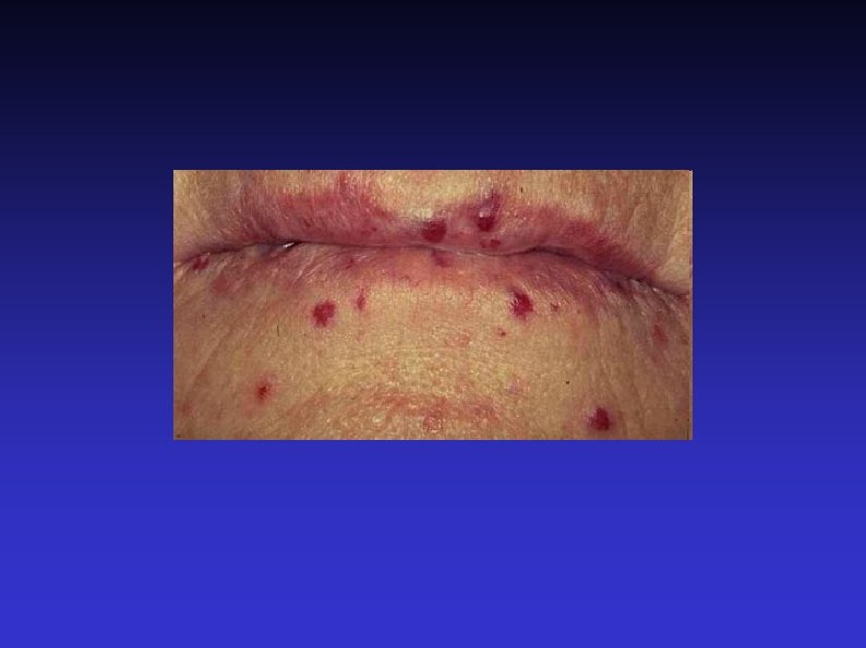
 Oral examination Lips, perioral lesions 1 or 2 tongue")
 Nasopharynx Unless large lesion, will need mirror, Hopkins rod")
 Indirect laryngoscopy With mirror or nasendoscope Can assess nasal")
 Examination of neck Head and neck cancers metastasise to")
 Skin 6) Airway 2) Swallow 7) Thyroid 3) Voice")
 Skin lesions Ulceration Scars and wounds Stoma Obvious large")
 Swallow Larynx should rise A goitre may rise, too")
 Voice As before. “Count to ten”.")
 Examine from behind Let patient know what you are")
 Anterior Triangle Mastoid Mental process Sternal notch Ramus and")
 Airway Trachea Larynx Trotter’s sign Hyoid bone Thyroglossal cyst")
 Thyroid Moves on swallowing Two lobes Describe any lumps")
 Posterior Triangle Mastoid Medial end of clavicle Junction of")
 Sternocleidomastoid As well as traditional names of nodes, can")
- Slides: 96
Examination of the Ears, Nose, Throat, and Neck Prof Isam Amine Head of ENT department Faculty of medicine Damascus university
HISTORY Chief complaint: Ear? Nose? Throat? Neck? HPI: Onset, frequency, duration Associated symptoms What has the patient already tried? Pertinent positives & negatives Always think: “Could this be related to underlying malignancy or something more serious? ” Previous work-up, testing, imaging, or interventions What has already been done or tried for this?
HISTORY Past Medical History: Allergies? Asthma? Neurologic or rheumatologic disorders? Past Surgical History: Head and neck procedures? Allergies- Aspirin Sensitivity? Meds- Is this problem medicationrelated? Social History - Smoker? Alcohol use? Family History- Does this run in the patient’s family? Remember: The patient may not know their full medical history. Often, you will have to ask specific and directed questions to get the information you are looking for
ENT REVIEW OF SYSTEMS Gen: fever/chills/weight changes Ear: tinnitus/ vertigo/ hearing loss/ otalgia/ otorrhea Nose: congestion/ rhinorrhea/ epistaxis/ decreased smell Throat: pain/ dysphagia/ odynophagia Larynx: hoarseness/ voice changes/ noisy breathing/ difficulty breathing / pain with speaking (odynophonia) Trachea: noisy or difficulty breathing Neck: lymphadenopathy/ new lumps or bumps/ pain/ swelling Face: sinus pain/ pressure/ swelling/ numbness
Examination of the Ear 1) Introduction 7) Otoscopic examination 2) Position the patient 8) Fistula test 3) Start with the better ear 9) Free field testing 4) Inspect the pinna 10) Tuning fork tests 5) Inspect the mastoid 11) Facial nerve 6) Inspect the external auditory meatus 12) Post nasal space
Examination of the Ear 1) Introduce yourself to the patient Any deafness? Communication
Examination of the Ear 2) Position the patient Away from the wall In chair Can walk around patient
Examination of the Ear 3) Start with the better side Start with the dry side Remove hearing aids
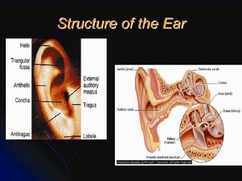
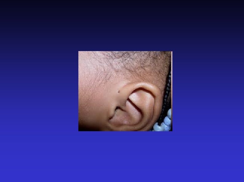
Examination of the Ear 4) Inspect the pinna Front and behind Skin condition Lesions Scars Pre-auricular area (common place for sinus) Condition of cartilage
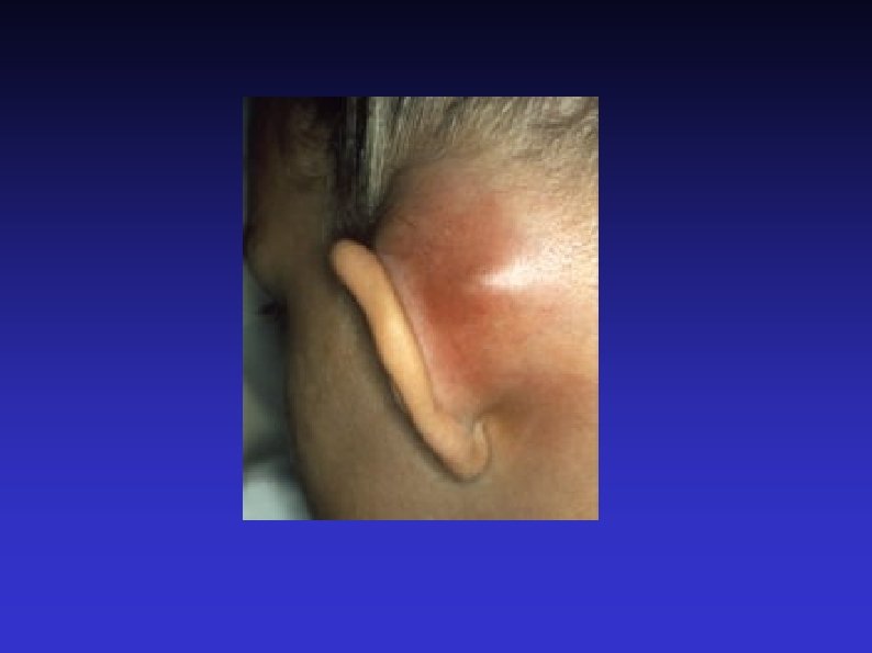
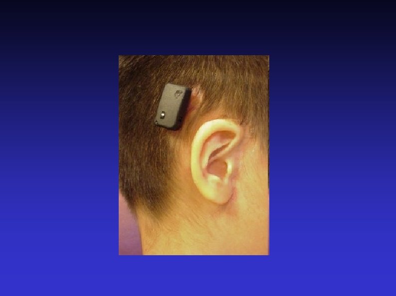
Examination of the Ear 5) Inspect the mastoid Mastoiditis is very, very rare Post auricular lymph nodes Scars BAHAs and Cochlear implants
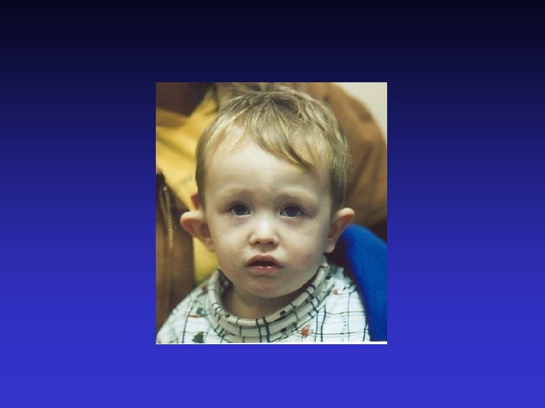
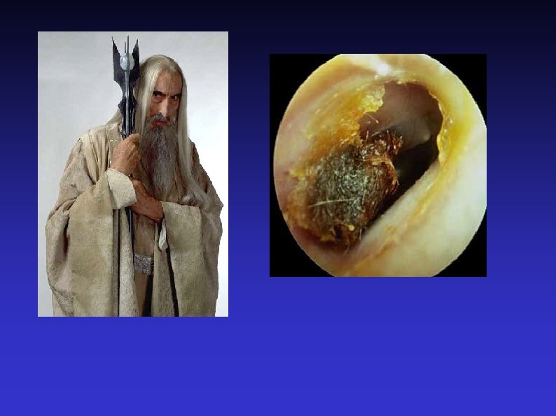
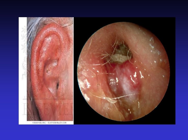
Examination of the Ear 6) Inspect the external auditory meatus Pull pinna upwards, outwards and backwards In infants downwards and backwards In children pull backwards Otorrhoea and otomycosis Canal stenosis Exostoses and osteomas
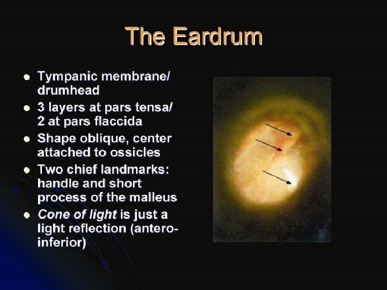
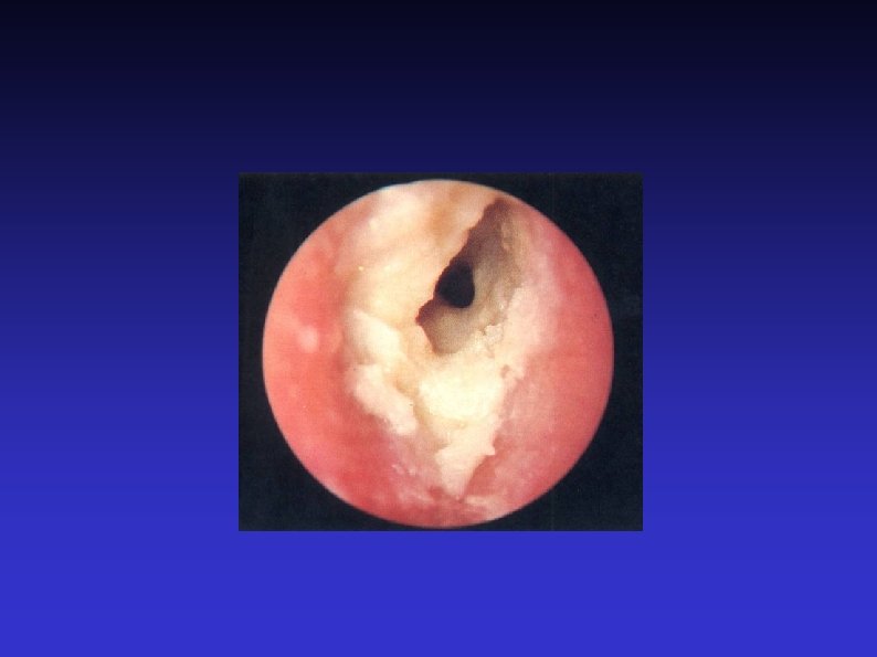
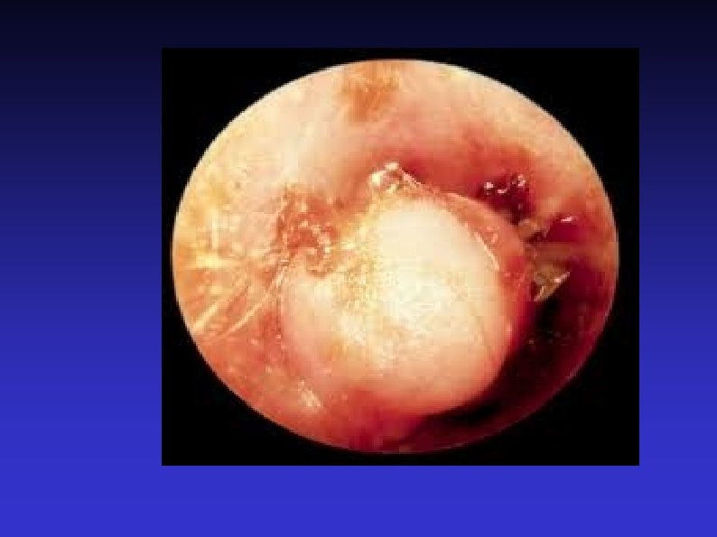
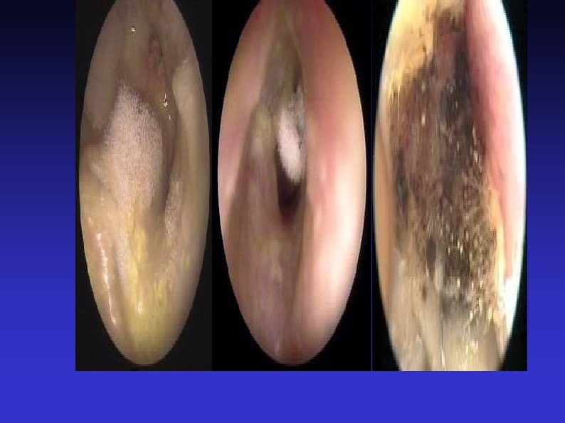
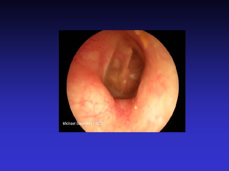
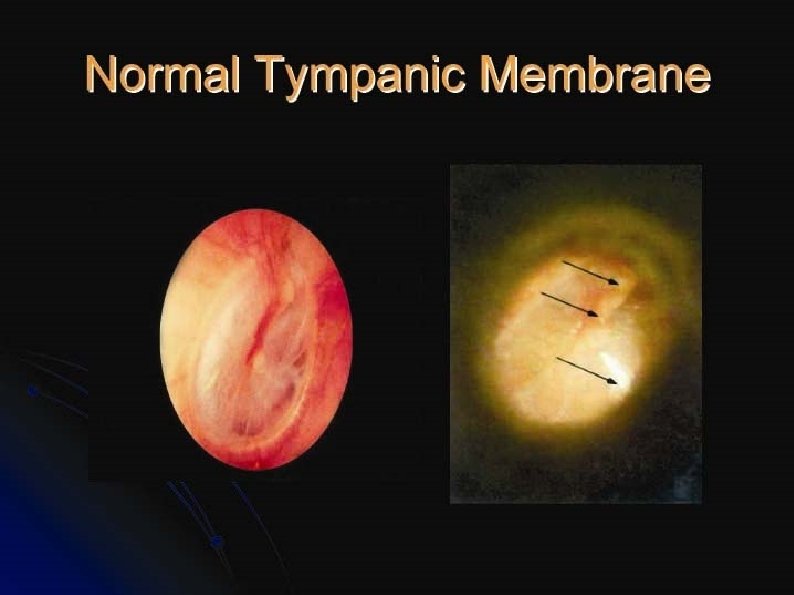
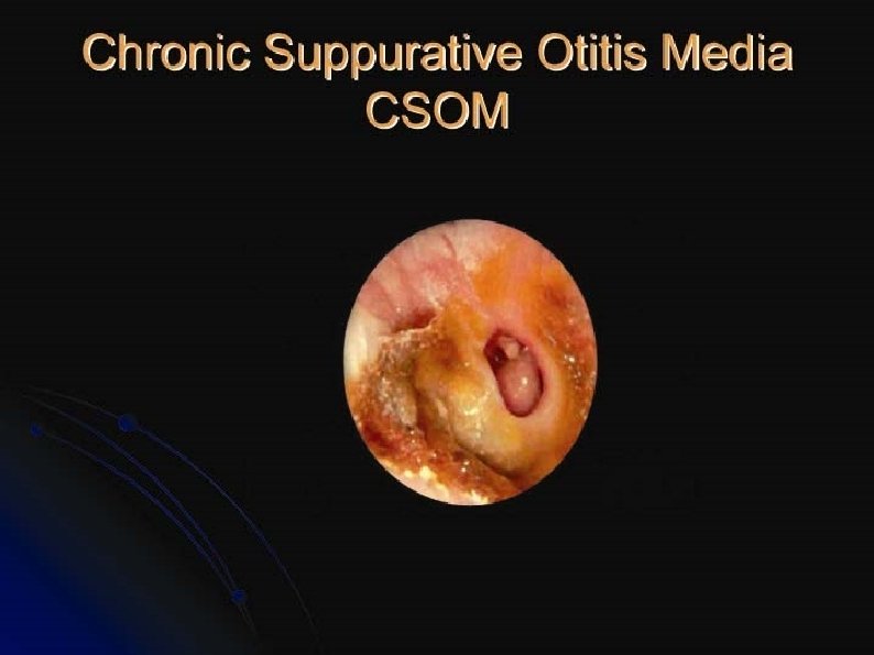
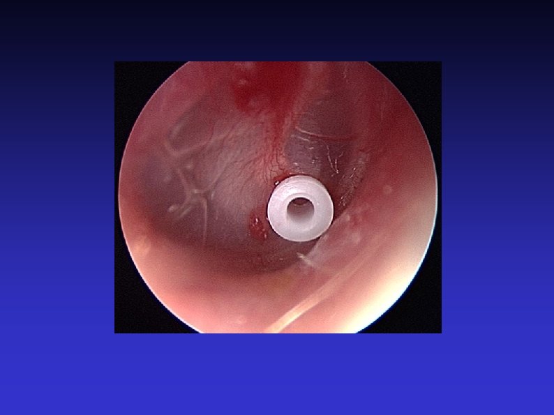
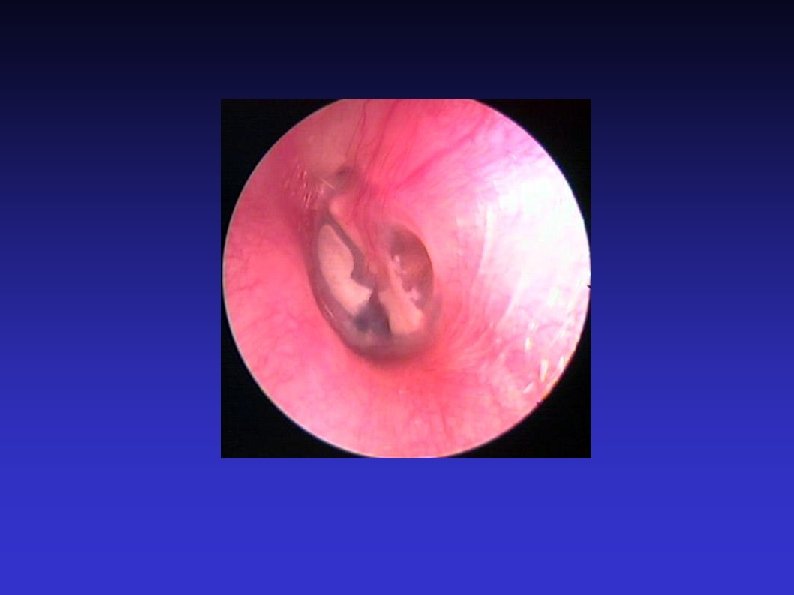
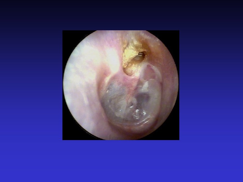
Examination of the Ear 7) Otoscopic examination The lateral process and handle of the malleus lie towards the centre of the tympanic membrane Four quadrants Perforation Central or marginal What can be seen through it Mastoid cavity Dry Wet, inflamed
Examination of the Ear 8) Fistula test A test for ENT doctors! Warn the patient A cholesteatoma has erroded part of a semi-circular canal Pressure in the EAM causes conjugate deviation of the eyes
Examination of the Ear 9) Free field testing Start with the better ear Use masking, such as a tragal rub Whispered voice, conversation voice, and shouted voice at 60 cm Use double number fingers or bisyllable words
Examination of the Ear 10) Tuning fork tests Traditionally 512 Hz (256 Hz may be better) Rinne and Weber (they were both German) Help differentiate between conductive and sensorineual hearing loss Limited reliability (around 80%) For Weber, incisors > vertex > forehead
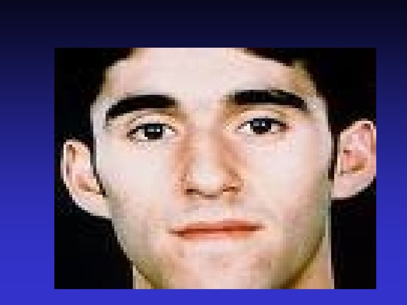
Examination of the Ear 11) Facial nerve Otoneurological conditions affect the cranial nerves closest to the vestibulocochlear nerve Cholesteatoma Malignant otitis externa (osteomyelitis) Vestibular Schwannoma (formerly acoustic neuroma) Other skull-base tumours
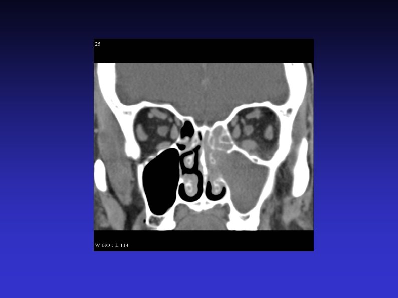
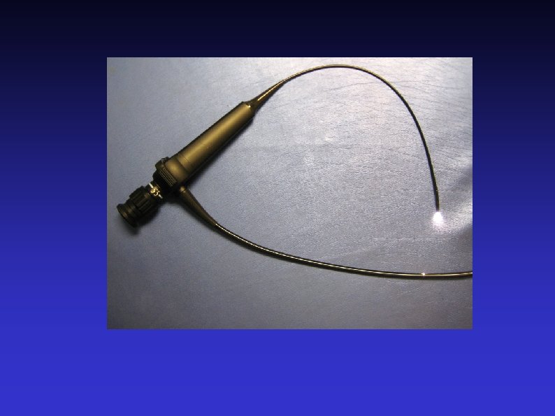
Examination of the Ear 12) Post nasal space Conditions affecting the middle ear can sometimes affect the post nasal space Hopkins rod, flexible endoscope, or angled mirror usually needed Massive lesions can sometimes be seen through the nose, or behind the soft palate
Examination of the nose 1) Introduce yourself 5) Anterior rhinoscopy 6) Oral examination 7) Post nasal space examination 8) Neck examination
Examination of the nose 1) Introduce yourself Any hyponasal speech?
Examination of the nose 2) Position the patient Head-mirror or headlight?
Examination of the nose 3) Inspect the external nose Compare nose to rest of face Size and shape Skin Swelling, bruising, ulcers Profile
Examination of the nose 4) Examine the nasal tip, vestibule, and assess the nasal airways Nasal tip Nostrils and air flow Mist test Condition of mucosa inside vestibule
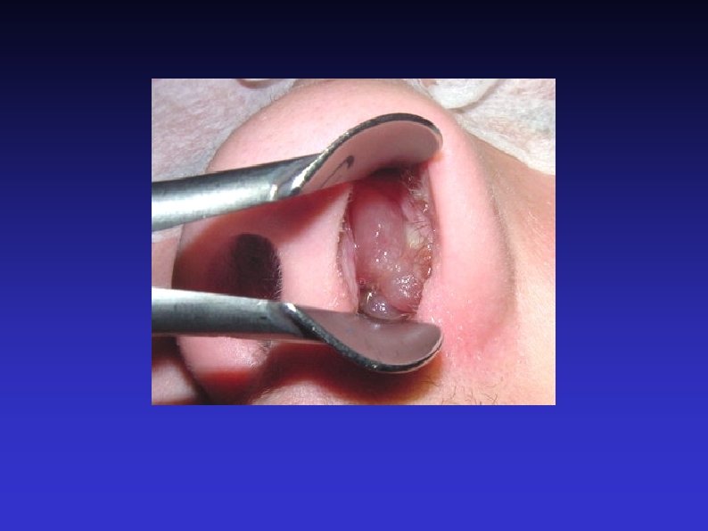
Examination of the nose 5) Anterior rhinoscopy Thudichum’s speculum vs otoscope Obvious lesions Mucosa Septum Turbinates (and osteomeatal complex)
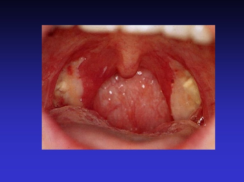
Examination of the nose 6) Oral examination Rotten teeth Alveolar process of the maxilla Palate and uvula
Examination of the nose 7) Post nasal space examination With mirror, Hopkins rod, or nasendoscope Not easy outside of ENT clinic Large masses, such as an antrochoanal polyp
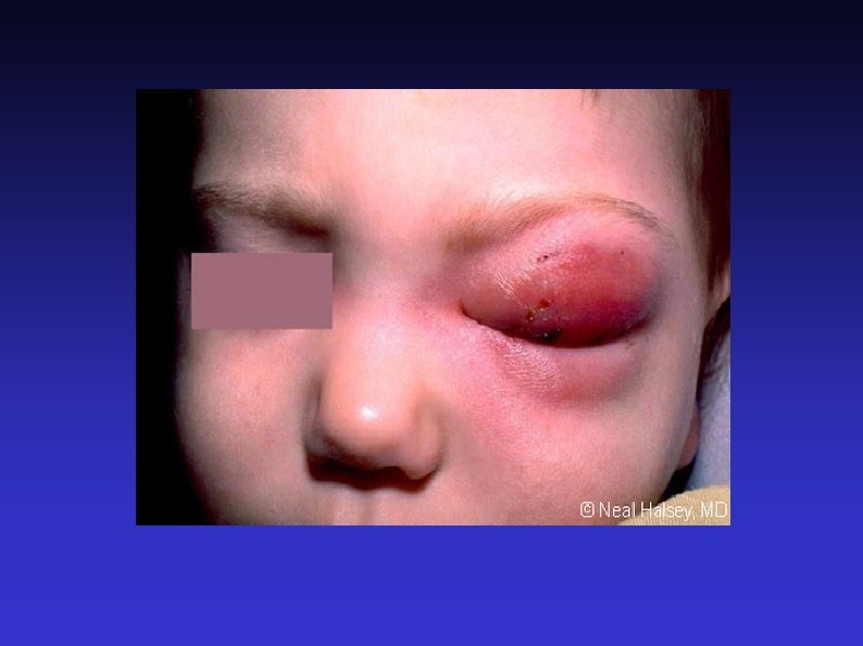
Examination of the nose 8) Neck examination Anterior nose drains to submandibular region Posterior drains to middle deep cervical
Examination of the throat 1) Introduce yourself 5) Nasopharynx 2) Position the patient 6) Indirect laryngoscopy 3) Assess speech 7) Examine the neck 4) Oral examination
Examination of the throat 1) Introduce yourself
Examination of the throat 2) Position the patient Headlamp, mirror or other light source Seated in chair with space to examine from all sides
Examination of the throat 3) Assess speech Stridor Hoarseness Hot potato Any other dysphonia
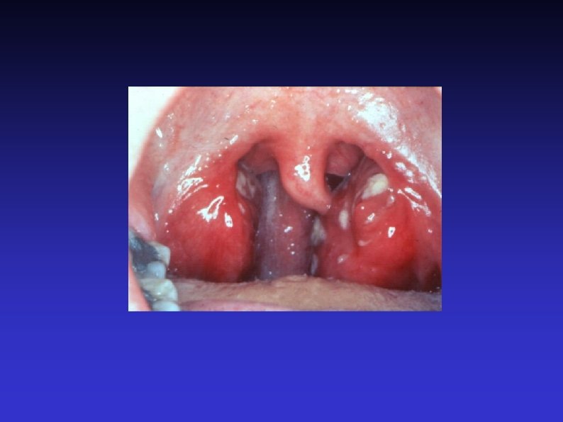
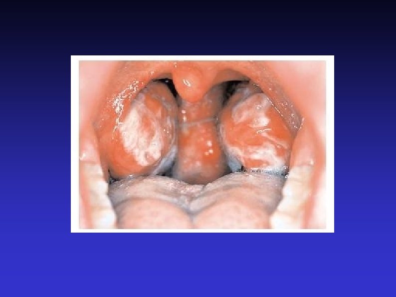
Examination of the throat 4) Oral examination Lips, perioral lesions 1 or 2 tongue depressors (or finger) Inspect tongue, buccal mucosa and oropharynx Salivary duct orifaces Say ‘Ahhh’ Finger examination of tongue, floor of mouth, cheeks and back of throat
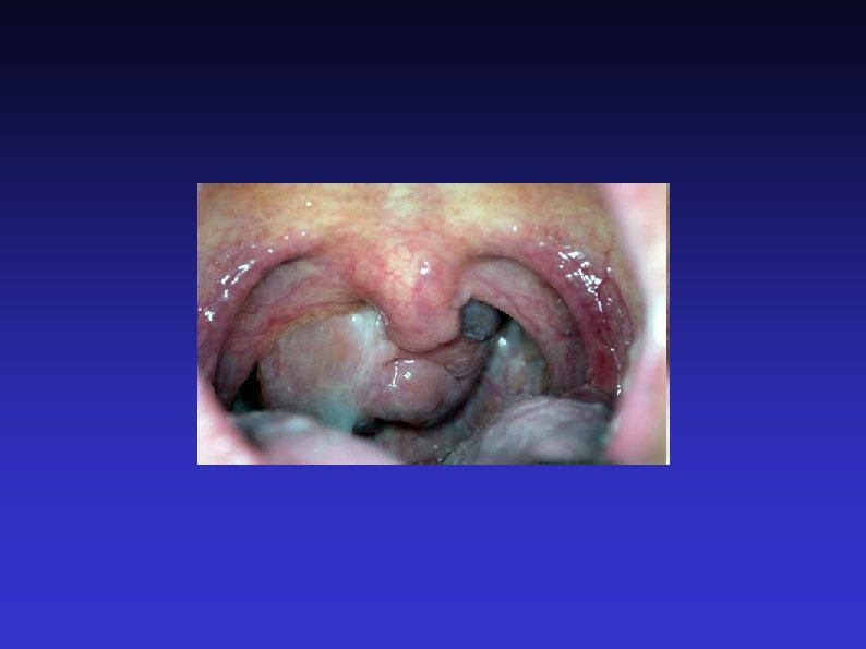
Examination of the throat 5) Nasopharynx Unless large lesion, will need mirror, Hopkins rod or nasendoscope
Examination of the throat 6) Indirect laryngoscopy With mirror or nasendoscope Can assess nasal cavity, nasopharynx, oropharynx, hypopharynx and larynx Can assess movement of cords, especially with strobe
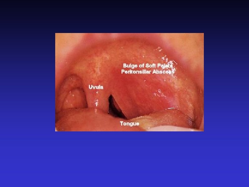
Examination of the throat 7) Examination of neck Head and neck cancers metastasise to neck nodes and to the lungs Tonsillar infections are the commonest cause of enlarged lymph nodes
Examination of the neck 1) Skin 6) Airway 2) Swallow 7) Thyroid 3) Voice 8) Posterior triangle 4) Examine from behind 9) Sternocleidomastoid 5) Anterior triangle
Examination of the neck 1) Skin lesions Ulceration Scars and wounds Stoma Obvious large masses
Examination of the neck 2) Swallow Larynx should rise A goitre may rise, too
Examination of the neck 3) Voice As before. “Count to ten”.
Examination of the neck 4) Examine from behind Let patient know what you are doing Tender areas Gentle One side at a time
Examination of the neck 5) Anterior Triangle Mastoid Mental process Sternal notch Ramus and border of mandible Midline Upper edge of SCM
Examination of the neck 6) Airway Trachea Larynx Trotter’s sign Hyoid bone Thyroglossal cyst (around half move with tongue protrusion)
Examination of the neck 7) Thyroid Moves on swallowing Two lobes Describe any lumps Percuss the sternum
Examination of the neck 8) Posterior Triangle Mastoid Medial end of clavicle Junction of trapezius and clavicle Lower edge of SCM Border of trapezius Clavicle
Examination of the neck 9) Sternocleidomastoid As well as traditional names of nodes, can be described as I to VI Easier to remember, and more relevant to metastatic spread