Examination of patient with periodontal diseases Dr Kadhim


- Slides: 19

Examination of patient with periodontal diseases Dr. Kadhim Jawad 1

General characteristics of periodontal diseases They can be discussed under the following three categories: Dr. Kadhim Jawad 2

A - Clinically: 1 - Color and texture alteration of the gingival tissues. 2 - Increased tendency to bleeding. 3 - Increased pocket depth. 4 - Tooth mobility or drifting. Dr. Kadhim Jawad 3

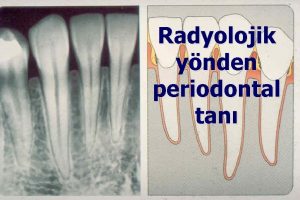
B - Radiographically: 1 - Widening of periodontal ligament space. 2 - Bone loss: horizontal, vertical or both. Dr. Kadhim Jawad 4

C - Histologically. 1 - Inflammatory cells infiltration. 2 - Pronounced loss of collagen. 3 - Loss of CT attachments in some cases. 4 - Apical migration of junctional epithelium. Dr. Kadhim Jawad 5

Examination should include all parts of the periodontium: 1 - Gingiva. 2 - PL & root cementum. 3 - Alveolar bone. In addition to the estimation of Oral hygiene status. Dr. Kadhim Jawad 6

1 -Examination of gingival tissues: Healthy gingiva. Diseased gingiva. GI scoring: Healthy(0), mild(1), moderate(2)&sever(3). Dr. Kadhim Jawad 7

2 -Examination of PL & RC: A-Pocket depth. B-Attachment level. C-Furcation involvement. D-Tooth mobility. Dr. Kadhim Jawad 8

A- Pocket depth. Dr. Kadhim Jawad 9

B- Loss of attachment. Dr. Kadhim Jawad 10

C-Furcation involvement. Dr. Kadhim Jawad 11

Furcation involvement. Degree I: horizontal loss of supporting tissues not exceeding 1/3 of tooth width. Degree II: exceeding 1/3 but not encompassing the total width of furcation area. Degree III: Through & through involvement. Dr. Kadhim Jawad 12

Dr. Kadhim Jawad 13

D- Tooth mobility: D I: mobility of the crown in horizontal direction between (0, 2 - 1 mm). D II: mobility of the in horizontal direction more exceeding 1 mm. D III: vertical mobility of the crown without or with horizontal mobility. Dr. Kadhim Jawad 14

Causes of tooth mobility other than PDs: 1 - Over loading of the tooth. 2 - Trauma from occlusion. 3 - Periapical lesion. 4 - After periodontal surgery. Dr. Kadhim Jawad 15

Factors affecting probing measurements: (over & under estimation). 1 - Thickness of the probe. (under). 2 - Malposition & angulation of the probe. (over). 3 - Crown anatomy. (over). 4 - Amount of pressure applied. (both). 5 - Degree of soft tissue inflammation. (both). Dr. Kadhim Jawad 16

3 - Examination of alveolar bone: This can be best made by taking proper radiographs to see and evaluate: A- alveolar bone height. B- alveolar bone crest outline. Radiographic evaluation should be always interpreted in view of the clinical picture because of problems of overlapping that may lead to omitting of buccal & lingual cortical plates. Dr. Kadhim Jawad 17

Dr. Kadhim Jawad 18

4 - oral hygiene status: In conjunction with the examination of periodontal structures, the state of patient oral hygiene should be determined. This can be easily estimated by using PLI. To determine the presence and extent of microbial dental plaque on tooth surface and dentogingival areas. Dr. Kadhim Jawad 19
 Yousif kadhim accident
 Objective examination of patient
 Boutonniere and swan neck deformity
 Obrunded
 Patient 2 patient
 Periodontal ligament
 Periodontal disease
 Periapical granuloma
 Akut streptokokal gingivitis
 Periodontal cep
 Ligamento periodontal
 General principles of periodontal surgery
 Periodontal dokuların embriyolojisi
 Periodontal instruments classification
 Elementos del periodonto
 Seccin 7
 Periodontal abscess
 Plaque index
 Kista residual
 Lima maestra de cada diente
