
Evolution of PACS as a Model to Predict

- Slides: 21
Evolution of PACS as a Model to Predict Adoption of Digital Pathology Joe Biegel VP, Product Management Mc. Kesson Medical Imaging Group October 23, 2007
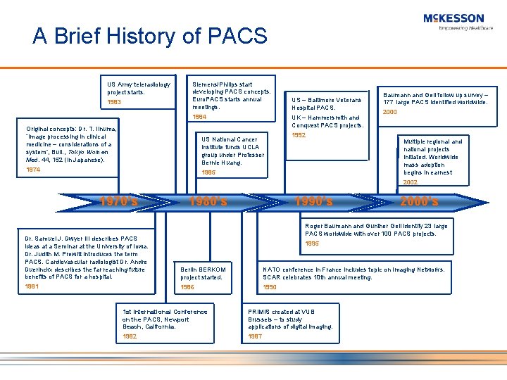
A Brief History of PACS US Army teleradiology project starts. 1983 Siemens/Philips start developing PACS concepts. Euro. PACS starts annual meetings. 1984 Original concepts: Dr. T. Iinuma, “Image processing in clinical medicine – considerations of a system”, Bull. , Tokyo Women Med. 44, 152 (in Japanese). UK – Hammersmith and Conquest PACS projects. US National Cancer Institute funds UCLA group under Professor Bernie Huang. 1974 US – Baltimore Veterans Hospital PACS. 1992 1985 Baumann and Gell follow up survey – 177 large PACS identified worldwide. 2000 Multiple regional and national projects initiated. Worldwide mass adoption begins in earnest 2002 1970’s 1980’s 1990’s 2000’s Roger Baumann and Gunther Gell identify 23 large PACS worldwide with over 100 PACS projects. Dr. Samuel J. Dwyer III describes PACS ideas at a Seminar at the University of Iowa. Dr. Judith M. Prewitt introduces the term PACS. Cardiovascular radiologist Dr. Andre Duerinckx describes the far reaching future benefits of PACS for a hospital. Berlin BERKOM project started. NATO conference in France includes topic on Imaging Networks. SCAR celebrates 10 th annual meeting. 1981 1986 1990 1995 1 st International Conference on the PACS, Newport Beach, California. PRIMIS created at VUB Brussels – to study applications of digital imaging. 1982 1987
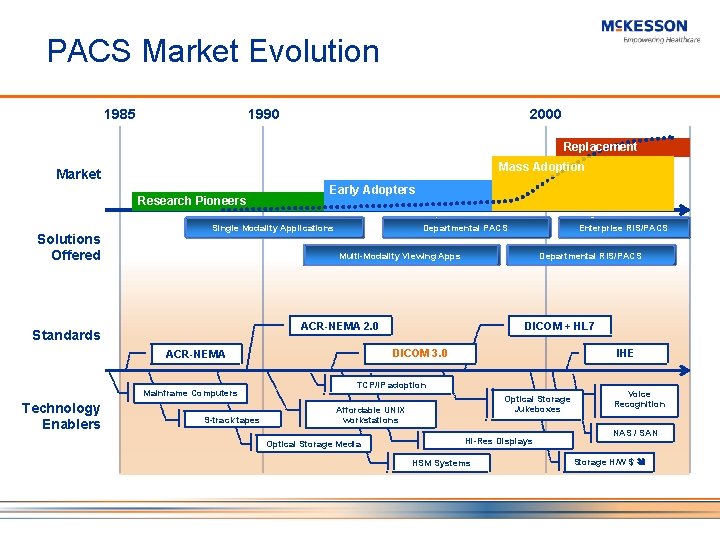
PACS Market Evolution 1985 1990 2000 Replacement Mass Adoption Market Research Pioneers Solutions Offered Early Adopters Single Modality Applications Departmental PACS Multi-Modality Viewing Apps Departmental RIS/PACS DICOM + HL 7 ACR-NEMA 2. 0 Standards DICOM 3. 0 ACR-NEMA Mainframe Computers Technology Enablers Enterprise RIS/PACS 9 -track tapes IHE TCP/IP adoption Optical Storage Jukeboxes Affordable UNIX workstations Optical Storage Media Hi-Res Displays HSM Systems Voice Recognition NAS / SAN Storage H/W $
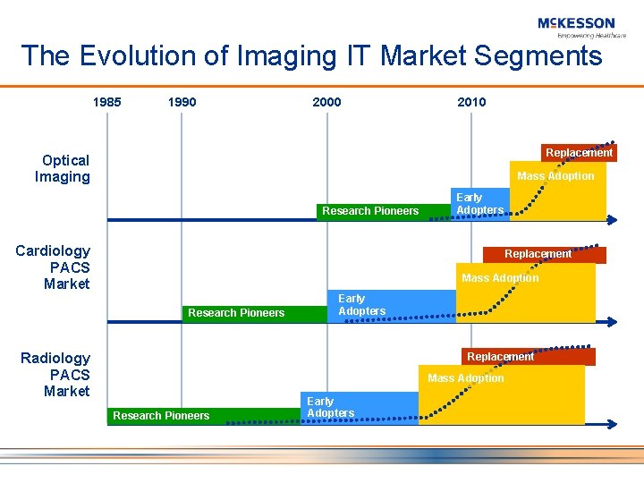
The Evolution of Imaging IT Market Segments 1985 1990 2000 2010 Replacement Optical Imaging Mass Adoption Research Pioneers Cardiology PACS Market Early Adopters Replacement Mass Adoption Research Pioneers Radiology PACS Market Early Adopters Replacement Mass Adoption Research Pioneers Early Adopters
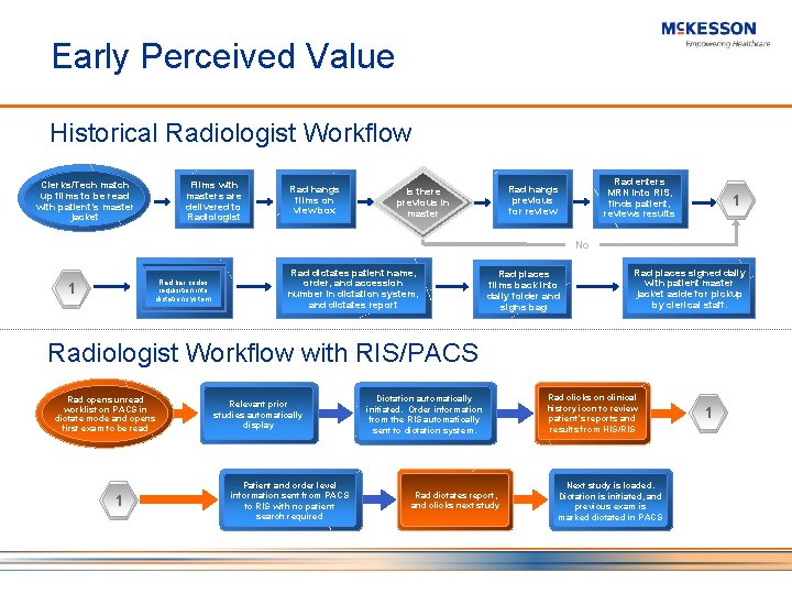
Early Perceived Value Historical Radiologist Workflow Films with masters are delivered to Radiologist Clerks/Tech match up films to be read with patient’s master jacket Rad hangs films on view box Rad enters MRN into RIS, finds patient, reviews results Rad hangs previous for review Is there previous in master 1 No Rad bar codes requisition into dictation system 1 Rad dictates patient name, order, and accession number in dictation system, and dictates report Rad places films back into daily folder and signs bag Rad places signed daily with patient master jacket aside for pickup by clerical staff. Radiologist Workflow with RIS/PACS Rad opens unread worklist on PACS in dictate mode and opens first exam to be read 1 Relevant prior studies automatically display Patient and order level information sent from PACS to RIS with no patient search required Dictation automatically initiated. Order information from the RIS automatically sent to dictation system. Rad dictates report, and clicks next study Rad clicks on clinical history icon to review patient’s reports and results from HIS/RIS Next study is loaded. Dictation is initiated, and previous exam is marked dictated in PACS 1
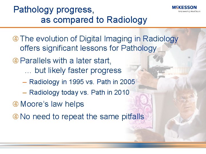
Pathology progress, as compared to Radiology The evolution of Digital Imaging in Radiology offers significant lessons for Pathology Parallels with a later start, … but likely faster progress ─ Radiology in 1995 vs. Path in 2005 ─ Radiology today vs. Path in 2010 Moore’s law helps No need to repeat the same pitfalls Source: Bruce Beckwith, MD – 2005 DICOM International Conference – Budapest, Hungary
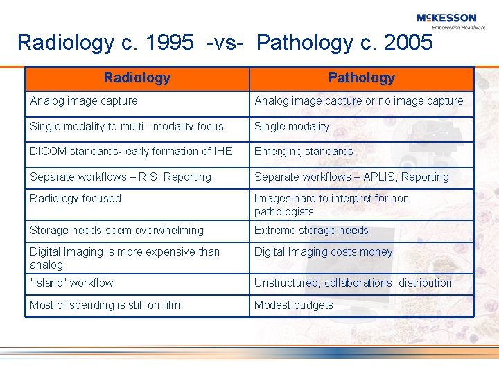
Radiology c. 1995 -vs- Pathology c. 2005 Radiology Pathology Analog image capture or no image capture Single modality to multi –modality focus Single modality DICOM standards- early formation of IHE Emerging standards Separate workflows – RIS, Reporting, Separate workflows – APLIS, Reporting Radiology focused Images hard to interpret for non pathologists Storage needs seem overwhelming Extreme storage needs Digital Imaging is more expensive than analog Digital Imaging costs money “Island” workflow Unstructured, collaborations, distribution Most of spending is still on film Modest budgets
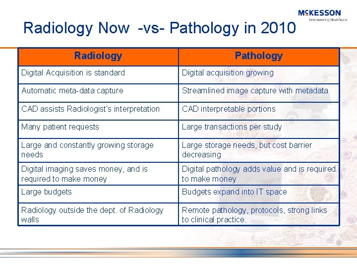
Radiology Now -vs- Pathology in 2010 Radiology Pathology Digital Acquisition is standard Digital acquisition growing Automatic meta-data capture Streamlined image capture with metadata CAD assists Radiologist’s interpretation CAD interpretable portions Many patient requests Large transactions per study Large and constantly growing storage needs Large storage needs, but cost barrier decreasing Digital imaging saves money, and is required to make money Digital pathology adds value and is required to make money Large budgets Budgets expand into IT space Radiology outside the dept. of Radiology walls Remote pathology, protocols, strong links to clinical practice.
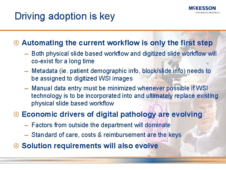
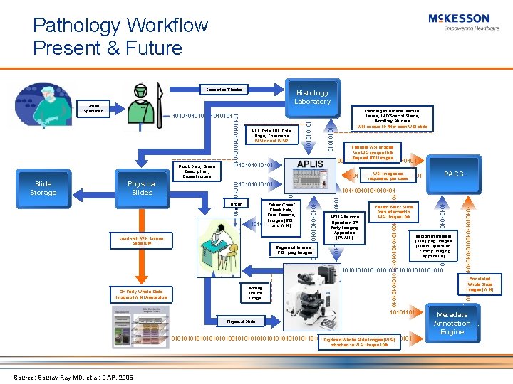
Driving adoption is key Automating the current workflow is only the first step ─ Both physical slide based workflow and digitized slide workflow will co-exist for a long time ─ Metadata (ie. patient demographic info, block/slide info) needs to be assigned to digitized WSI images ─ Manual data entry must be minimized whenever possible if WSI technology is to be incorporated into and ultimately replace existing physical slide based workflow Economic drivers of digital pathology are evolving ─ Factors from outside the department will dominate ─ Standard of care, costs & reimbursement are the keys Solution requirements will also evolve
Pathology Workflow Present & Future Cassettes/Blocks 1010101 PACS 10110010101 Region of Interest (ROI) jpeg images (Direct Operation 3 rd Party Imaging Apparatus) 1010101010101010 3 rd Party Whole Slide Imaging (WSI) Apparatus Analog Optical Image 10101101 Physical Slide 01010101010101010101010101010101010101 Digitized Whole Slide Images (WSI) attached to WSI Unique ID# Source: Sourav Ray MD, et al: CAP, 2006 1010101101001010101010 Patient Block Slide Data attached to WSI Unique ID# 1010101010 APLIS Remote Operation 3 rd Party Imaging Apparatus (TWAIN) 101010110100101010101010 Region of Interest (ROI) jpeg Images 1010101010 Patient/Case/ Block Data, Prior Reports, Images (ROI) 10101011 and WSI) Load with WSI Unique Slide ID# Request WSI Images Via WSI unique ID# Request ROI Images WSI Images as 101100101010101 requested per case 1010101010 Order s 10101 Physical Slides Pathologist Orders: Recuts, Levels, IHC/Special Stains, Ancillary Studies WSI unique ID # for each WSI slide 10110010101010101 0101010 Slide Storage H&E Data, IHC Data, Flags, Comments WSI or not WSI? 10101 Block Data, Gross Description, Gross Images 10110101010101 101010110 Gross Specimen Histology Laboratory Annotated Whole Slide Images (WSI) Metadata Annotation Engine
Future Pathology Workflow The vision of automating the current workflow is a noble goal, but it is only the first step ─ The true value of Digital Pathology will have to be defined in terms of faster, improved Patient Care Going Digital will change the practice of Pathology and will lead to a new, yet undefined workflows ─ Standards, IHE will help: necessary but insufficient to drive wide-scale adoption
DICOM in Pathology Visible Light Supplement 15 ─ Included support for gross images, microscopic images, x, y. z position, lab accession numbers Working Group 26 ─ Evolving to include whole slide imaging Growing number of imaging products targeting pathology are DICOM compliant US Veteran’s Administration: ─ Pathology imaging vendors must be DICOM compliant
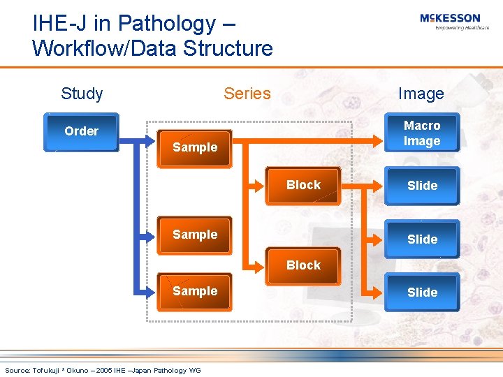
IHE-J in Pathology – Workflow/Data Structure Study Series Image Macro Image Order Sample Block Sample Slide Block Sample Source: Tofukuji * Okuno – 2005 IHE –Japan Pathology WG Slide
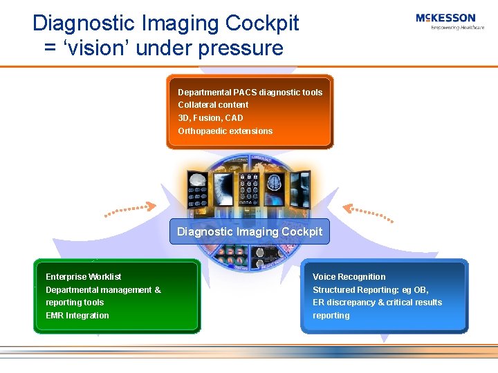
Diagnostic Imaging Cockpit = ‘vision’ under pressure Departmental PACS diagnostic tools Collateral content 3 D, Fusion, CAD Orthopaedic extensions Diagnostic Imaging Cockpit Enterprise Worklist Voice Recognition Departmental management & Structured Reporting: eg OB, reporting tools ER discrepancy & critical results EMR Integration reporting
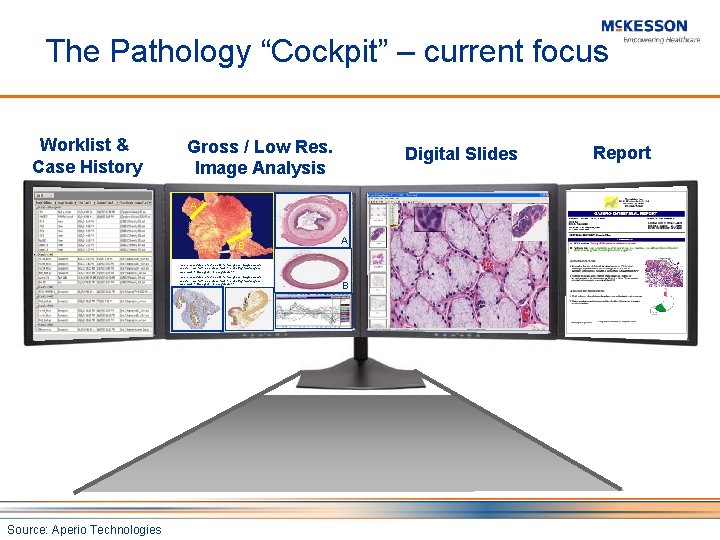
The Pathology “Cockpit” – current focus Worklist & Case History Gross / Low Res. Image Analysis Digital Slides A B A This is an example text bocks for the gross image shown above. This text is so small that it is unlikey that anyone can read it. Can you? I really doubt it. Source: Aperio Technologies B Report
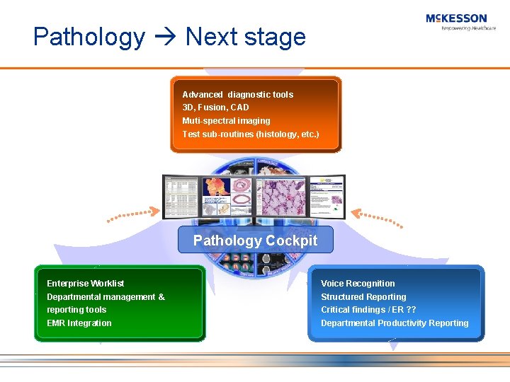
Pathology Next stage Advanced diagnostic tools 3 D, Fusion, CAD Muti-spectral imaging Test sub-routines (histology, etc. ) Pathology Cockpit Enterprise Worklist Voice Recognition Departmental management & Structured Reporting reporting tools Critical findings / ER ? ? EMR Integration Departmental Productivity Reporting
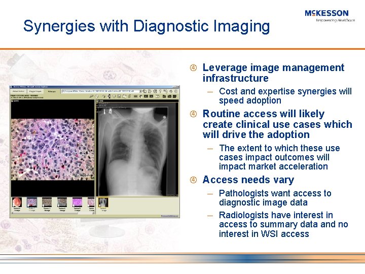
Synergies with Diagnostic Imaging Leverage image management infrastructure ─ Cost and expertise synergies will speed adoption Routine access will likely create clinical use cases which will drive the adoption ─ The extent to which these use cases impact outcomes will impact market acceleration Access needs vary ─ Pathologists want access to diagnostic image data ─ Radiologists have interest in access to summary data and no interest in WSI access
Value Points = Business Control Points Modality = Data Acquisition Storage = Physical Storage Devices (H/W) Image Access Points = Clinical Application(s) Workflow = Order $ Data = Data-mining CAD – address the volume – may be more formative than in Radiology Financial benefit needs to become more clear
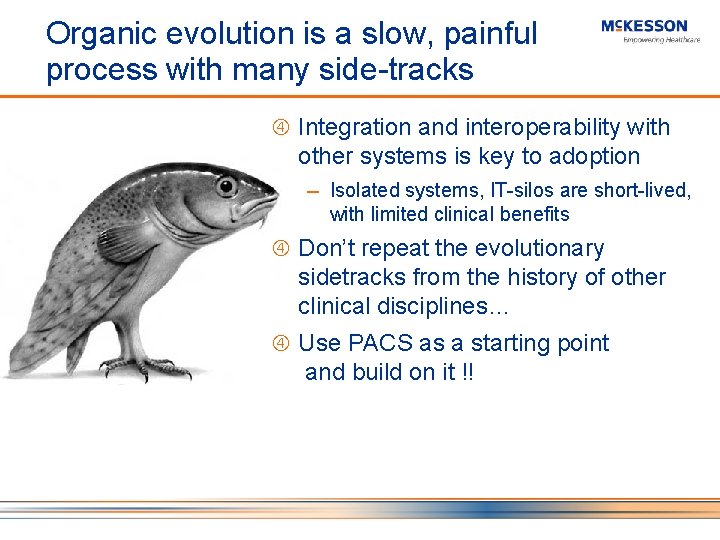
Organic evolution is a slow, painful process with many side-tracks Integration and interoperability with other systems is key to adoption ─ Isolated systems, IT-silos are short-lived, with limited clinical benefits Don’t repeat the evolutionary sidetracks from the history of other clinical disciplines… Use PACS as a starting point and build on it !!
Summary Pathologists need PACS now value is immediate ─ Pathologists should have PACS access now – the patient record needs to contain relevant imaging data ─ If you want to accelerate the adoption-curve of digital pathology: ─ ‘Hook’ your digital Pathology plans to your PACS It is likely that early workflows will leverage other specialties, BUT these will inevitably be transient solutions with the “real” Digital Pathology workflow to follow ─ Pathology dept specific workflow solutions are needed and must have digital path imaging as part of the workflow now.
Questions?