Evaluation of the Febrile Patient Fevers and Fevers

Evaluation of the Febrile Patient Fevers and Fevers of Unknown Origin A case-based approach Richard Serrao MD Assistant Professor of Medicine, BUSM Sections of Internal Medicine and Infectious Diseases

The febrile patient �Fevers: Relatively straightforward approach Definitions Fever vs hyperthermia vs hyperpyrexia Mechanisms Initial approach �Fevers of unknown origin: More complex, more fun to chase Definition Spectrum Fun Cases and categories

What is a fever? �Elevation in body temperature above normal range from increase in temperature regulatory set point : 99. 5– 100. 9 °F Mouth >99. 9 Axilla/otic >99. 0 Anus >99. 5 -100. 9 �Hyperpyrexia: typically not infectious 104– 106. 7 °F Set points vary depending on clinical setting

Fever versus hyperthermia � Fever: resetting of thermostatic set-point in the anterior hypothalamus and the resultant initiation of heat-conserving mechanisms until the internal temperature reaches the new level If acute (and less commonly chronic), infection unless proven otherwise � Hyperthermia: an elevation in body temperature that occurs in the absence of resetting of the hypothalamic thermoregulatory center Usually not mediated by infectious diseases The difference between these two is mechanism
 �Pyrogens (endogenous and exogenous) trigger fevers via release")
Mechanisms of fever (in 3 sentences) �Pyrogens (endogenous and exogenous) trigger fevers via release of prostaglandin E 2 hypothalamic stimulation vasoconstriction, then shivering temp rise �Endogenous: IL 1, 6, 8, TNF, IFNa, b, g arachidonic acid pathway activated Can be released in collagen vascular, malignancy �Exogenous: i. e. LPS binds to lipopolysaccharide binding protein release of IL-1 Typically infectious
 endotoxin of GNRs binds")
Mechanisms of fever: Examples of Common Bacterial pyrogens �Lipopolysaccharide (LPS) endotoxin of GNRs binds to LPS-binding protein and is transferred to CD 14 on macrophages, which stimulates the release of TNFα. �Staphylococcus aureus enterotoxins �Staphylococcus aureus toxic shock syndrome toxin (TSST) Both Staphylococcus toxins are superantigens and activate T cells leading to the release of interleukin (IL)-1, IL-2, TNFα and TNFβ, and interferon (IFN)-gamma in large amounts �Group A and B streptococcal toxins Exotoxins induce human mononuclear cells to synthesize not only TNFα but also IL 1 and IL-6
: Mechanisms of hyperthermia/hyperpyrexia Excessive heat production: exertional hyperthermia, thyrotoxicosis, pheochromocytoma,")
Fever types (by severity): Mechanisms of hyperthermia/hyperpyrexia Excessive heat production: exertional hyperthermia, thyrotoxicosis, pheochromocytoma, cocaine, delerium tremens, malignant hyperthermia Disorders of heat dissipation: heat stroke, autonomic dysfunction Disorders of hypothalamic function: neuroleptic malignant syndrome, CVA, trauma All non infectious!


General approach to Fevers �Chase the symptom Headache, meningismus, pharyngitis, dysuria, diarrhea, abdominal pain, etc. ▪ Makes it obvious! �Start with the basics: ua/urine culture, CXR, blood cultures �Follow the pattern �Look at everything for patients in whom the pattern is inconsistent �Most fevers diagnosed early (i. e. not an FUO yet) will have an infectious cause…

…ergo: The preliminary work-up �History �PE �CBC with diff �Blood cultures (3 sets drawn from different sites over at least several hours OFF abx) �Chem 7 and LFTs �Hepatitis serologies if LFTs abnormal �UA, urine culture �CXR Most patients will have the answer here

“A patient with a fever that has not had a basic work up yet does not an FUO make”
Petersdorf and Beeson 1961 “Fever > 38. 3 (101) on")
Fever of Unknown Origin (FUO)Petersdorf and Beeson 1961 “Fever > 38. 3 (101) on several occasions, persisting without diagnosis for at least 3 weeks in spite of at least 1 week’s investigation in hospital. ”

Categories of FUO/updated criteria Featurea Nosocomial Neutropenic HIV-associated Classic Patient’s situation Hospitalized, acute care, no infection when admitted Neutrophil count Confirmed HIVeither <500/µL or positive expected to reach that level in 1 -2 days All others with fevers for ≥ 3 weeks Duration of illness while investigated 3 daysb or 3+ outpatient visits Examples Septic thrombophlebitis, sinusitis, C. difficile colitis, drug fever Perianal infection, MAIc infection, aspergillosis, TB, noncandidemia Hodgkin’s lymphoma, drug fever a. All require temperatures of ≥ 38. 3°C (101°F) on several occasions. at least 2 days’ incubation of microbiology cultures. c. M. avium/M. intracellulare. b. Includes 3 daysb (or 4 weeks as outpatient) Infections, malignancy, inflammatory diseases, drug fever Modified from DT Durack, AC Street, in JS Remington, MN Swartz (eds): Current Clinical Topics in Infectious Diseases. Cambridge, MA, Blackwell, 1991.

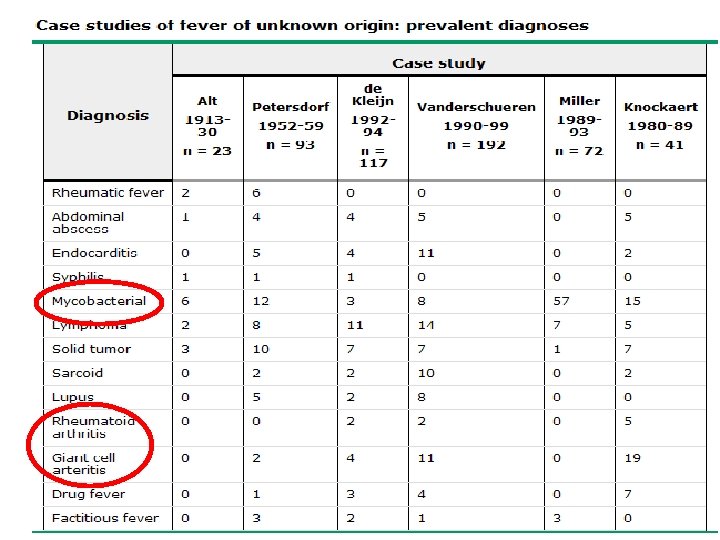
The changing spectrum � Over decades, the etiology has been shifting � 3 classic categories are consistent: Infections Malignancies Connective tissue diseases � True FUOs are uncommon � Excessive use of empiric antibiotics can mask/delay diagnosis of occult abscesses/infections; drug fevers increasing � MDR organisms, lengthy ICU and chemo regimens changing the flora

The changing spectrum � Undiagnosed FUOs has gone from 75% to <10%, but the fraction of FUOs that are undiagnosed has increased � More connective tissue diseases are being diagnosed � Extrapulm TB, solid tumors and intrabdominal abscesses are less prevalent due to CT scanning CT guided IR has replaced ex-laps for diagnosis � Better blood culturing techniques has reduced IE as an etiology for FUO Culture negative or fastidious organisms (bartonella) remain as FUOs The longer a patient has a fever, the less likely an ID cause will be found

Case #1 � 72 y. o, male with cirrhosis, diabetes and is on chronic steroids for RA who is s/p partial colectomy for recurrent diverticular disease
 abscess � usually located in the abdomen or pelvis � cirrhosis,")
Case #1: (Occult) abscess � usually located in the abdomen or pelvis � cirrhosis, steroid or immunosuppressive medications, recent surgery, and diabetes are predisposing factors � arise in locations of disrupted barrier in bowel wall (appy, tics, IBD). � develop in subphrenic, omental, pouch of Douglas, pelvic, and retroperitoneal locations. � Pyogenic liver abscesses in setting of biliary tract disease, appendicitis or diverticulitis. � Amebic liver abscesses can look like pyogenic abscesses; check e histolytica serologies. � Splenic abscesses occur from hematogenous seeding: look for concominant endocarditis � Perinephric abscesses can have normal UA/urine culture

When an infection is the cause of FUO by disease � � � � � Intraabdominal abscess (liver, splenic, psoas, etc) Appendicitis Cholecystitis tubo-ovarian abscess Intracranial abscess Sinusitis dental abscess Chronic pharyngitis Tracheobronchitis Things that are hidden, lung abscess may give false negative Septic jugular phlebitis blood cultures, or have an mycotic aneurysm Endocarditis unrevealing (missed on) intravenous catheter infection exam vascular graft infection Wound infection Osteomyelitis infected joint prosthesis Pyelonephritis prostatitis We’re getting better at finding these things!

We’re getting better at finding infections and cancers thereby reducing the total FUOs seen, but leaving many undiagnosed cases
;")
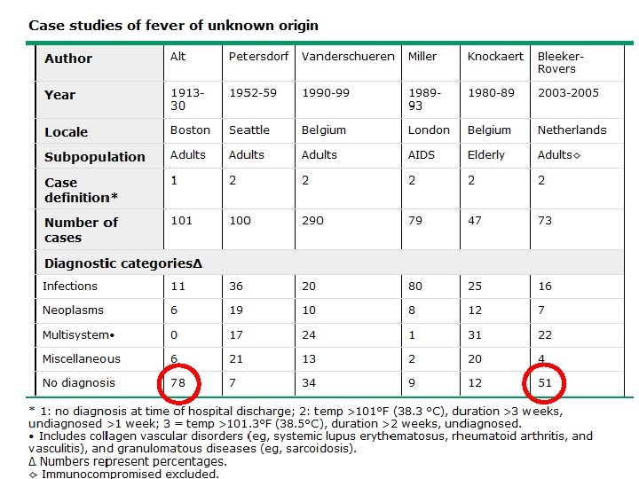
True FUOs rare � 2800 hospital beds over 2 year period (2003 - 2005); only 73 cases of FUOs found Noninfectious inflammatory diseases (vasculitis, SLE, PMR) — 22% Infection — 16% Malignancy — 7% Miscellaneous — % No diagnosis — 51% Bleeker-Rovers et al. Medicine (Baltimore). 2007 Jan; 86(1): 26 -38.

Case #2 � 45 y. o G 5, P 5 woman from subsaharan Africa with HIV who presents with daily fevers as high as 103. � She has no cough, SOB, headache. She is on no meds. � Her LFTs are mildly elevated with mild hepatomegaly and ascites. HAV/HBV/HCV negative. � CXR is negative. � CBC demonstrates microcytic anemia with teardrop cells. � She is not on HAART and has a CD 4 of 376, VL 28, 000

Case #2 �Ascites fluid is lymphocytic predominant. Blood cultures, ascitic fluid: no growth. �BM aspirate: oraclesyndicate. twoday. net

Case #2: Tuberculosis as an FUO � single most common infection in most FUO series. � Can be extrapulmonary, miliary, or occur in the lungs of patients with significant preexisting pulmonary disease, or immunodeficiency � pulmonary tuberculosis in AIDS patients is subtle; CXR normal in up to 20% � Readily treatable � PPD positive in <50% � Sputum + in only 25% � May require lymph node, BM or liver biopsy � Isolator blood cultures need incubation for >16 days

Geography and FUOs � Developing world TB Typhoid Amebic liver abscesses AIDS � Returning traveler Malaria Brucellosis Kala azar Filariasis Schistosomiasis Lassa fever
 infections")
Worldwide “FUOs” � Ease of travel can increase risk for non endemic (autochthonous) infections to be present in the recent traveler given long incubation period malaria, brucellosis, kala azar, filariasis, schistosomiasis, Lassa fever � the most common infections among series of FUOs have not changed over the century: typhoid fever, TB, amebic abscesses, malaria. � FUOs are more often caused by an atypical presentation of a common entity than by a rare disorder

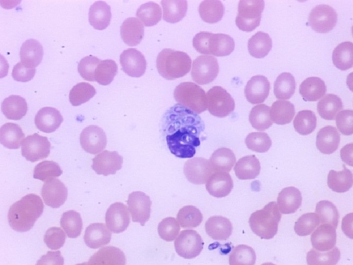
Case: Fever in the states � 56 y. o. male HIV+, CD 4 332, VL ND on HAART presents in Nov with fever 104. 0 q day, shaking chills, myalgias and malaise. Mild diffuse HA. Evanescent rash noted on left shoulder �Recent hiking in NH �PE: flushed, ill but not toxic. No focality �Pancytopenia (new), AST 150, ALT 161, TB 0. 4
 �Always look at peripheral smear in presumptive BM disorders �Intraleukocytic/intraerythrocytic inclusions �<60%")
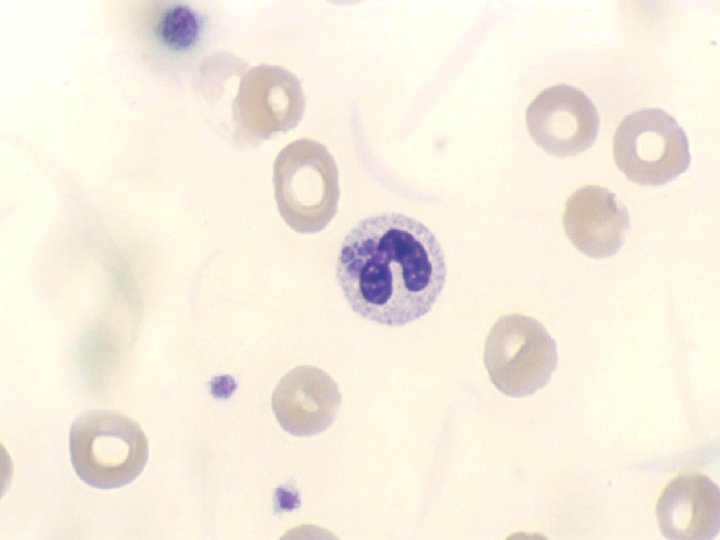
Anaplasmosis (Ehrlichiosis) �Always look at peripheral smear in presumptive BM disorders �Intraleukocytic/intraerythrocytic inclusions �<60% recall tick exposure �Search for coexistent lyme and babesiosis

When an infection is the cause of FUO by pathogen �Tuberculosis, Mycobacterium avium complex, syphilis, Q fever, legionellosis �Salmonellosis (including typhoid fever), listeriosis, ehrlichiosis, �Actinomycosis, nocardiosis, Whipple’s disease �Fungal (candidaemia, cryptococcosis, sporotrichosis, aspergillosis, mucormycosis, Malassezia furfur) �Malaria, babesiosis, toxoplasmosis, schistosomiasis, fascioliasis, toxocariasis, amoebiasis, infected hydatid cyst, trichinosis, trypanosomiasis �Cytomegalovirus, HIV, Herpes simplex, Epstein. Barr virus, parvovirus B 19
")
Subpopulations of FUOs � HIV/AIDS: 79% infections ▪ 50% mycobacterial (2/3 atypical mostly MAC) 8% malignancies: NHL; disseminated KS rare 9% no diagnosis � Neutropenia � Most due to bacteremia Fungi as source of FUO increases after 7 days (fungemia, then aspergillus) Confounders include: cancer, drugs, blood products Fever when neutropenia resolves: hepatosplenic candidiasis Age: Children: 30% self limited viral syndrome Age >65: 30% caused by PMR, vasculitis, GCA, sarcoid; 22% infections; 12% cancers

Case #3: FUO in AIDS � 55 y. o. HCV/HIV+, CD 4 5, VL 115, 000 noncompliant with HAART. CC: confusion, fever, anisocoria �Head CT: mass lesion with hemorrhage; CT body: no lymph node enlargement �CSF: normal �Pyrimethamine/sulfadiazine started empirically for toxo

Case #3: FUO in AIDS �Within 2 days, >20 SC erythematous tender nodules on shins and buttocks
 �Rheumatic fever �Sarcoidosis �Drug induced �Post-streptococcal �Mycoplasma �Coccidioidomycosis �Other")
Fever with erythema nodosum (panniculitis) �Rheumatic fever �Sarcoidosis �Drug induced �Post-streptococcal �Mycoplasma �Coccidioidomycosis �Other fungal �TB/nontuberculous mycobacteria �SLE �Vasculitis �syphilis

Case #3: FUO in AIDS: Skin Biopsy

Case #3: FUO in AIDS: Culture results: Mycobacterium avium intracellulare �Blood and skin grew Mycobacterium avium intracellulare �Patient recovered with Clarithromycin, rifampin and ethambutol Tandon R, Kim K, Serrao R. Disseminated mycobacterium avium-intracellulare infection in a person with AIDS with cutaneous and CNS lesions. The AIDS Reader. 2007; 17(11): 555 -560

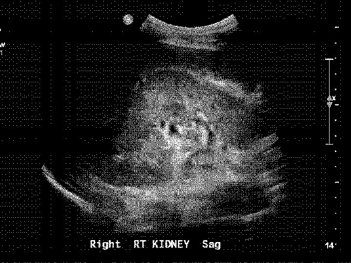
FUO in AIDS case 2 � 36 y. o. male with ? PMH of PCP PNA 1 yr PTA and several suicide attempts with benzos/percocets �BIB family members who noted patient to be lethargic, c/o chills and RUQ pain �Exam notable for T 102. 6, HR 110, 100/55, O 2 sat 90% RA, lethargic, conjunctival injection, thrush, ? Nuchal rigidity, dry crackles, mod RUQ tenderness, hepatomegaly, +/- guarding �Admitted to ICU with dx of acute hepatic failure secondary to probable tylenol overdose (sister saw empty bottles in room) �Grew up in Chicago
, 11/33, Plts 90 � 134/3. 3/102/9/34/2. 3 AG 23 �LFTs:")
�WBC: 12 (90 P), 11/33, Plts 90 � 134/3. 3/102/9/34/2. 3 AG 23 �LFTs: AST 2344, ALT 2688, Alk Phos 113 �PT 15, PTT 42 �DIC screen + �Acetominophen level, pancultures pending �CXR: diffuse interstitial infiltrates

�Patient started on ceftriaxone 2 g iv q 12, vanco 1 g q 12, ampicillin 2 q 8 and high dose Bactrim. �N-acetylcysteine administered empirically �STAT RUQ ultrasound read as hepatomegaly with TNTC punctate lesions throughout parenchyma of liver; no bil dil; no stones

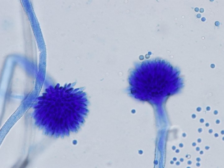
Disseminated histoplasmosis

Disseminated histoplasmosis
 with transaminase elevations, unexplained")
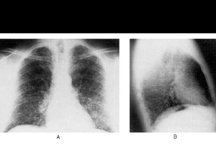
Disseminated histoplasmosis �Consider in immunocompromised host (AIDS, solid organ transplant) with transaminase elevations, unexplained fever, reticulonodular pulmonary infiltrates from endemic area

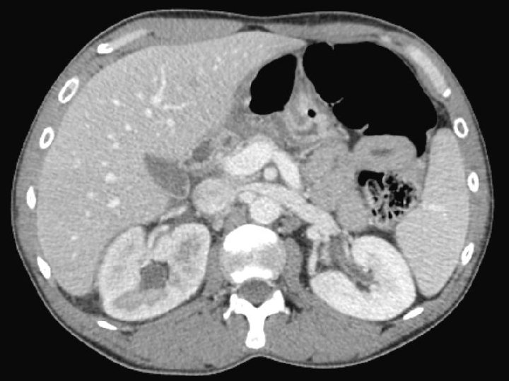
Case #4 � 35 y. o. recuperating counts after prolonged neutropenia for AML who has been on cefepime and vanco and defervesced early. �C/o RUQ tenderness, LFT abnormalities, febrile �CT abdomen reveals multiple punctate small echogenic lesions

CT Scan shows multiple hypodense lesions in the liver and spleen. Curr Probl Diagn Radiol, November/December 2004

Case #4: Hepatosplenic candidiasis �Complication of prolonged neutropenia that required systemic antimicrobials when fever abates �Due to duration of systemic antimicrobials (as compared to aspergillus which is due to duration of neutropenia) �Treat with fluconazole/echinocandins if suspect glabrata

Case #5 � 71 y. o. male c/o fevers, night sweats, weight loss for 3 months. Generalized muscle aches and difficulty getting up from chair �PMH: sulfa allergy, noted to be anemic 1 yr ago, negative blood loss w/u �PPD neg �Labs anemic & leukopenic with monocytosis. ANA, DS DNA, and LFTS nl �ESR 102, elevated serum ferritin
 arteritis. Left panel: Granulomatous and lymphocytic inflammation")
Temporal artery biopsy in giant cell (temporal) arteritis. Left panel: Granulomatous and lymphocytic inflammation of the adventitia and medial wall of the temporal artery. Right panel: Elastic tissue stain showing disruption of the elastica (arrows) due to immunologically mediated destruction of the elastica. Courtesy of Cynthia Magro, MD via uptodate. com

DDx of FUO in elderly � Temporal arteritis/PMR/vasculitis/sarcoid: 30% � Infectious: 22% Endocarditis Cryptogenic intraabdominal abscess Extrapulm TB � Malignancy: 12% Lymphoma Hepatoma Renal cell ca Unusual to cause FUO: colon ca, leukemia, MDS


Approach to FUO: Clues �Clues on exam and labs: 11 diagnostic clues/patient in history and physical 3/patient in laboratory tests 81% of these clues are misleading �Ask about: Travel Animal exposure (pets, living on a farm, occupational) Immunosuppression Drug and toxin history Localizing symptoms Bleeker-Rovers CP et al Medicine. 2007 Jan; 86(1): 26 -38.
 � Intermittent: fever for a certain period, cycling back")
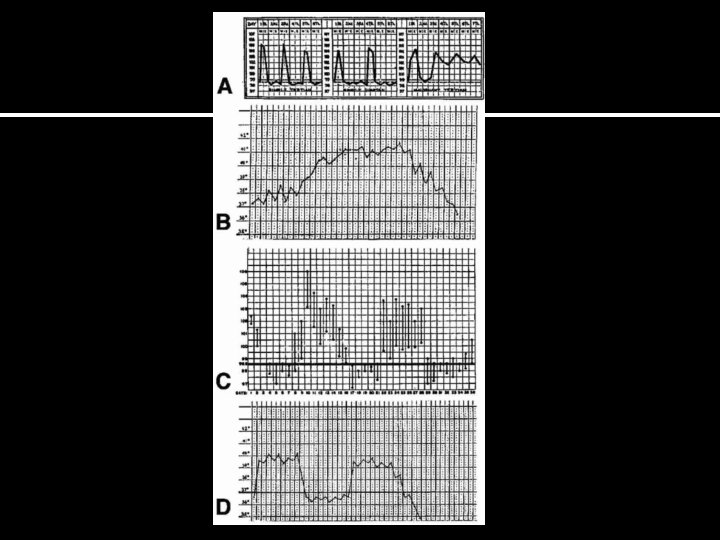
Clues: Fever types (by curves) � Intermittent: fever for a certain period, cycling back to normal: Malaria, kalaazar, sepsis � Continuous: Temperature remains above normal throughout the day and does not fluctuate more than 1 °C in 24 hours: � lobar pneumonia, typhoid, UTI, brucella or typhus Pel-Ebstein fever: high for one week, low for the next: � Quotidian fever, with a periodicity of 24 hours, typical of Plasmodium falciparum or Plasmodium knowlesi Tertian fever (48 hour periodicity), typical of Plasmodium vivax or Plasmodium ovale malaria Quartan fever (72 hour periodicity), typical of plasmodium malariae Hodgkins lymphoma Remittent fever: Temperature remains above normal throughout the day and fluctuates more than 1 °C in 24 hours Infective endocarditis
 Borreliosis")
Old school curves A. B. C. D. Malaria Typhoid fever Hodgkins (Pel Ebstein) Borreliosis (relapsing fever) Specificity of these curves is still generally poor Hirschmann Clin Infect Dis. 1997 Mar; 24(3): 291 -300; quiz 301 -2 www. bioscience. org
 �Hyperpyrexia >106. 7 Intracranial hemorrhage most common Sepsis Kawasaki")
Clues: Fever types (by severity) �Hyperpyrexia >106. 7 Intracranial hemorrhage most common Sepsis Kawasaki syndrome Neuroleptic malignant syndrome Drug fever Serotonin syndrome Thyroid storm

Clues: ID associated relative bradycardia �Typhoid fever �Malaria �Typhus �Yellow fever �Leptospirosis �Dengue �RMSF �Q fever

Clues: Fever after travel �Malaria �Typhoid fever �Acute HIV �Kala-azar �Amebic liver abscess �Tuberculosis �Brucellosis �meloidosis

Case #5: interesting clue 27 y. o. female presents with sore throat, fever and diffuse myalgias/arthralgias -WBC 14. 3, ESR 67, CPK slightly incr. Neg CXR, blood cxs neg -CT abd/pelvis: splenomegaly. -RF, ANA, ANCA neg -Unprotected sex 2 months ago http: //www. dailystrength. org/groups//media/611476
 returns at 121")
Case #5 �HIV 1, 2 Ab negative �HIV-RNA PCR (viral load) returns at 121 copies
 �Arthralgias, fevers, elevated ferritin and salmon colored evanescent rash")
Still’s disease (Juvenile Rheumatoid Arthritis) �Arthralgias, fevers, elevated ferritin and salmon colored evanescent rash � 1. 5 cases per 100, 000 -1, 000 population �Bimodal distribution: 15– 25 and second peak between ages of 36– 46 �Diagnosis of exclusion
 � Major criteria: fever")
Diagnostic criteria: need 5 (at least 2 of them major) � Major criteria: fever >39 C for > 1 week Arthralgias or arthritis for at least two weeks, Nonpruritic salmon colored rash (usually over trunk or extremities while febrile), Leukocytosis ( 10, 000/micro. L or greater), with granulocyte predominance � Minor criteria: Sore throat, Lymphadenopathy, Hepatomegaly or splenomegaly, Abnormal liver function tests, Negative tests for antinuclear antibody and rheumatoid factor What infectious disease does this sound like? Mono syndrome: HIV/EBV/CMV

Collagen vascular disease as FUO �SLE and ANCA+ disorders rare �Polymyalgia rheumatica �Temporal arteritis �Giant cell arteritis �Adult still’s disease �Behcets �Kikuchi’s disease Again, most of these in older patients
 � 55 y. o. female presents with 4")
Case #6: interesting clue (? ) � 55 y. o. female presents with 4 months of persistent night sweats, fevers �Visited a farm with kids and watched a goat give birth 5 months ago
 � � � � Caused by coxiella burnetii")
Q fever (and culture neg endocarditis) � � � � Caused by coxiella burnetii (serologies): “parturient cats, ” sheep, goats, cattle; aerosolization/contact with body fluids/ingestion of infected milk; ticks as reservoirs; ILI, painless hepatitis, atypical pna, prolonged FUO; dx: serologies Cultures are negative in 2 -5% in all cases of BE especially in partially treated Culture negativity common with Coxiella Burnetii, tropheryma whippeli, brucella, mycoplasma, chlamydia, histoplasma, legionella, bartonella HACEK: haemophilus spp, actinobacillus, cardiobacterium, eikenella and kingella: alert lab to incubate cultures for 7 -21 days Peripheral manifestations are rare TEE reveals veg in 90% of SBE presenting as FUO Treated with tetracycline

The algorithmic approach to workup/labs/imaging

Mourad, et al. Arch Intern Med. 2003; 163: 545

Laboratory work up: False positive rate=rate of a helpful result � ESR and crp: TB, osteo, CA, CVD >100 � LDH: lymphoma, PJP � PPD or quantiferon: TB � HIV ab (consider viral load): HIV � 3 blood culture sets over several hours (prior to antibiotics): bacteremia � RF: rheumatoid arthritis � CPK: PMR, polymyositis � Heterophile ab (children, young adults): mono (EBV) � SPEP: MM � ANA: autoimmune process � CT of abdomen and chest: r/o lymphadenopathy, nodules

Utility of ESR: good negative predictive value � ESR > 100 Malignancy (lymphoma, myeloma, metastatic colon or breast): 58% Infections: endocarditis, TB, osteo: 25% Collagen vascular disease: 25% � Less serious causes causing ESR > 100 Drug hypersensitivity Thrombophlebitis Renal disease (nephrotic syndrome) � Normal ESR suggests that a significant inflammatory process, of whatever origin, is absent can be seen in GCA, however Zacharski et al JAMA. 1967 Oct 23; 202(4): 264 -6.

Utility of CT, tagged WBC and PET Scanning �Useful to pick up lymphadenopathy, granulomas Lymphomas, tuberculous, fungal �Gallium-67 and/or indium-111: highly sensitive for whole body but not specific �PET scan data is limited in FUO �Start with CT/ultrasounds, then tagged wbc/PET later

Additional tests � Dictated by history Subtle nervous system signs: LP Travel to midwest or southwest: investigate cocci, histo, blasto Trauma: LENI � Biopsies: Liver biopsy generally low yield for miliary tb, granulomatous hepatitis or sarcoid Lymph nodes: better for lymphoma, cat scratch Temporal artery: low yield in minimal clinical signs BM: generally high yield in thrombocytopenic/anemic, otherwise low in immunocompetent � Therapeutic antibiotic trials: do not do Bleeker-Rovers CP et al. Medicine (Baltimore). 2007; 86(1): 26
")
Outcomes �Treatment of fever alone is generally not necessary except in hyperpyrexia (>106. 7) �Outcome depends on underlying diagnosis �Children: 88% with infections have no sequelae �Adults: most who remain undiagnosed after extensive evaluation have a good prognosis

Pattern recognition: recognition a couple more classic and interesting cases

Fever in patient with “spider bites” and back pain � 21 y. o. male presents for the 3 rd time to local ER for back pain. H/o narcotic abuse. Sent out after toradol injections and ibuprofen. Each visit noted to have temps of 99. 8. Multiple “spider bites” near buttocks

Vertebral discitis �Strongly consider in IVDU �Plain films notoriously negative �Blood cultures can have high yield if concominant endocarditis �Detailed neurologic exam necessary �Urgent imaging if focal neuro deficits �Need to rule out osteomyelitis, epidural abscess

Sterile pyuria � 46 y. o. male with poorly controlled DM, chronic active HCV, and active IVDU presents with 1 month of dysuria and eventual fevers �Several outpatient cultures negative �Admitted with acute B obstructive uropathy, pyelo, and ureteral “clots”

Bilateral renal aspergillus obstructive uropathy �Suspect TB in patient at risk for tuberculous pyelo as top cause for ID diff for sterile pyuria �In non-endemic area, IVDU/HCV/DM suspect aspergillus

Fever and bullous eruption � 78 y. o. female with COPD/CHF intubated x 1 week in ICU. She is on vancomycin, cefepime. She has mild eosinophilia. Her hands look like this:

http: //www. jaoa. org/content/104/4/157/F 4. expansion

Drug allergies �Vancomycin mediated linear Ig. A: appears like bullous pemphigoid �Responds to drug cessation/steroids �Generally requires biopsy

Drug fevers � � � 30% of hospitalized patients suffer from adverse drug reactions; outpatients can develop drug fever years later after starting a drug Eosinophilia and rash are only seen in 25% of drug fevers Most common drugs: Antimicrobials: sulfonamides, penicillins, nitrofurantoin, vancomycin, antimalarials H 1 and H 2 blocking agents Antiepileptics: barbiturates, phenytoin Iodide contrast media NSAIDS Antihypertensive drugs: hydralazine, methyldopa Antiarrythmics: quinidine, procainamide Antithyroid Contaminants such as quinine (in heroin or cocaine) Rarely cause: digoxin, aminoglycosides 25% of AIDS patients develop drug fever usually manifest as nausea and vomiting Diagnosis: therapeutic trial off drug: wait at least a week before ruling in/out

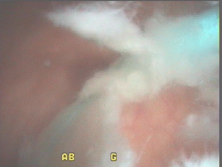
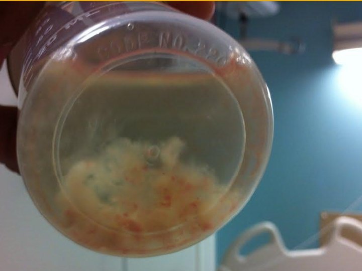
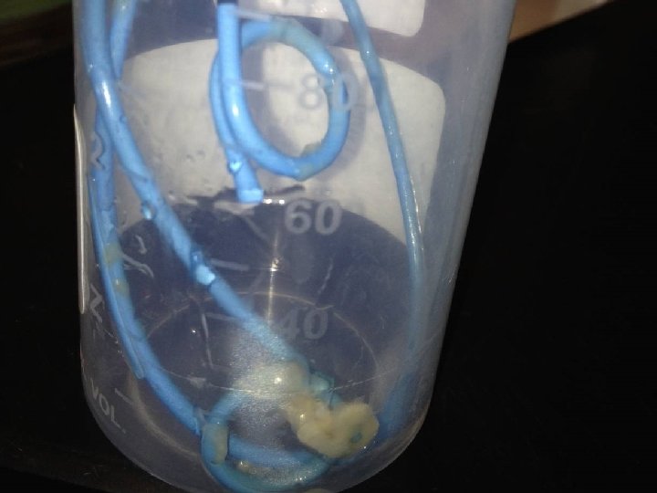
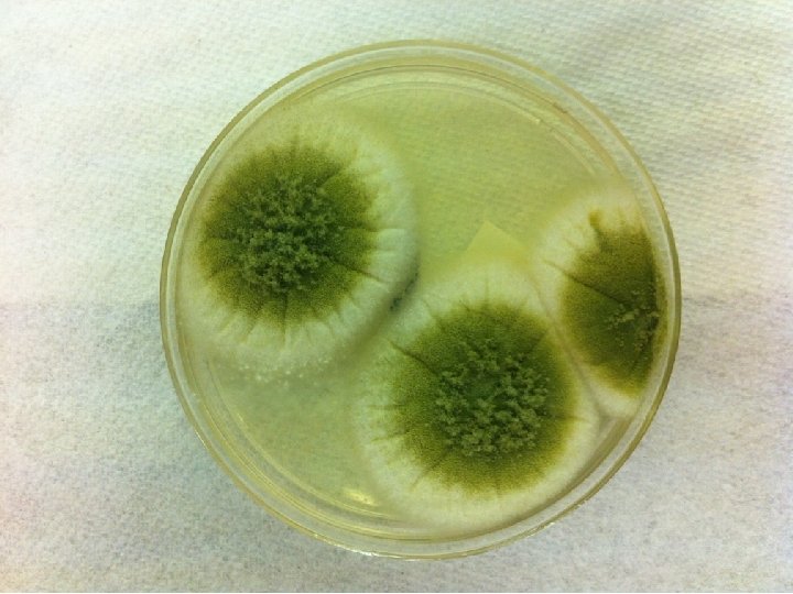
The “at-risk” patient � 82 y. o. male s/p urgent partial colectomy for diverticular abscess. NPO, NGT, on vanco and zosyn, TPN, vented, triple lumen left IJ. Yeast in sputum, febrile daily with negative cxr, UA, abdominal CT

Fungemia �Strongly consider empiric or pre-emptive treatment in patients on broad spectrum antibiotics, TPN, intrabdominal surgery, central lines, critically ill �If fungemic, check ophtho and TTE to rule out endophthalmitis/endocarditis. �Consider line/mucosa/bowel as source �Discontinue antimicrobials asap to minimize pressure for fungemia

The culture negative “impetigo” � 25 y. o. female presents with intermittent fevers for past week and painful facial lesions �Temp 103 �Hg. B 9. 2, WBC 2 (50% pmns), platelets 60 �BM Bx: myelodysplastic syndrome �Patient remains febrile despite broad spectrum abx and systemic antifungals for 1 week

Biopsy reveals PMNs, neg gram stain, neg culture Dermis. net

Neutrophilic dermatosis: Sweet’s syndrome �Acute onset of fever, leukocytosis, erythematous plaques infiltrated by neutrophils with no evidence of vasculitis or infection �Responds to steroids

Final word on esoteric causes Factitious fevers: psych patients, munchausens Disordered heat homeostasis: follows hypothalamic dysfunction (CVA, anoxic brain injury) or abnormal heat dissipation (skin conditions such as ichthyiosis); hyperthyroidism � Dental abscess: 20 case reports in literature: defervesced after removal of decayed teeth; consider concominant brain abscess, meningitis, mediastinal abscess, endocarditis � Other infections (previously described): with pulm manifestations: Q fever, leptospirosis, psittacosis, tularemia, meliodosis; without pulm manifestations: secondary syphilis, disseminated gonococcal infections, chronic meningococcemia, whipple’s disease, yersiniosis � Alcoholic hepatitis: fever, hepatomegaly, jaundice, anorexia � �

Final word on esoteric causes �PE: 14% �Hematoma �Hyperthyroidism and subacute thyroiditis �Pheochromocytoma �Heriditary: FMF, tumor necrosis factor receptor-1 -associated periodic syndrome (TRAPS), hyper-Ig. D syndrome, Muckle-Wells syndrome, and familial cold autoinflammatory

Thank you and, please, don’t get burned
- Slides: 97