Evaluation of Plain Abdominal Radiograph Plain xray of

Evaluation of Plain Abdominal Radiograph

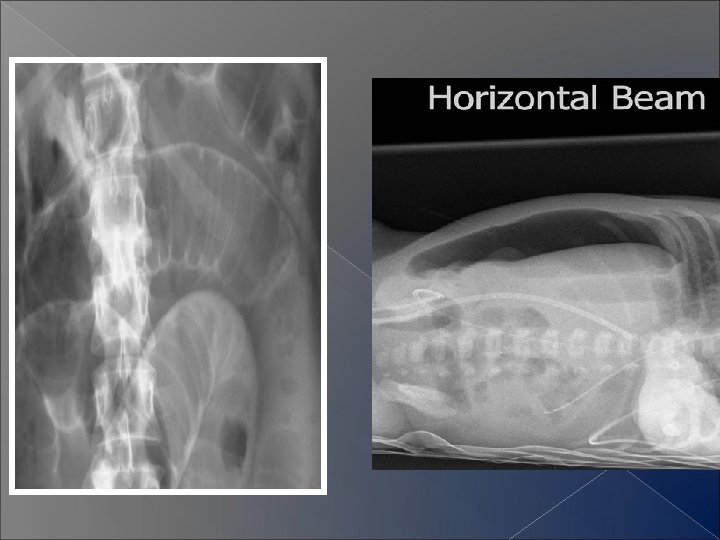
Plain x-ray of the abdomen � Standard ü supine plain films: AP ü erect AP ü Lt. lateral decubitus

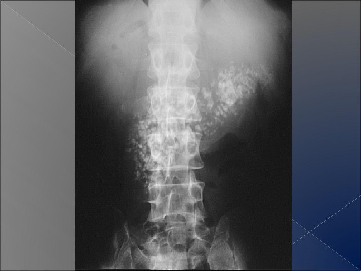
What to examine? � gas pattern : intraluminal , intramural � Exrtraluminal � Soft gas tissue masses � Calcification

Normal gas pattern 1. 2. 3. 4. 5. What segment ? The caliber ? The most distal point ? The gut mucosa ? Air - fluid levels ?

Normal gas pattern � The stomach: � Location � Pattern � fluid level

Normal gas pattern Small bowel Caliber � Location � Pattern � fluid levels �

Normal gas pattern � Large bowel Caliber � Location � Pattern � fluid levels �

How to distinguish between normal small & large bowel gas pattern � The Location � Assessment of mucosal pattern : 1. Plicae circularis 2. Colonic haustra � Number of loops � Lower ileum & sigmoid colon ? ?

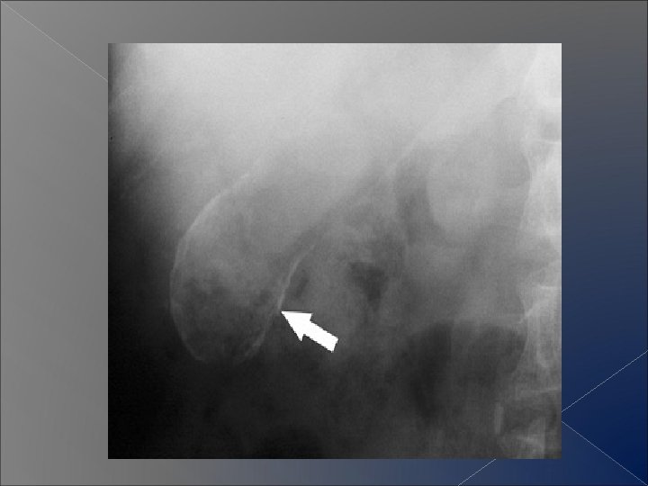
Normal fluid levels � Usually seen in erect position Ø Stomach Ø Duodenum Ø Small bowel Ø Large bowel

Dilatation of bowel � Mechanical � Paralytic � Acute obstruction ileus ischemia � Inflammatory bowel disease

Mechanical small bowel obstruction -Causes : -Features:

Mechanical large bowel obstruction -Causes : -Features:

Generalized Paralytic ileus � Causes : � Features of dilated large & small bowel � Gas is seen within the rectum

Localized ileus � Causes : � Features

Closed loop obstruction � Sigmoid volvulus :

Toxic dilatation of the colon � Causes � Features � Barium enema is CONTRAINDICATED

Free intraperitoneal air � ERECT FILMS , additional views � Causes � Features

Intramural gas � Pneumatosis

Intramural gas � Bowel wall ischaemia
 , Ø the pattern")
Abdominal calcification � Assess : Ø the location (two views) , Ø the pattern Ø the shape � Common calcifications

Patterns of abdominal alcifications � Rim like � Linear � Lamellar � Cloudy

Rim like calcification � Causes

Linear calcification � Causes

Lamellar or laminar calcification � Causes

Amorphous or cloudy calcification

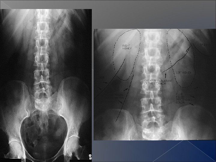
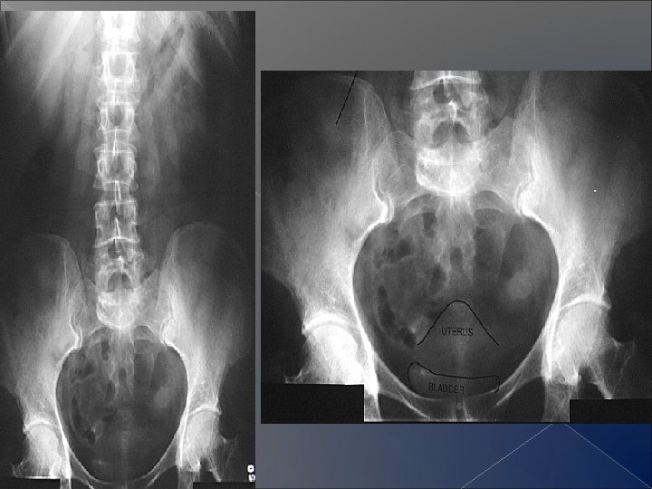
Soft tissue masses in the abdomen � In normal subject 1. Lateral & inferior edge of liver Spleen Both kidneys Psoas muscles UB Uterine indentation 2. 3. 4. 5. 6.
- Slides: 31