Evaluation and Management of Spinal Cord Emergency and


 • SCI are critical emergencies that must be recognized and")


 – Breast –")


 • • • Unsteady walking Disuse of hands Numbness")

 will present itself")


 is the most common spinal cord")


 Hyper-reflexia Pathological reflexes Increased")
 • Motor")


- Slides: 74
Evaluation and Management of Spinal Cord Emergency and Cervical Spondylotic Myelopathy James J. Lehman, DC, MBA, FACO Associate Professor of Clinical Sciences University of Bridgeport College of Chiropractic Director Community Health Clinical Education University of Bridgeport
Learning Objectives • Recognize signs of spinal cord emergency.
Learning Objectives • Correlate anatomy and the patients’ signs and symptoms in order to identify cervical spondylotic myelopathy (CSM).
Spinal Cord Injuries (SCI) • SCI are critical emergencies that must be recognized and treated early to increase the possibility of preventing permanent loss of function. • Spinal cord emergencies: False reassurance from reflexes. Acad Emerg Med 1998.
History and Clinical Presentation • …can provide the most important information in the assessment of a possible emergency. • Spinal cord emergencies: False reassurance from reflexes. Acad Emerg Med 1998.
Red Flags • Night pain/sweats/fever • Unexpected weight loss • Bowel and bladder dysfunction • Long tract signs • Signs of neurogenic claudication • Weakness and paresthesias in extremities
Cervical Spine Fracture/Dislocation • Suspect upper cervical spine instability • History of roll-over MVA or blow to head
Rust’s Sign • May grab head upon removal of cervical collar • May use hand to lift head when rising from supine position
Rust’s Sign • Patient is attempting to stabilize the head with slight traction and reduce pain • Patient presents guarded movements • Imaging studies must proceed any provocative testing • CT scan is indicated with acute trauma to the cervical spine when radiographic examination is negative.
Odontoid Fracture CT Scan
Rust Sign • Rear-end MVA • Patient is unable to rise from supine posture without holding hand behind head • Suspect moderate to severe sprain/strain James J. Lehman, DC, MBA, DABCO
Recognizing Spinal Cord Injuries Differential Diagnosis Spinal cord compression secondary to vertebral fracture or space occupying lesion.
Recognizing Spinal Cord Injuries Differential Diagnosis Spinal cord compression secondary to vertebral fracture or space occupying lesion.
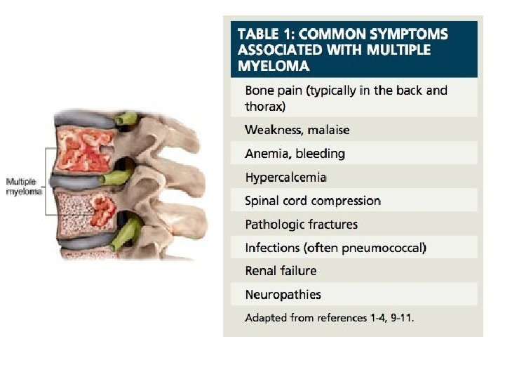
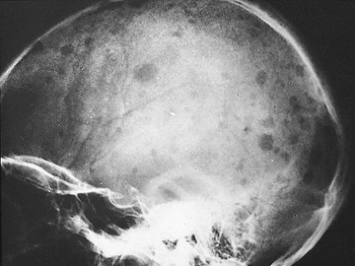
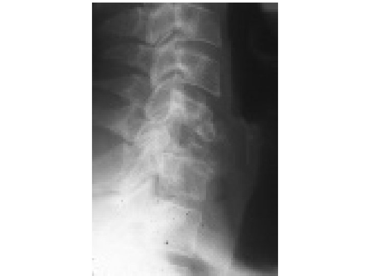
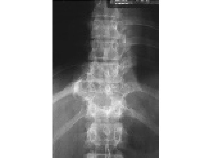
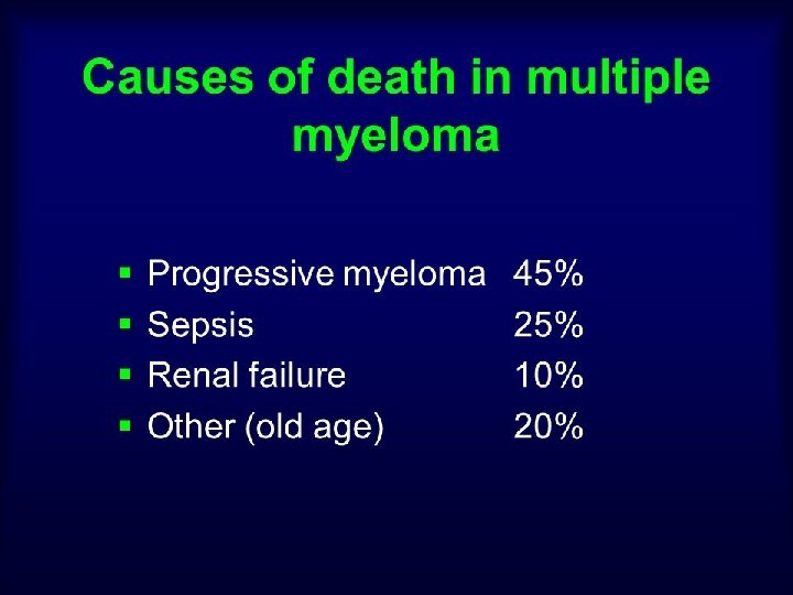
Neoplastic Disease • Metastatic disease most common spinal tumor (25 X) – Breast – Lung – Prostate – Kidney – Lymphoma • Primary spinal tumor is most often multiple myeloma in adults
Multiple Myeloma • What clinical signs would you expect when a patient presents with multiple myeloma?
CRAB Criteria
Spinal Infection “Discitis” • Acute onset of neck or back pain • HX of infection • Current fever • Elevated SED rates or WBC count • Frequently misdiagnosed in ER • MRI indicated
Staph Infection of Spine and Osteomyelitis • Recent spinal procedure • Focal severe pain not relieved with rest
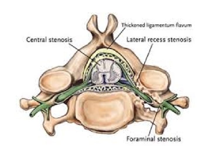
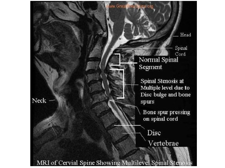
Central Canal Spinal Stenosis
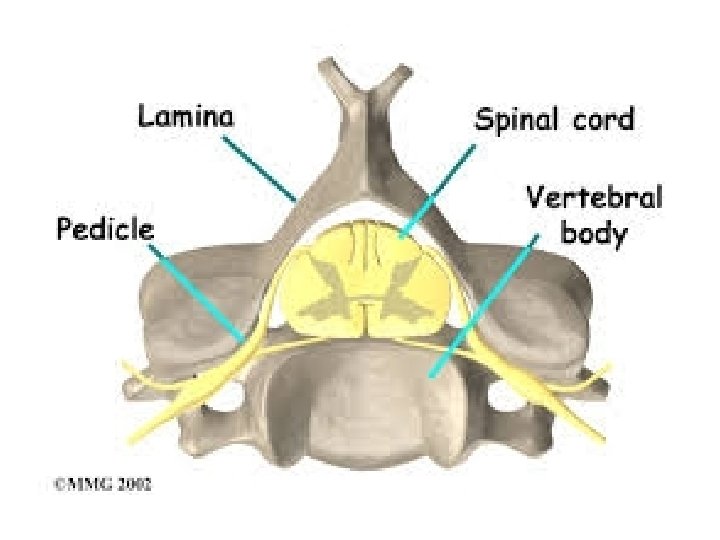
Vertebral Anatomy
Cervical Vertebra • • Vertebral artery Spinal cord Zygapophyseal joints Spinal nerve roots James J. Lehman, DC, MBA, FACO
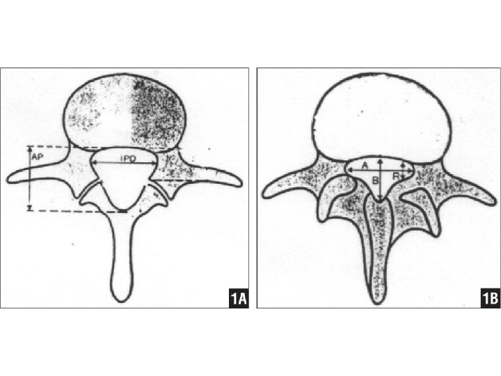
Spinal Canal Measuring Pavlov’s ratio Ratio of sagittal dimension of canal and vertebral body Less than 0. 82 is significant for stenosis Removes effects of radiographic magnification
Congenital Spinal Stenosis
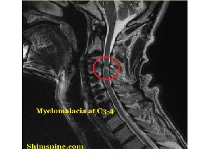
Myelomalacia • Post-traumatic softening of spinal cord due to hemorrhage or inadequate circulation to cord. • Initially, LMNL signs followed by UMNL signs • Often subtle signs initially • Frequently, misdiagnosed • MRI indicated • Spinal decompression might be of benefit
Diagnosis is the key to successful treatment… James J. Lehman, DC, MBA, FACO
Primum Non Nocere • CT scan showing C 5 pedicle and facet fracture. James J. Lehman, DC, MBA, FACO
Signs of Cervical Myelopathy (CSM) • • • Unsteady walking Disuse of hands Numbness in the arms and hands Atrophy Twitching reflexes or muscles.
Unsteady walking • Everyday walking is a complex process that is a combination of brain and spinal cord input. • With cervical cord compression, an unsteady walking pattern can develop. • Some people can develop a wide based gait. Early signs of this can be tested in the office with tandem gait observation.
Disuse of hands • Another sign of cervical spondylotic myelopathy (CSM) will present itself with hand dysfunction. Ask if patient has a difficult time holding a cup of coffee or buttoning their clothes.
Numbness in the arms and hands • As the cervical spinal cord is compressed, the spinal nerves will be impacted. This can lead to numbness in the arms and hands (paresthesias). • Unlike carpal tunnel syndrome, cervical myelopathy will often involve numbness throughout the arm and hand. • Cervical radiculopathy can cause numbness in the arms and hands but is usually limited to specific dermatomes.
Atrophy • With continued compression of the cervical spinal cord, the innervation to the muscles in the arms and hands can be diminished. As a result, the muscles can diminish in size and demonstrate atrophy of the involved musculature.
Twitching reflexes of muscles • Hyperreflexia is described by the patient as a twitching of the muscles. • The upper and lower extremity reflexes are mediated by the corticospinal tract. • The loss of inhibition results in hyperreflexia and the presence of pathological reflexes.
Cervical Spondylotic Myelopathy: A Common Cause of Spinal Cord Dysfunction in Older Persons WILLIAM F. YOUNG, M. D. , Temple University Hospital, Philadelphia, Pennsylvania Am Fam Physician. 2000 Sep 1; 62(5): 1064 -1070.
Cervical Spondylotic Myelopathy • Cervical spondylotic myelopathy (CSM) is the most common spinal cord disorder in persons more than 55 years of age in North America and perhaps in the world.
Cervical Spondylotic Myelopathy • The aging process results in degenerative changes in the cervical spine that, in advanced stages, can cause compression of the spinal cord.
Cervical Spondylotic Myelopathy • Symptoms often develop insidiously and are characterized by neck stiffness, arm pain, numbness in the hands, and weakness of the hands and legs.
Cervical Spondylotic Myelopathy • The differential diagnosis includes any condition that can result in myelopathy, such as multiple sclerosis, amyotrophic lateral sclerosis and masses (such as metastatic tumors) that press on the spinal cord.
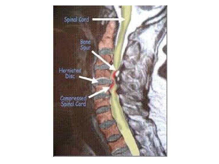
Cervical Spondylotic Myelopathy • The diagnosis is confirmed by magnetic resonance imaging that shows narrowing of the spinal caused by osteophytes, herniated discs and ligamentum flavum hypertrophy.
Cervical Spondylotic Myelopathy • Choice of treatment remains controversial, surgical procedures designed to decompress the spinal cord and, in some cases, stabilize the spine are successful in many patients. WILLIAM F. YOUNG, M. D Cervical Spondylotic Myelopathy: A Common Cause of Spinal Cord Dysfunction in Older Persons. Am Fam Physician. 2000 Sep 1; 62(5): 1064 -1070.
Cervical Cord Compression • Cervical compression tests and active cervical flexion may elicit signs of myelopathy rather than radiculopathy James J. Lehman, DC, MBA, FACO
Lhermitte’s Sign Patient may report an electrical shock-like sensation shooting down the spine and any combination of extremities with certain head movements or postures, especially active cervical flexion.
Lhermitte’s Sign The sign suggests a lesion of the dorsal columns of the cervical cord or of the caudal medulla.
Lhermitte’s Sign Although often considered a classic finding in multiple sclerosis, it can be caused by compression of the spinal cord in the neck from any cause such as cervical spondylosis, disc herniation, tumor, and Arnold-Chiari malformation.
Delayed onset Lhermitte's sign has been reported following head and/or neck trauma. This occurs ~2 1/2 months following injury, without associated neurological symptoms or pain, and typically resolves within 1 year. Chan RC. & Steinbock P. (1984). "Delayed onset of Lhermitte's sign following head and/or neck injuries. Report of four cases. ". J Neurolosurg 60 (3): 609– 12.
Upper Motor Neuron Lesion • Injury of upper motor neurons is common because of the large amount of cortex occupied by the motor areas, and because motor pathways extend all the way from the cerebral cortex to the lower end of the spinal cord.
Upper Motor Neuron Lesion • Damage to the descending motor pathways anywhere along this trajectory gives rise to a set of symptoms called the upper motor neuron syndrome. • Damage to Descending Motor Pathways: The Upper Motor Neuron Syndrome. Neuroscience. 2 nd edition. Purves D, Augustine GJ, Fitzpatrick D, et al. , editors. Sunderland (MA): Sinauer Associates; 2001.
Upper Motor Neuron Lesion • Damage to the motor cortex or the descending motor axons in the internal capsule causes an immediate flaccidity of the muscles on the contralateral side of the body and face.
Upper Motor Neuron Lesion • This initial period of “hypotonia” after upper motor neuron injury is called spinal shock, and reflects the decreased activity of spinal circuits suddenly deprived of input from the motor cortex and brainstem. • Damage to Descending Motor Pathways: The Upper Motor Neuron Syndrome. Neuroscience. 2 nd edition. Purves D, Augustine GJ, Fitzpatrick D, et al. , editors. Sunderland (MA): Sinauer Associates; 2001.
Lower Motor Neuron Lesion • The symptoms that arise from damage to the lower motor neurons of the brainstem and spinal cord are referred to as the “lower motor neuron syndrome. ”
Lower Motor Neuron Lesion • Damage to lower motor neuron cell bodies or their peripheral axons results in paralysis (loss of movement) or paresis (weakness) of the affected muscles.
Lower Motor Neuron Lesion • In addition to paralysis and/or paresis, the lower motor neuron syndrome includes a loss of reflexes (areflexia) due to interruption of the efferent (motor) limb of the sensory motor reflex arcs.
Lower Motor Neuron Lesion • Damage to lower motor neurons also entails a loss of muscle tone, since tone is in part dependent on the monosynaptic reflex arc that links the muscle spindles to the lower motor neurons
Lower Motor Neuron Lesion • A somewhat later effect is atrophy of the affected muscles due to denervation and disuse.
Lower Motor Neuron Lesion • The muscles involved may also exhibit fibrillations and fasciculations, which are spontaneous twitches characteristic of single denervated muscle fibers or motor units, respectively.
Lower Motor Neuron Lesion • These phenomena arise from changes in the excitability of denervated muscle fibers in the case of fibrillation, and from abnormal activity of injured α motor neurons in the case of fasciculations.
Lower Motor Neuron Lesion • These spontaneous contractions can be readily recognized in an electromyogram, providing an especially helpful clinical tool in diagnosing lower motor neuron disorders • Purves D, Augustine GJ, Fitzpatrick D, et al. , editors. The Lower Motor Neuron Syndrome. Neuroscience. 2 nd edition. Sunderland (MA): Sinauer Associates; 2001.
Characteristics of Motor Neuron Lesions • • • Upper (UMNL) Hyper-reflexia Pathological reflexes Increased tone Spastic paralysis Clonus • • Lower (LMNL) Hypo-reflexia Flaccid paralysis/paresis Loss of tone Atrophy Fasciculations Fibrillations Reaction of degeneration
Three Part Peripheral Nervous System Examination • Deep Tendon Reflex Testing (Myotatic) • Motor Testing for Strength • Sensory Testing
Active Learning Task • • Form groups of 4 -5 learners Select spokesperson Create a putative SOAP note (10 minutes) Present your case (2 minutes)
Engaged Learning Exercise • Create a putative SOAP note for a patient presenting with cervical spondylotic myelopathy (CSM). • Please write a narrative for subjective data gleaned from history • List examination procedures and anticipated objective findings • Assessment is CSM • Plan should include your management
References • • • Spinal cord emergencies: False reassurance from reflexes. Acad Emerg Med 1998. WILLIAM F. YOUNG, M. D Cervical Spondylotic Myelopathy: A Common Cause of Spinal Cord Dysfunction in Older Persons. Am Fam Physician. 2000 Sep 1; 62(5): 1064 -1070 Chan RC. & Steinbock P. (1984). "Delayed onset of Lhermitte's sign following head and/or neck injuries. Report of four cases. ". J Neurolosurg 60 (3): 609– 12. Damage to Descending Motor Pathways: The Upper Motor Neuron Syndrome. Neuroscience. 2 nd edition. Purves D, Augustine GJ, Fitzpatrick D, et al. , editors. Sunderland (MA): Sinauer Associates; 2001. Purves D, Augustine GJ, Fitzpatrick D, et al. , editors. The Lower Motor Neuron Syndrome. Neuroscience. 2 nd edition. Sunderland (MA): Sinauer Associates; 2001.
Suggested Readings • Cervical Spondylotic Myelopathy. http: //www. aafp. org/afp/2000/0901/p 1064. html • Recognizing Spinal Cord Emergencies. http: //www. aafp. org/afp/2001/0815/p 631. html