Etiological Agents of nfective Endocarditis Staphylococcus Streptococcus Enterococcus

 is an infection of the endocardium,")


 are capable of")


 These bacteria responsible from acute")


 • Normal oral-intestinal flora. . Common causes")


 there is")
 there are persistently positive blood cultures (two positive cultures from")


- Slides: 37
Etiological Agents of İnfective Endocarditis: Staphylococcus, Streptococcus, Enterococcus and the HACEK Group
What is infective endocarditis? • İnfective Endocarditis (IE) is an infection of the endocardium, which is the inner lining of your heart chambers and heart valves. • Generally occurs when bacteria, fungi or other microorganisms from another part of your body, such as flora spread through your bloodstream and attach to damaged areas in your heart.
• If it's not treated quickly, endocarditis can damage or destroy your heart valves and can lead to life-threatening complications. • Treatments for endocarditis include antibiotics and, in certain cases, surgery.
Who are at risk ? • As there are many ways to develop endocarditis, people who have usually damaged heart valves, • Artificial heart valves • Other heart defects are at risk of endocarditis • Once established, IE can involve almost any organ system in the body and can be fatal if left untreated.
How infective endocarditis occur? • When microorganisms enter the bloodstream (Bacteremia) are capable of attaching to and colonizing valve tissue; • Creation of the infected mass or ‘vegetation’ by ‘burying’ of the proliferating organism within a protective matrix of serum molecules (for example, fibrin) and platelet
How endocarditis occur? • Catheters • Everyday oral activities. brushing the teeth, or other dental procedures, Poor dental hygiene • Intravenous (IV) illegal drug use. Contaminated needles and syringes are a special concern for people who use illegal intravenous (IV) drugs, such as heroin or cocaine. • Needles used for tattoos and body piercing • Damaged heart valves. Certain medical conditions, such as rheumatic fever or infection,
• mitral valve vegetation caused by bacterial endocarditis. http: //en. wikipedia. org/wiki/Infective_endocarditis
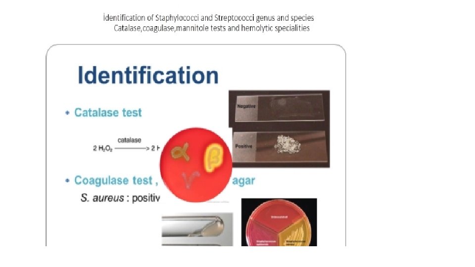
Which microorganism cause infective endocarditis? • The Gram-positive cocci of the Staphylococcus, Streptococcus, and Enterococcus account for 80– 90% of infective endocarditis. • Staphylococcus aureus species is the most frequently isolated microorganism associated with infective endocarditis in high-income countries and is reported in up to 40% of cases. • S. aureus and has coagulase enzyme. Coagulase test is help to distinguish from coagulase negative streptococci (CNS) • CNS cause subacute disease. It accounts for approximately % 30 cases.
• Streptococcal infective endocarditis caused by the oral viridans group remains most common in low-income countries. • In subacute disease % 50 -60 of cases approximately depends on Streptococcus viridans • Most clinical signs and symptoms are mediated immunologically • Streptococcus intermedius group may be responsible from acute or subacute disease that accounts for % 15 of Streptococcal IE • Abiotrophia species ( formerly known as variant Streptococci) % 5 of subacute cases
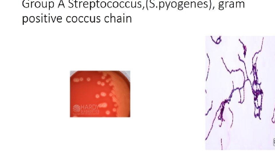
• Group A, Group A Streptococci (S. Pyogenes) These bacteria responsible from acute disease Repeat Sore throat infection Develop Post-streptococcal Diseases (Rheumatic heart disease) • Group B Streptococci Patients who have cancer , diabetes, alcoholism and pregnant are at risk for acute disease. Mortality rate is %40 • Group C, G Streptococci
• Group D Streptococci , Enterococci account for 10% of cases overall. • The source is gastrointestinal and genitourinary tract, and most cases are subacute • Non enterococcal group D Streptococci • The clinical course is subacute and infection reflects underlying abnormalities of the large bowel ( ulcerative colitis, polyps, cancer)
1 • Gram-positive cocci. facultative anaerobes, gram positive cocci, / chains/clusters or pairs cocci. Catalase positive /Staphylococci group. • Catalase negative/ Streptococci & Enterococci groups. • Streptococci subdivided into groups according their hemolytic reaction on blood agar in vitro & by serotypes according to surface cell wall specific carbohydrate antigens.
1 A • Viridans streptococci Group (VGS) • Normal oral-intestinal flora. . Common causes of dental caries. . Oral abscesses Gingivitis Deposit dextran, adhesins, Fibronectin-binding protein. • St. mutans, St. mitis accounted for many cases, and tend to be less susceptible to penicillins.
• A group of fastidious gram-negative bacteria can cause rarely endocarditis : • Gram negative bacteria: Brucella, Salmonella, Haemophilus, Cardiobacterium, Eikenella, • Pseudomonas aeruginosa Usually acute disease occur. When it involves the right side of the heart in IE
Yeast & Filamentous Fungi • Fungal endocarditis, usually Candida or Aspergillus, is rare but often fatal, arising in patients who are immunosuppressed or after cardiac surgery, mostly on prosthetic valves. • Most cases of fungal endocarditis occur in patients who are receiving prolonged antibiotics or intravenous nutrition through central vascular catheters
Yeast & Filamentous Fungi • The most common species is Candida albicans, followed by other less common Candida spp. ( C. glabrata, C. krusei, C. tropicals). • Candida part of human normal flora. . Oral-intestinal-Urinary tract (Vagina). . Infection often followed often using catheters or respiratory intubation. • Endocarditis due to Histoplasma capsulatum / Aspergillus species is very rare. . Immuno-suppressed patients.
The HACEK group of bacteria • • • The HACEK bacteria (Haemophilus aphrophilus, Aggregatibacter actinomycetemcomitans, Cardiobacterium hominis, Eikenella corrodens, Kingella kingea ) • These organisms usually cause subacute disease and about 5% of cases of IE • Complications may be congestive heart failure and massive arterial emboli from large biofilm vegetations in heart valves.
The HACEK group of bacteria • Haemophilus species, Aggregatibacter species, Cardiobacterium hominis, Eikenella corrodens, and Kingella species) are fastidious Gram-negative bacilli that have long been recognized as a cause of infective endocarditis • Common features of the HACEK group are frequent colonization of the oropharynx slow growth and enhanced growth in the presence of carbon dioxide.
İs there a problem HACEK group of bacteria ? • This group of bacteria may not be detected in routine blood culture systems unless enriched blood media are used in diagnosis. • The reported incidence of IE caused by gram-negative bacteria ranges from 1. 3 % to 10%, with HACEK group contributing to the majority of the cases.
The HACEK group of bacteria • The among the most commonly reported pathogens of HACEK group are Haemophilus spp • However, the taxonomy of this group has recently been updated, and today; • The genus Aggregatibacter spp also includes subspecies classically known as Actinobacillus actinomycetemcomitans, Haemophilus aphrophilus, and Haemophilus segnis.
The HACEK group of bacteria With the current classification, Aggregatibacter spp might be the dominant etiology of HACEK-related IE HACEK-IE tends to occur in young and middle-aged adults with previous dental procedures and underlying heart disease
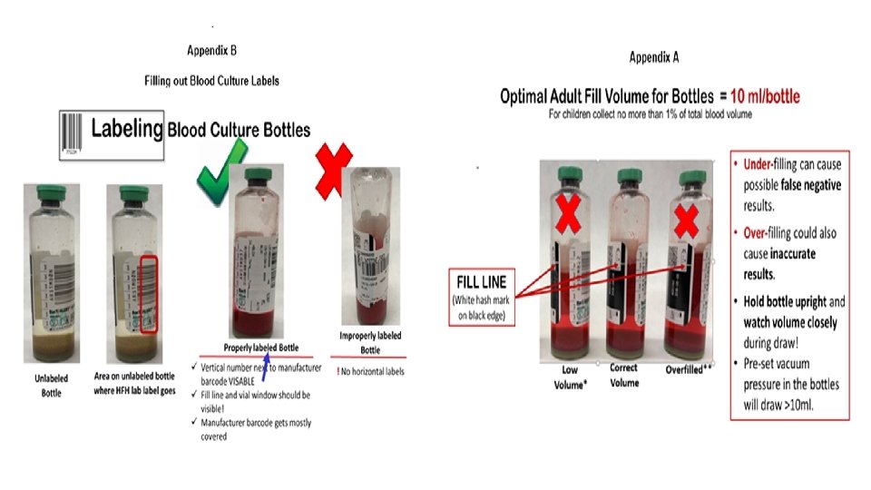
Diagnosis • Blood culture is the most important initial laboratory test in the workup of IE. Bacteremia is usually continuous and the majority of patients with IE have positive blood cultures • If endocarditis is suspected 3 blood cultures (10 m. L each) should be obtained within 24 hours (if the clinical symptoms like Acute Bacterial Endocarditis (ABE), 2 cultures within the first 1 to 2 hour)
Diagnosis • Sampling should be obtained from a peripheral vein rather than from a central venous catheter (because of the risk of contamination and misleading interpretation), using a meticulous sterile technique • When endocarditis is present and no prior antibiotic therapy was given, all 3 blood cultures usually are positive because the bacteremia is continuous • At least 1 culture is positive in 99 %
• Premature use of empiric antibiotic therapy should be avoided in patients with aquired or congenital valvular or shunt lesions to avoid culture –negative endoarditis • If prior antimicrobial therapy was given blood cultures should still be obtained but they may be negative
Automated Blood Culture System
Aerobic, anaerobic and paediatric bottles
After the incubation, the inoculation to blood agar from bottle
• A positive blood culture is a major criterion when 1) there is growth on two occasions of a microorganism typical for IE (eg, viridans group Streptococcus, Staphylococcus aureus, Enterococcus and HACEK group),
• OR 2) there are persistently positive blood cultures (two positive cultures from samples 12 h apart or three positive cultures drawn 1 h apart) of a microorganism consistent with IE, such as S. epidermidis, • OR 3) Coxiella burnetii (Q fever) grows from a single blood culture, or there is serologic evidence of C. burnetii (Ig. G titer 1: 800).
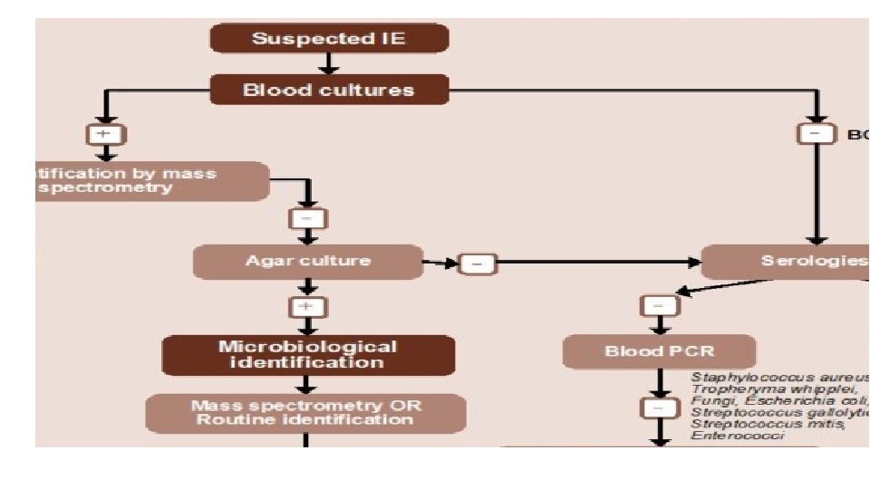
• Blood cultures are negative in 2% to 40% of cases of endocarditis, with some studies reporting higher blood culture-negative rates. • The causes of so-called “culture-negative endocarditis” has two categories: 1 - Negative blood cultures due to concomitant or antecedent antibacterial therapy, 2 - The presence of an organism that does not grow in routine blood cultures, with the former being more common.
• For etiologies of culture-negative endocarditis are Coxiella burnetii and Bartonella species, Tropheryma whipplei Cutibacterium acnes. • A rare cause of endocarditis, may cause culture-negative endocarditis due to the requirement for prolonged blood-culture incubation for growth of some strains. • In addition, some strains may not grow in blood cultures. Mycoplasmal endocarditis, while rare, is primarily caused by Mycoplasma hominis and is usually diagnosed using a nucleic acid amplification test.
• Non-Blood-Culture Tests : • Serologic tests • Microscopic examination with special stains, (i. e. , the periodic acid– Schiff stain for T. whipplei) • Direct fluorescence antibody techniques • Nucleic Acid Amplification test, common use Polymerase Chain Reaction (PCR) to recover unique microbial DNA or DNA encoding the 16 S or 28 S ribosomal unit.
References • Procop G. W, Church D. L, Hall G. S, Janda W. M, Koneman E. W, Schreckenberger P. C, Woods G. L. Koneman’s Color Atlasand Textbook of Diagnostic Microbiology. 7 th edition, Lippincott Williams and Wilkins, China, 2017 • Brooks Geo. F, Carroll Karen C, Butel Janet S, Morse Stephen A, Mietzner Timothy A. : Medical Microbiology (Jawetz, Melnick & Adelberg’s) • Twenty-sixth edition, Mc Graw Hill Companies, New york, 2013 • https: //www. escardio. org/Guidelines/Clinical-Practice-Guidelines/Infective-Endocarditis-Guidelines-on-Prevention. Diagnosis-and-Treatment-of • https: //www. asm. org/Articles/2019/February/Who-are-the-HACEK-organisms • http: //en. wikipedia. org/wiki/Infective_endocarditis • https: //pedclerk. bsd. uchicago. edu/page/infective-endocarditis • https: //www. idsociety. org/practice-guideline/laboratory-diagnosis-of-infectious-diseases/ • http: //simple-cardio. blogspot. com/2012/06/peripheral-signs-of-infective. . • https: //www. mayoclinic. org/diseases-conditions/endocarditis/symptoms-causes/syc-20352576#dialog. Id 36371491 • https: //www. google. com/search? q=blood+culture&tbm=isch&ved=2 a • From Li JS, Sexton DJ, Mick N, et al. Proposed modifications to the Duke Criteria for the diagnosis of infective endocarditis. Clin Infect Dis. 2000; 30: 633– 638. Used with permission from the University of Chicago Press. • https: //upload. wikimedia. org/wikipedia/commons/thumb/d/d 8