Ethanol Ablation in Locally Recurrent PTC Presented by

Ethanol Ablation in Locally Recurrent PTC Presented by: B. Rezvankhah MD
 Conclusion")
Agenda: Locoregional Recurrent PTC management Ethanol ablation (Literatures review) Conclusion

Locoregional Recurrent PTC management PTC is highly curable in 85% of cases because of its rather innocent biologic behavior. Therefore, the shift in emphasis from disease survival to recurrence is appropriate. The risk of structural disease recurrence is a continuum of risk that ranges from <1% in very low-risk patients to >50% in high-risk patients Recurrence in the neck can be either in lymph nodes or in the thyroid bed. A recurrence in the thyroid bed results in increased rates of morbidity and mortality Recurrent metastatic adenopathy is common and seen in 9– 20% of papillary thyroid carcinoma patients in long-term follow-up The prognostic significance of nodal metastases on long-term survival remains unclear

Treatment Modalities: Reoperation ± RAI therapy Radio Frequency Ablation Ethanol Ablation Active surveillance

Reoperation Surgery is favored for locoregional recurrences, (i. e. , cervical lymph nodes and/or soft tissue tumor in the neck) when distant metastases are not present( except for possible palliation of symptoms or prevention of airway or aerodigestive obstruction)
 Therapeutic compartmental central and/or lateral neck dissection in a previously")
RECOMMENDATION 71(ATA 2015) Therapeutic compartmental central and/or lateral neck dissection in a previously operated compartment, sparing uninvolved vital structures, should be performed for patients with biopsy-proven persistent or recurrent disease for central neck nodes >8 mm and lateral neck nodes >10 mm in the smallest dimension that can be localized on anatomic imaging. (Strong recommendation, Moderate-quality evidence)

Complications: Total Thyroidectomy and LD; Incidence of Permanent Hypoparathyroidism: 1. 4 % Temporary Hypoparathyroidism: 11. 59% Permanent unilateral VCP 1. 4% Reoperation; Incidence of permanent Hypoparathyroidism : up to 9. 5%, with a considerably higher incidence of temporaryhypoparathyroidism (up to 46. 3%). Permanent vocal fold paralysis : up to 6. 4% Bilateral VCP : 0% Liebert el al. THYROID. 2015

!Although, most series suggest surgery results in a high clearance rate of structural disease in over 80% of patients and basal Tg decreases by 60%– 90% after compartmental dissection for recurrent nodal disease in modern series, but only 30%– 50% of patients have unmeasurable basal Tg after such surgery ! In addition, compartmental surgical dissections may not be feasible in the setting of compartments that have been previously explored due to extensive scarring, and only a more limited or targeted lymph node resection may be possible.

RAI Therapy It is being increasingly recognized that radioiodine remnant ablation rarely prevents the discovery of postoperative lymph node metastases in PTC Published studies assessing the role of adjunctive radioactive iodine (RAI) in the treatment of recurrent LNM are scarce, but (Yim JH et al. 2011)recently found no improvement in either serum Tg levels or recurrence-free survival, if reoperated patients also received adjuvant RAI therapy. It causes important long-term side effects including secondary malignancy (doses exceeding 600 m. Ci) The Thyroid: A Fundamental and Clinical Text. 2013
 (C) Suspicious lymph nodes less than 8– 10 mm")
Active surveillance RECOMMENDATION 65(ATA 2015) (C) Suspicious lymph nodes less than 8– 10 mm in smallest diameter may be followed without biopsy with consideration for FNA or intervention if there is growth or if the node threatens vital structures. (Weak recommendation, Low-quality evidence) ü Small, postoperative thyroid bed nodules (defined as < 11 mm) occur in as many as a third of patients undergoing surgery, with or without adjuvant therapy. Only a small percent ( < 10%) of these nodules will prove to be malignant lymph nodes, and even fewer will progress over time. Rondeau G et al. THYRIOD. 2011
 is a minimally invasive procedure that uses")
Radio Frequency Ablation Radiofrequency ablation (RFA) is a minimally invasive procedure that uses electrical pulses to heat a probe that is placed in a tumor It is generally conducted in the outpatient setting, using either local anesthetics or conscious sedation anesthesia It does not directly stimulate nerves or heart muscle and therefore can often be used without the need for general anesthetic
 Conclusion")
Agenda: Locoregional Recurrent PTC management Ethanol ablation(Literatures review) Conclusion

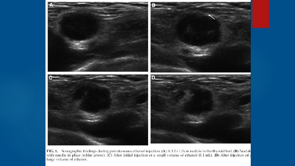
Ethanol Ablation Noninvasive treatment regimen that has been used to treat parathyroid tumors, liver cancer, hyperfunctioning thyroid nodules, and thyroid cysts The mechanism of EA for treatment of R-PTC was thought to causes ischemic tumor necrosis by inducing thrombosis of intralesional vessels & protein denaturation The lymph node showed complete replacement of the nodal architecture with foreign body material reactions and fibrosis

A conventional 3 -cm, 25 -gauge needle is attached to a tuberculin syringe containing up to 1 m. L of 95% ethanol. The skin and soft tissues between the skin and lymph node were anesthetized with 1% lidocainethe Injections were repeated between three and 10 times The volume of ethanol used in each treated node ranged from 0. 1 to 2 m. L

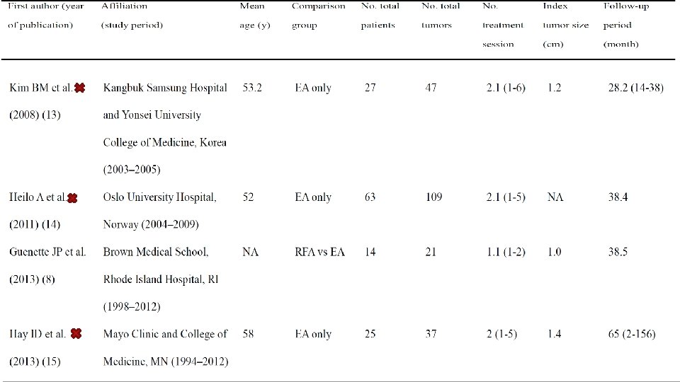
AJR 2002 Fourteen patients who had undergone previous thyroidectomy for PTC presented with 29 metastatic lymph nodes on sonographic F/U between May 1993 and April 2000 6 men & 8 women (ranged 27 to 83 years old). The mean diameter of the treated lymph nodes was 8. 7 mm (range, 4. 4– 17. 2 mm). The mean number of neck surgeries per patient was 2. 1 Mean sonographic follow-up was 18 months (range, from 2 months to 6 years 5 months)

Inclusion criteria: Presence of biopsy-confirmed metastatic papillary thyroid carcinoma in cervical lymph nodes Five or fewer involved lymph nodes that were amenable to percutaneous ethanol injection. Patients had to be poor surgical candidates, to have expressed a preference for no further surgery, or to have been unresponsive to previous radioiodine therapy. All patients had exhibited poor response to previous iodine-131 ablative therapy(Mean total iodine-131 dose per patient was 7, 548 MBq).

! Lymph node metastases larger than 3 cm, exhibit extra nodal extension, or metastasis present in more than five lymph nodes are more significant from a prognostic standpoint Machens et al. World J Surgery. 2002

Treatment goal: Decrease in size of the lymph nodes on follow-up images AND No residual evidence of perfusion Treatment failure: Injected nodes either enlarged or showed evidence of persistent perfusion on color Doppler sonography

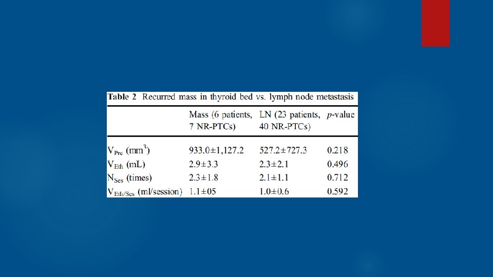
Results: Nine lymph nodes in seven patients disappeared during the followup period The mean volume of LN prior to EA was 492 mm 3 and decreased to a mean volume of 76 mm 3 after 1 year and 20 mm 3 after 2 years Six nodes were re-treated 2– 12 months after initial percutaneous ethanol injection because of persistent flow on color Doppler sonography (n = 4), stable size (n = 1), or increased size (n = 1).

… Two patients developed four new metastatic nodes during the follow-up period that were amenable to percutaneous ethanol injection. Two patients developed innumerable metastatic nodes that precluded retreatment with percutaneous ethanol injection

Complication: Most patients experienced brief discomfort at the injection site, likely related to local extravasation of a small amount of ethanol into the surrounding cervical soft tissues No patient experienced transient or permanent hoarseness or vocal cord paralysis
 27 patients (47 LN)were treated by EA for NR-PTCs between September")
Eur Radiol (2008) 27 patients (47 LN)were treated by EA for NR-PTCs between September 2003 and December 2005. 5 men and 22 women, and the patient’s average was 53. 2 years (range, 19– 80 years). The mean F/U was 28. 2± 6. 3 months(range, 14– 38).

According to the AJCC staging: I in 2, II in 5, III in 10, IV-A in 10. The mean number of neck surgeries was 2. 1 (range, 1– 4). Overall, 47 NR-PTCs were treated by EA. (Among them, 10 NRPTCs in 7 patients were newly detected during follow-up.
 The presence of three or fewer NR-PTCs and no recurrence beyond")
Inclusion criteria: (1) The presence of three or fewer NR-PTCs and no recurrence beyond the neck at the PEI point in time, (2) The NR-PTCs were not close to vessels to avoid the potential risk of ethanol intravasation (3) The patients with NR-PTCs were poor surgical candidates (the patients with high risk for general anesthesia due to a medical condition or for surgery due to repeated neck dissection) and/or preferred not to have further surgery.
 = (V")
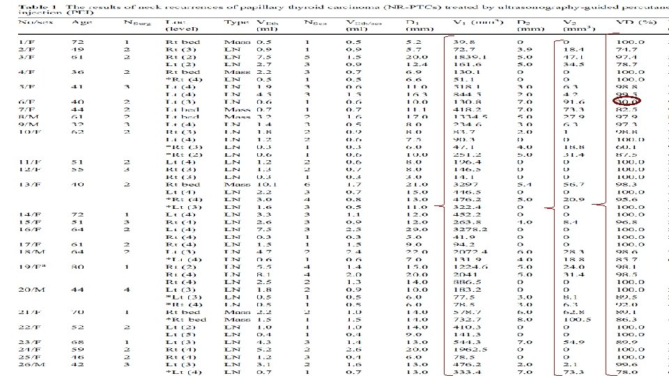
Percent volume decrease of each NR-PTC was calculated as: VD (%) = (V 1 – V 2)/V 1× 100 Decreased volume of the lesion and absence of intra lesional power. Doppler signals were considered a sign of successful results of the treatment Ultrasonography was performed at 3– 6 -month intervals for evaluation of PEI effectiveness
 significantly decreased in volume from a mean of")
Results: All 47 NR-PTCs (n=47) significantly decreased in volume from a mean of 678. 8± 871. 4 mm 3 to a mean of 15. 7± 24. 7 mm 3 (average volume decrease, 93. 6± 12. 6%; p<0. 05) during the follow-up Twenty-six (55. 3%) NR-PTCs in 16 patients remained visible on the last follow-up ultrasonography. They showed a progressive decrease in volume without tumor vascularity on power-Doppler study, or decreased volume without tumor vascularity, and then stable volume to the last follow-up ultrasonography
 From June")
J Clin Endocrinol Metab, 2011 63 patients (27 men and 42 women) From June 2004 through December 2009, were retrospectively included in this study 109 neck lymph nodes in total(in 63 patients) 57 patients had undergone central neck lymph node dissection. (Many had also undergone lateral neck node dissection All 63 patients had been treated with 131 I (median, two times; range, 1– 10 times) with median accumulated dose of 131 I of 284 m. Ci (10. 5 GBq)

Information to classify the tumor was not available for 12 patients, and in six patients no information was available about the lymph nodes. All 63 patients had localized disease and had no distant metastases

! Metastasis was confirmed in cytological smears and/or by demonstration of raised levels of. FNAB-Tg in needle washouts The initial cytology was positive in 91 lymph nodes. FNAB-Tg confirmed malignancy in the other 18(although smears from 12 of the 18 cases were suspicious of malignancy. Cytology was benign in two cases, and four did not yield diagnostic material. Four cases without elevated FNAB-Tg (0. 1 g/liter) demonstrated malignant cells in the cytological smears.
 Carcinoma cells identified in FNAB specimens and/or elevated level")
The inclusion criteria: 1) Carcinoma cells identified in FNAB specimens and/or elevated level of FNAB-Tg; 2) No radiological findings suspicious of malignancy inferior to the neck; 3)Record of previous treatment with 131 I; 4)Detection of up to three metastatic lymph nodes in previously dissected neck compartments.

Treatment was considered effective when reduction in volume and/or reduction of lymph node vascularization were demonstrated Criteria for satisfactory treatment: 1)Complete lymph node disappearance; 2)Reduction of the anteroposterior lymph node diameter to 4 mm or less without visible vascularization; 3)Normalization of lymph node size and appearance, and in some nodes scar formation; 4)No detectable malignant cells in the FNAB specimens and no detectable Tg in needle washouts.

Malignant lymph nodes were localized to levels VI and VII, containing 46 and three lymph nodes, respectively. Another 37 lymph nodes were detected in level IV, 11 in level III, four in level Va, two in level Vb, three in level IIa, one in level IIb, and finally two had a parapharyngeal location.

Results:
 with stage III or IVA")
Surgery 2013 25 PTC patients(37 LN proven by biopsy) with stage III or IVA disease (mean age 58 years) since 1994 All patients had previously been treated by both primary definitive surgery and postoperative RAI therapy. 16 were situated in the central compartment (level VI) and 21 in the lateral neck (levels III, IV, and V) The average of F/U was more than 5 years

The present study is the only one devoted solely to the management of NNM in patients >45 years with advanced localized PTC (p. TNM stages III and IVA), and it has the longest follow-up of any series of PTC patients treated with UPEA.

 Elimination of significant nodal blood flow (‘‘tumor perfusion’’) by Doppler")
Treatment goals: (1) Elimination of significant nodal blood flow (‘‘tumor perfusion’’) by Doppler ultrasonography. (2) Reduction in volume of treated NNM, ideally to the point of disappearance on rescanning. (3) When possible, concomitant reduction in circulating serum Tg levels on THST.
 decreased in size. None had significant Doppler flow.")
Results: 35 of 37 NNM (95%) decreased in size. None had significant Doppler flow. Seventeen (46%) disappeared on rescanning. Serum Tg fell in 19 of 22 (86%) without Tg-auto. AB. None of the UPEA-treated NNM, followed on average for 5. 4 years, required further intervention. Six patients (24%) subsequently developed 18 ‘‘new’’ recurrences. Of the 18, 15 (83%) were managed successfully by UPEA rather than operation. None of the 25 patients developed permanent hoarseness or have died from PTC.
 The")
… The average number of treatment sessions /patient was 2 (range, 1 -5) The largest diameter of treated nodes ranged from 4 to 21 mm (mean, 14) 2 of 3 ablated NNM in one patient showed, at last follow-up, apparently increased volumes. However, the nodes on ultrasonography seemed to be avascular, and the patient’s serum Tg level was undetectable(<0. 1 ng/m. L) on THST, at 110 months

NOTE: No decrease in recurrence rate No data for survival due to short F/U(except Hay et al. SURGERY. 2013) Locally mild pain due to the procedure relieved over few minutes (Transient Hoarseness in 2 patients, No permanent complication)

JAMA Otolaryngol Head Neck Surg. 2015

Outcomes : Retreatment success Change in Tg, Recurrence rates, Complication rates, Follow-up duration. Successful treatment by PEI : shrinkage of the lesion by 50% or greater on followup & no evidence of new growth within the lesion. Failure of PEI : any growth in a PEI-treated PTC lesion PEI was used to treat 270 metastatic PTC lesions in 168 patients (11. 4%). Reoperation was used to treat 1449 patients (88. 6%)

Results: Based on the analysis of the pooled data: surgery was successful in 94. 8%of studied cases compared with 87. 5%success rate for PEI treatment (OR, 2. 58; 95%CI, 1. 55 - 4. 31; P <. 001) The PEI pooled risk of recurrence at the site of the treated lesion or elsewhere in the neck was 11. 9%; the recurrence rate for patients who underwent a reoperation was 12. 7%(OR, 1. 07; 95%CI, 0. 65 -1. 77; P =. 78) Surgery was associated with a 3. 5%pooled risk of complications, while PEI incurred a pooled risk of 1. 2%(OR, 2. 9; 95%CI, 0. 72 -12. 3; P =. 08) Post-PEI Tg serum levels ranged from 0. 2 to 2. 8 ng/m. L compared with reoperation postoperative Tg serum levels of 0. 3 to 127. 5 ng/m. L (P =. 01) The mean Tg serum levels before procejure ranged from 0. 7 to 9. 2 ng/m. L in PEI compared with those of reoperation ranged from 3. 2 to 431 ng/m. L (P =. 02)
. The investigators")
! There are differences in patient populations undergoing the different treatments(selection bias). The investigators leading the studies examining PEI tend to select poor surgical candidates with a limited number of cervical lesions. ! Comparison of complications of the 2 techniques is also complicated. The PEI procedure was chiefly used to treat lateral neck lymph nodes, where risk of RLN injury is minimal !The benefit of PEI in a single treated lesion may not be permanent, and patients may require repeated treatments. (the recurrence risk of 11. 9%). In contrast, the median disease-free period following adequately aggressive reoperations can be as long as 7. 25 years.

In conclusion : surgery is the indisputable standard of care for recurrent PTC recurrences. While PEI is a minimally invasive, it cannot provide definitive treatment for recurrent PTC

THYROID 2016 The inclusion criteria: fewer than four recurrent tumors in one patient, and no recurrent tumor beyond the neck at the time of procedure Studies that contained data on eight or more consecutive patients were included Confirmation of diagnosis: US-guided FNA or Tg measurement of needle washout prior to the procedure Outcomes: evaluation of volume reduction, complete disappearance, changes in serum level of Tg, recurrence, and complications

The percent reduction in volume was defined as: VRR =[(initial volume – final volume) · 100)/initial volume. VRR >50% was defined by a lesion VRR of >50% on the final follow-up US Recurrence was defined as an identification of new tumors or persistent ablation zones with pathologic confirmation of malignancy 10 eligible studies(270 patients and 415 thyroi d nodules) RFA : 201 locally recurrent thyroid cancers in 141 patients (52. 2%) EA : 214 locally recurrent thyroid can cers in 129 patients (47. 8%)
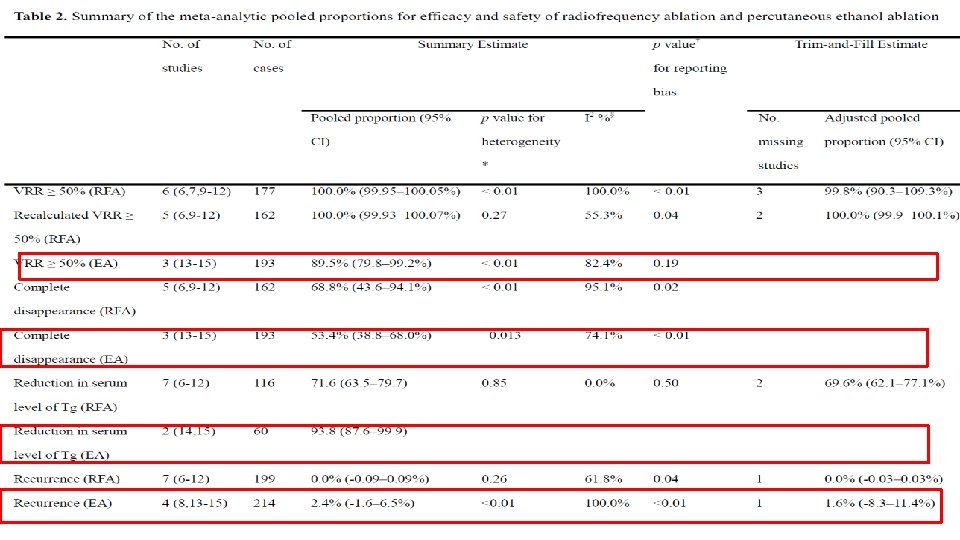

The present meta-analysis shows that the pooled proportion of VRR >50% for RFA was higher than that for EA (89. 5%; I 2 =82. 4%; p=0. 2764); The pooled proportion of complete disappearance after RFA (68. 8%) was higher than that after EA (53. 4%; p=0. 3384); The pooled proportion of recurrence after RFA (0. 0%) was lower than that after EA (2. 4%, adjusted 1. 6%; p=0. 9766). The pooled proportion of the reduction in serum level of Tg after RFA was 71. 6%, and after EA it was 93. 8% ( p<0. 0001). The pooled proportion of complications of both RFA (5. 8%, adjusted 1. 6%) and EA (1. 6%) was low ( p=0. 8479)

! Although both RFA and EA reduced the serum Tg levels, the Tg remained elevated after treatment. This is most likely explained by the fact that although macroscopic tumors detected on US were completely treated, microscopic tumor foci in other areas of the neck could remain. This is a limitation of local tumor control methods
 Conclusion")
Agenda: Locoregional Recurrent PTC management Ethanol ablation(Literatures review) Conclusion

Conclusion Advantages: Ø EA can be repeated many times without increased technical difficulty. Ø Its cost is much less than reoperation. Ø It avoids the morbidity, hospitalization, and general anesthesia. Ø The most common complications are discomfort and pain at the treatment site( No permanent vocal chenges) Disadvantages: Ø It can’t improve recurrence rate. Ø It could be substituted by active surveillance in many cases.

Thanks for Your Kind Attention
- Slides: 57