
Essentials of Human Anatomy Urinary System 1 General

- Slides: 31
Essentials of Human Anatomy Urinary System 1
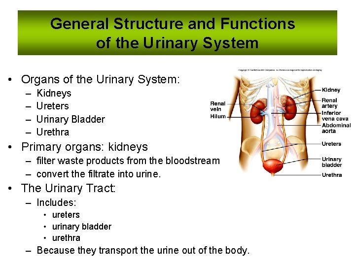
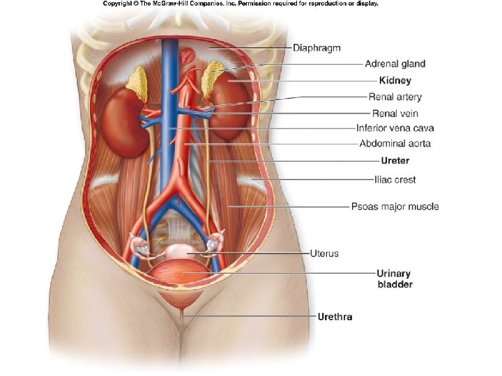
General Structure and Functions of the Urinary System • Organs of the Urinary System: – – Kidneys Ureters Urinary Bladder Urethra • Primary organs: kidneys – filter waste products from the bloodstream – convert the filtrate into urine. • The Urinary Tract: – Includes: • ureters • urinary bladder • urethra – Because they transport the urine out of the body.
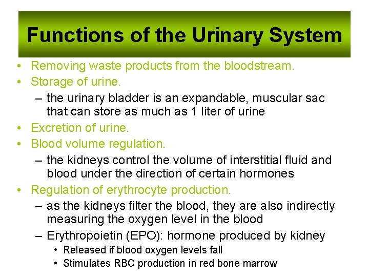
Functions of the Urinary System • Removing waste products from the bloodstream. • Storage of urine. – the urinary bladder is an expandable, muscular sac that can store as much as 1 liter of urine • Excretion of urine. • Blood volume regulation. – the kidneys control the volume of interstitial fluid and blood under the direction of certain hormones • Regulation of erythrocyte production. – as the kidneys filter the blood, they are also indirectly measuring the oxygen level in the blood – Erythropoietin (EPO): hormone produced by kidney • Released if blood oxygen levels fall • Stimulates RBC production in red bone marrow
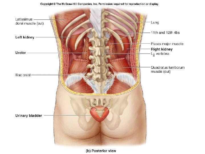
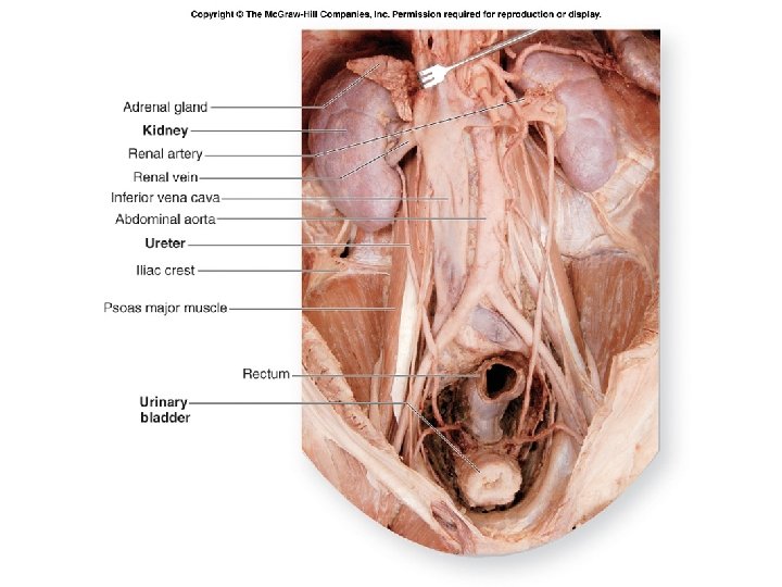
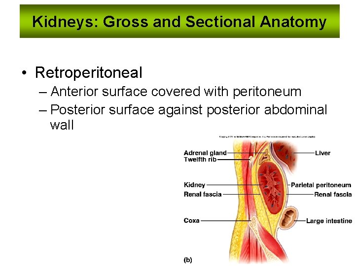
Kidneys: Gross and Sectional Anatomy • Retroperitoneal – Anterior surface covered with peritoneum – Posterior surface against posterior abdominal wall
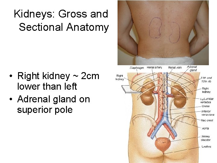
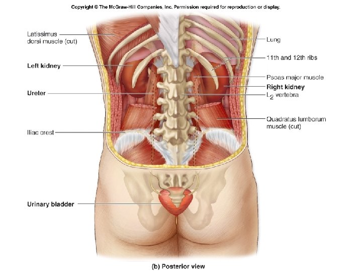
Kidneys: Gross and Sectional Anatomy • Right kidney ~ 2 cm lower than left • Adrenal gland on superior pole
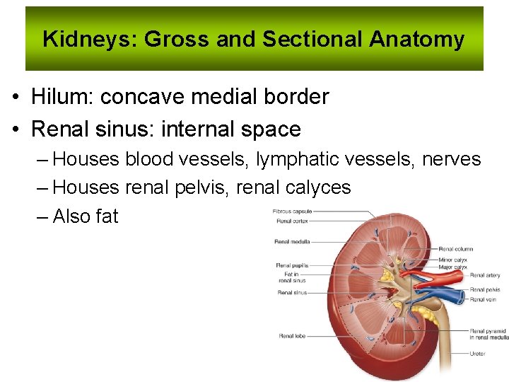
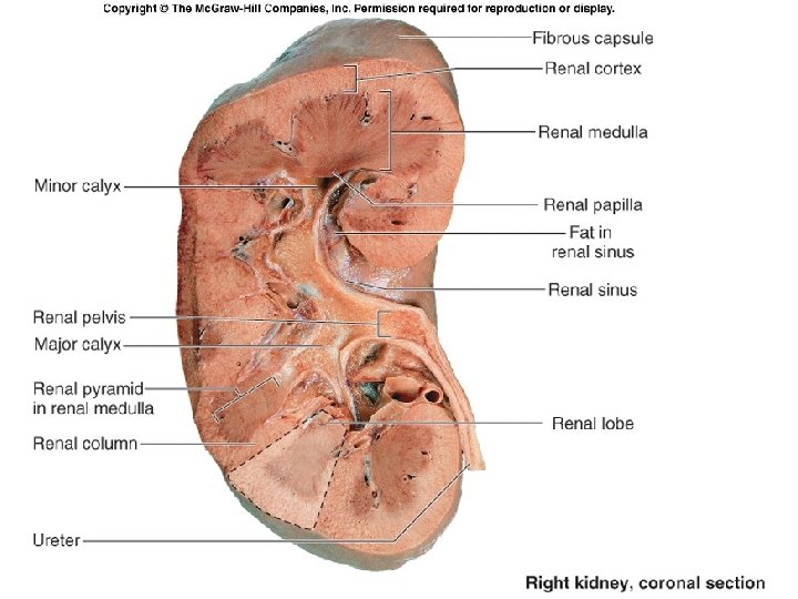
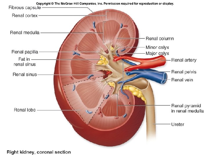
Kidneys: Gross and Sectional Anatomy • Hilum: concave medial border • Renal sinus: internal space – Houses blood vessels, lymphatic vessels, nerves – Houses renal pelvis, renal calyces – Also fat
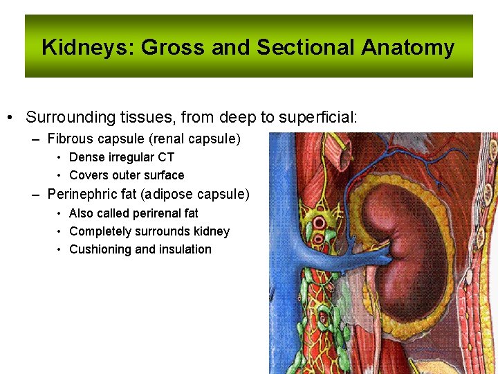
Kidneys: Gross and Sectional Anatomy • Surrounding tissues, from deep to superficial: – Fibrous capsule (renal capsule) • Dense irregular CT • Covers outer surface – Perinephric fat (adipose capsule) • Also called perirenal fat • Completely surrounds kidney • Cushioning and insulation
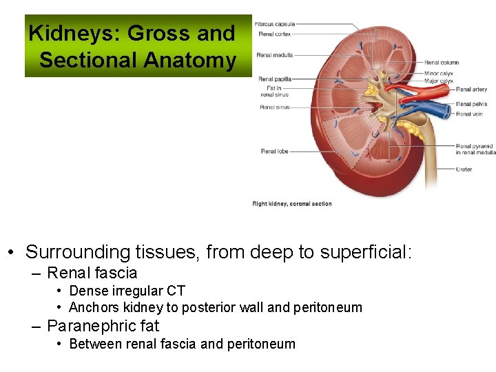
Kidneys: Gross and Sectional Anatomy • Surrounding tissues, from deep to superficial: – Renal fascia • Dense irregular CT • Anchors kidney to posterior wall and peritoneum – Paranephric fat • Between renal fascia and peritoneum
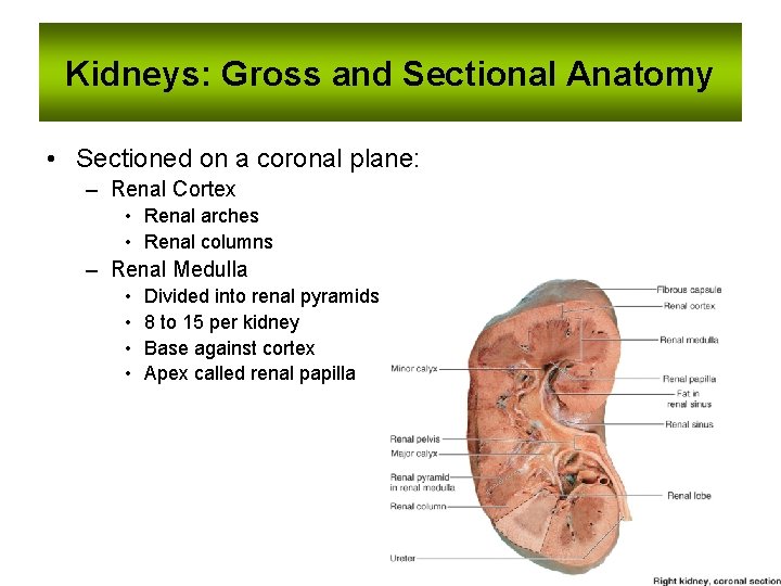
Kidneys: Gross and Sectional Anatomy • Sectioned on a coronal plane: – Renal Cortex • Renal arches • Renal columns – Renal Medulla • • Divided into renal pyramids 8 to 15 per kidney Base against cortex Apex called renal papilla
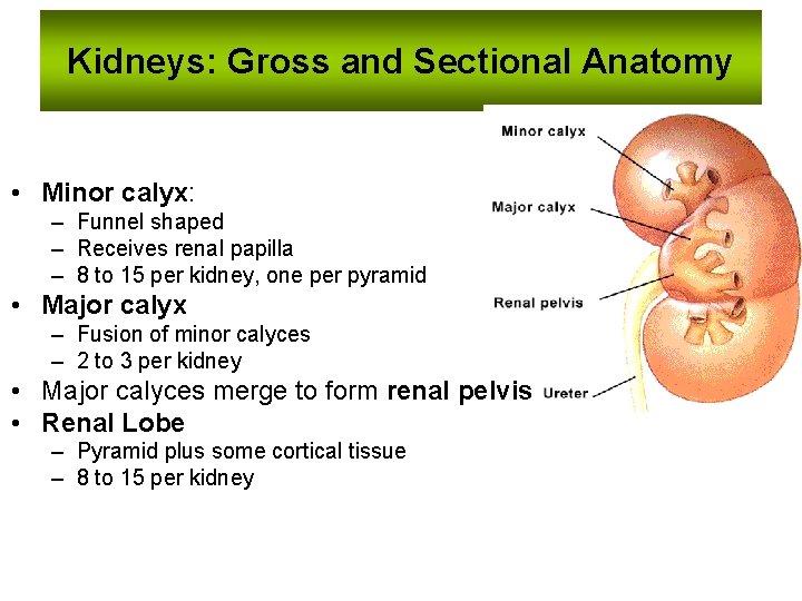
Kidneys: Gross and Sectional Anatomy • Minor calyx: – Funnel shaped – Receives renal papilla – 8 to 15 per kidney, one per pyramid • Major calyx – Fusion of minor calyces – 2 to 3 per kidney • Major calyces merge to form renal pelvis • Renal Lobe – Pyramid plus some cortical tissue – 8 to 15 per kidney
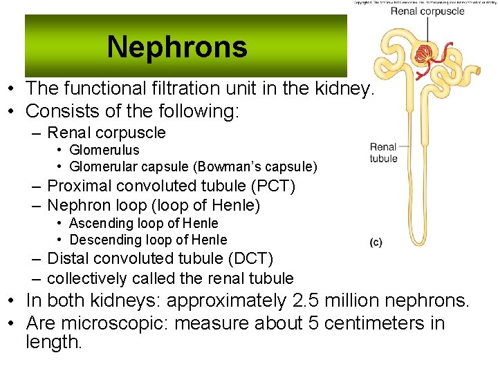
Nephrons • The functional filtration unit in the kidney. • Consists of the following: – Renal corpuscle • Glomerulus • Glomerular capsule (Bowman’s capsule) – Proximal convoluted tubule (PCT) – Nephron loop (loop of Henle) • Ascending loop of Henle • Descending loop of Henle – Distal convoluted tubule (DCT) – collectively called the renal tubule • In both kidneys: approximately 2. 5 million nephrons. • Are microscopic: measure about 5 centimeters in length.
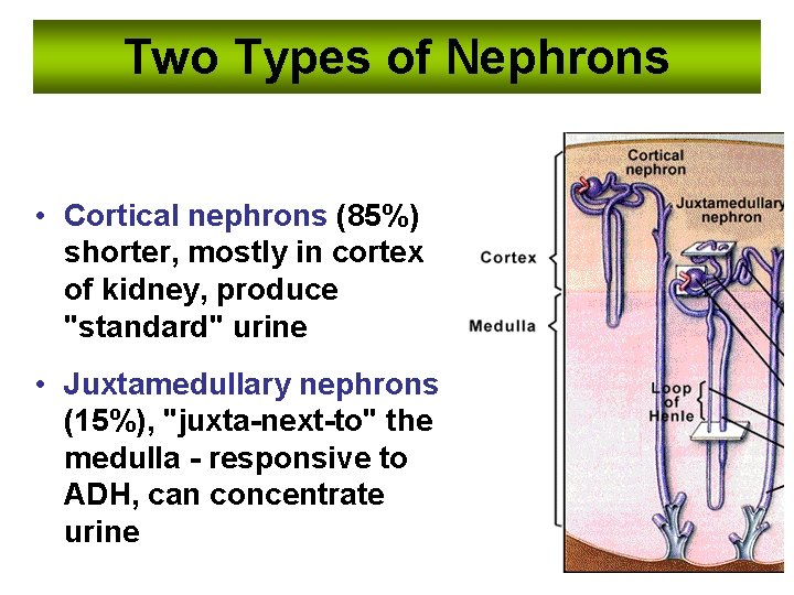
Two Types of Nephrons • Cortical nephrons (85%) shorter, mostly in cortex of kidney, produce "standard" urine • Juxtamedullary nephrons (15%), "juxta-next-to" the medulla - responsive to ADH, can concentrate urine
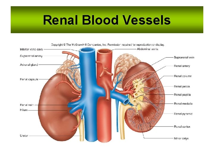
Renal Blood Vessels
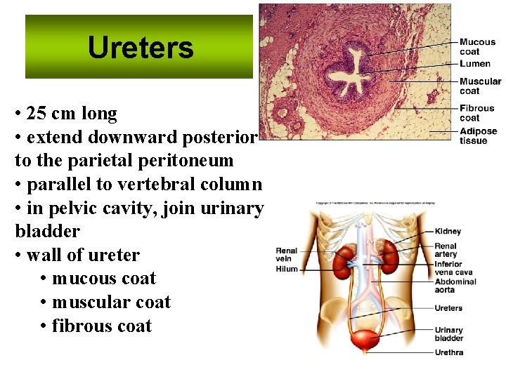
Ureters • 25 cm long • extend downward posterior to the parietal peritoneum • parallel to vertebral column • in pelvic cavity, join urinary bladder • wall of ureter • mucous coat • muscular coat • fibrous coat
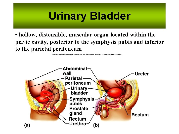
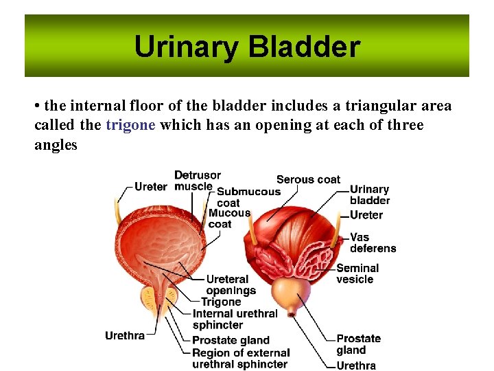
Urinary Bladder • hollow, distensible, muscular organ located within the pelvic cavity, posterior to the symphysis pubis and inferior to the parietal peritoneum
Urinary Bladder • the internal floor of the bladder includes a triangular area called the trigone which has an opening at each of three angles
Micturition (Urination) • The expulsion of urine from the bladder. • Initiated by a complex sequence of events called the micturition reflex. • The bladder is supplied by both parasympathetic and sympathetic nerve fibers of the autonomic nervous system.
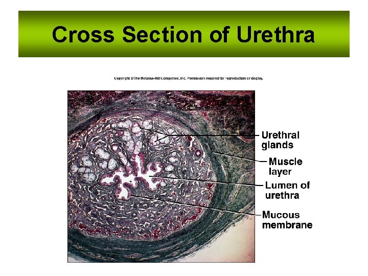
Urethra • Fibromuscular tube – exits the urinary bladder through the urethral opening – at anteroinferior surface • conducts urine to the exterior of the body. • Tunica mucosa: is a protective mucous membrane – houses clusters of mucin-producing cells called urethral glands. • Tunica muscularis: primarily smooth muscle fibers – help propel urine to the outside of the body. • Two urethral sphincters: – Internal urethral sphincter • restrict the release of urine until the pressure within the urinary bladder is high enough – External urethral sphincter • and voluntary activities needed to release the urine are activated.
Urethra • The internal urethral sphincter – involuntary (smooth muscle) – superior sphincter surrounding the neck of the bladder, where the urethra originates. – a circular thickening of the detrusor muscle – controlled by the autonomic nervous system • The external urethral sphincter – – – inferior to the internal urethral sphincter formed by skeletal muscle fibers of the urogenital diaphragm. a voluntary sphincter controlled by the somatic nervous system this is the muscle children learn to control when they become “toilet-trained”
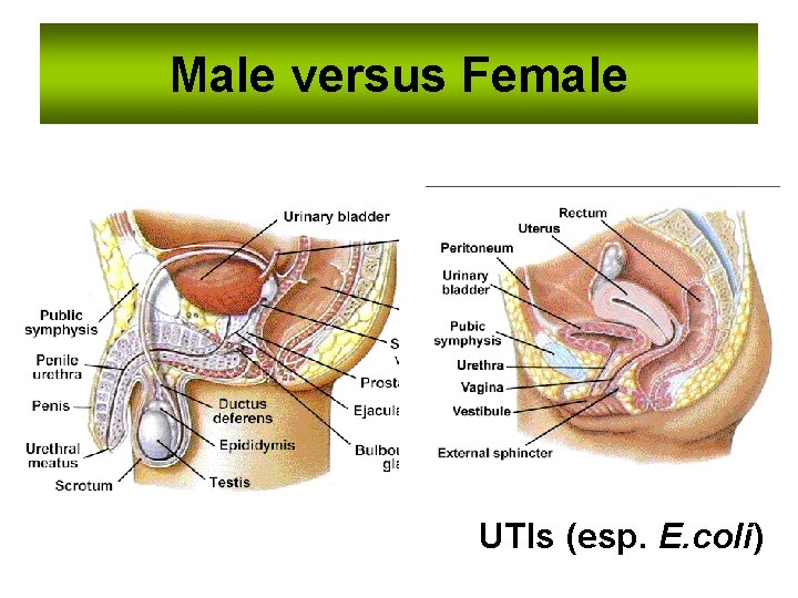
Male versus Female UTIs (esp. E. coli)
Cross Section of Urethra
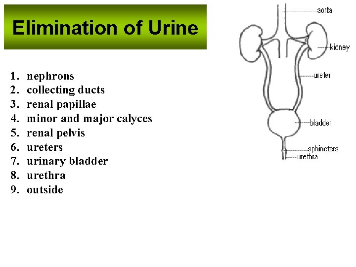
Elimination of Urine 1. 2. 3. 4. 5. 6. 7. 8. 9. nephrons collecting ducts renal papillae minor and major calyces renal pelvis ureters urinary bladder urethra outside
• Kidneys may sustain 90% loss of nephrons and still not show apparent symptoms!!! • 2 -4 % of population only have 1 kidney!
THE END