
Essentials of Human Anatomy Physiology Seventh Edition Elaine


































- Slides: 111
Essentials of Human Anatomy & Physiology Seventh Edition Elaine N. Marieb Chapter 3 Cells and Tissues Slides 3. 1 – 3. 19 Lecture Slides in Power. Point by Jerry L. Cook Copyright © 2003 Pearson Education, Inc. publishing as Benjamin Cummings
Anatomy Aug 24, 2015 • SAP 1 d: DESCRIBE HOW STRUCTURE AND FUNCTION ARE RELATED IN TERMS OF CELL AND TISSUE TYPE • WARM UP: Write the following statements with the correct answer. 1. _____ are the smallest unit of life. 2. ___ is the control center of the cell. 3. ___ is the site for ribosome production. 4. ___ is the thread-like network of DNA. • CLASSWORK: Notes on cell organelles; draw/label plasma membrane • CLOSING: Why do you think there are so many different items in the plasma membrane?
Cells and Tissues · Carry out all chemical activities needed to sustain life · Cells are the building blocks of all living things · Tissues are groups of cells that are similar in structure and function Copyright © 2003 Pearson Education, Inc. publishing as Benjamin Cummings Slide 3. 1
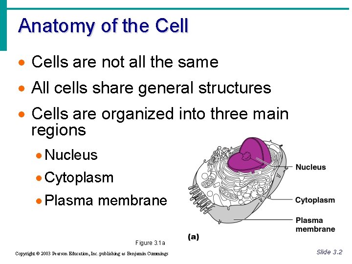
Anatomy of the Cell · Cells are not all the same · All cells share general structures · Cells are organized into three main regions · Nucleus · Cytoplasm · Plasma membrane Figure 3. 1 a Copyright © 2003 Pearson Education, Inc. publishing as Benjamin Cummings Slide 3. 2
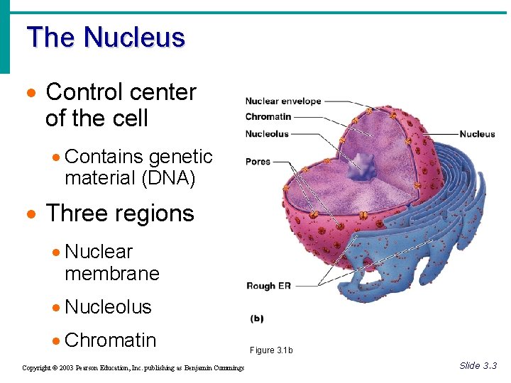
The Nucleus · Control center of the cell · Contains genetic material (DNA) · Three regions · Nuclear membrane · Nucleolus · Chromatin Copyright © 2003 Pearson Education, Inc. publishing as Benjamin Cummings Figure 3. 1 b Slide 3. 3
Nuclear Membrane · Barrier of nucleus · Consists of a double phospholipid membrane · Contain nuclear pores that allow for exchange of material with the rest of the cell Copyright © 2003 Pearson Education, Inc. publishing as Benjamin Cummings Slide 3. 4
Nucleoli · Nucleus contains one or more nucleoli · Sites of ribosome production · Ribosomes then migrate to the cytoplasm through nuclear pores Copyright © 2003 Pearson Education, Inc. publishing as Benjamin Cummings Slide 3. 5
Chromatin · Composed of DNA and protein · Scattered throughout the nucleus · Chromatin condenses to form chromosomes when the cell divides Copyright © 2003 Pearson Education, Inc. publishing as Benjamin Cummings Slide 3. 6
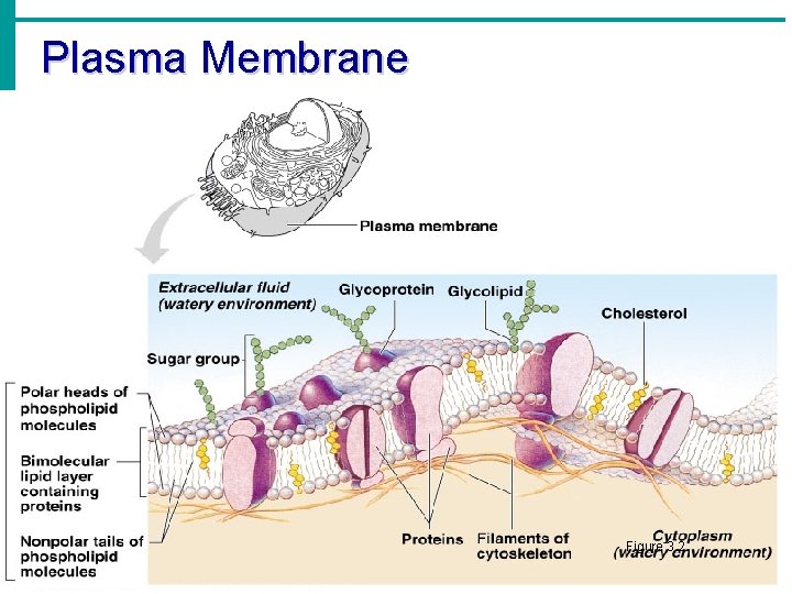
Plasma Membrane · Barrier for cell contents · Double phospholipid layer · Hydrophilic heads · Hydrophobic tails · Other materials in plasma membrane · Protein · Cholesterol · Glycoproteins Copyright © 2003 Pearson Education, Inc. publishing as Benjamin Cummings Slide 3. 7 a
Plasma Membrane Figure 3. 2 Copyright © 2003 Pearson Education, Inc. publishing as Benjamin Cummings Slide 3. 7 b
Anatomy Aug 25, 2015 • SAP 1 d: DESCRIBE HOW STRUCTURE AND FUNCTION ARE RELATED IN TERMS OF CELL AND TISSUE TYPE • WARM UP: Write and answer the following questions in your notebook. 1. What are the two main parts of the plasma membrane? 2. What does hydrophilic and hydrophobic mean? 3. List 4 items associated with the plasma membrane • CLASSWORK: Notes on cell organelles; Cell color sheet • CLOSING: Why do you think there are so many different organelles in a cell?
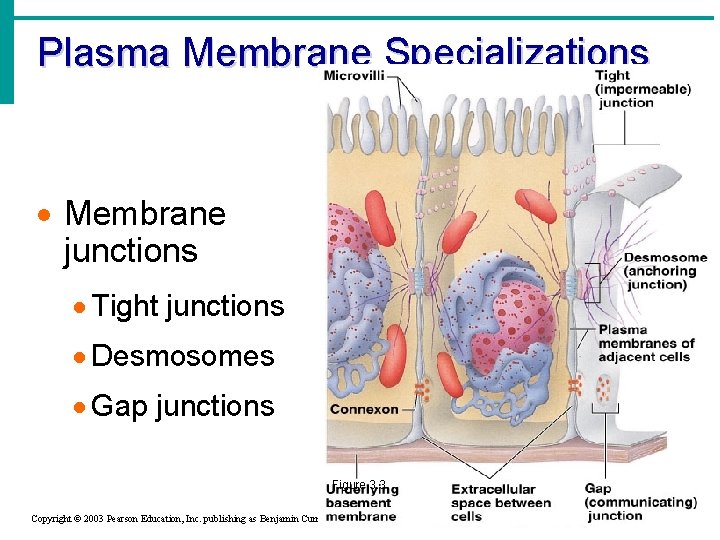
Plasma Membrane Specializations · Microvilli · Finger-like projections that increase surface area for absorption Figure 3. 3 Copyright © 2003 Pearson Education, Inc. publishing as Benjamin Cummings Slide 3. 8 a
Plasma Membrane Specializations · Membrane junctions · Tight junctions · Desmosomes · Gap junctions Figure 3. 3 Copyright © 2003 Pearson Education, Inc. publishing as Benjamin Cummings Slide 3. 8 b
Cytoplasm · Material outside the nucleus and inside the plasma membrane · Cytosol · Fluid that suspends other elements · Organelles · Metabolic machinery of the cell · Inclusions · Non-functioning units Copyright © 2003 Pearson Education, Inc. publishing as Benjamin Cummings Slide 3. 9
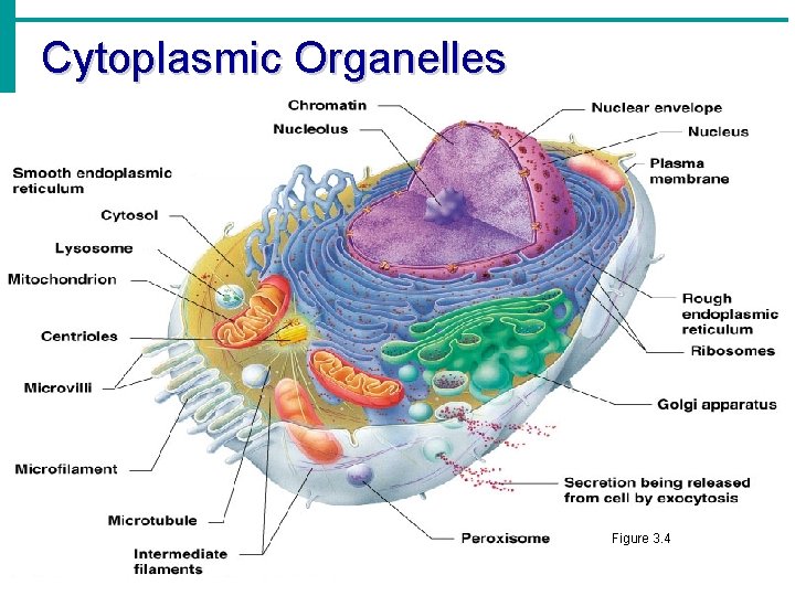
Cytoplasmic Organelles Figure 3. 4 Copyright © 2003 Pearson Education, Inc. publishing as Benjamin Cummings Slide 3. 10
Cytoplasmic Organelles · Ribosomes · Made of protein and RNA · Sites of protein synthesis · Found at two locations · Free in the cytoplasm · Attached to rough endoplasmic reticulum Copyright © 2003 Pearson Education, Inc. publishing as Benjamin Cummings Slide 3. 11
Cytoplasmic Organelles · Endoplasmic reticulum (ER) · Fluid-filled tubules for carrying substances · Two types of ER · Rough Endoplasmic Reticulum · Studded with ribosomes · Site where building materials of cellular membrane are formed · Smooth Endoplasmic Reticulum · Functions in cholesterol synthesis and breakdown, fat metabolism, and detoxification of drugs Copyright © 2003 Pearson Education, Inc. publishing as Benjamin Cummings Slide 3. 12
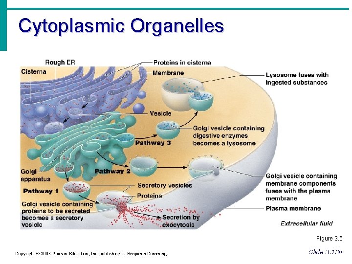
Cytoplasmic Organelles · Golgi apparatus · Modifies and packages proteins · Produces different types of packages · Secretory vesicles · Cell membrane components · Lysosomes Copyright © 2003 Pearson Education, Inc. publishing as Benjamin Cummings Slide 3. 13 a
Cytoplasmic Organelles Figure 3. 5 Copyright © 2003 Pearson Education, Inc. publishing as Benjamin Cummings Slide 3. 13 b
Cytoplasmic Organelles · Lysosomes · Contain enzymes that digest nonusable materials within the cell · Peroxisomes · Membranous sacs of oxidase enzymes · Detoxify harmful substances · Break down free radicals (highly reactive chemicals) · Replicate by pinching in half Copyright © 2003 Pearson Education, Inc. publishing as Benjamin Cummings Slide 3. 14
Cytoplasmic Organelles · Mitochondria · “Powerhouses” of the cell · Change shape continuously · Carry out reactions where oxygen is used to break down food · Provides ATP for cellular energy Copyright © 2003 Pearson Education, Inc. publishing as Benjamin Cummings Slide 3. 15
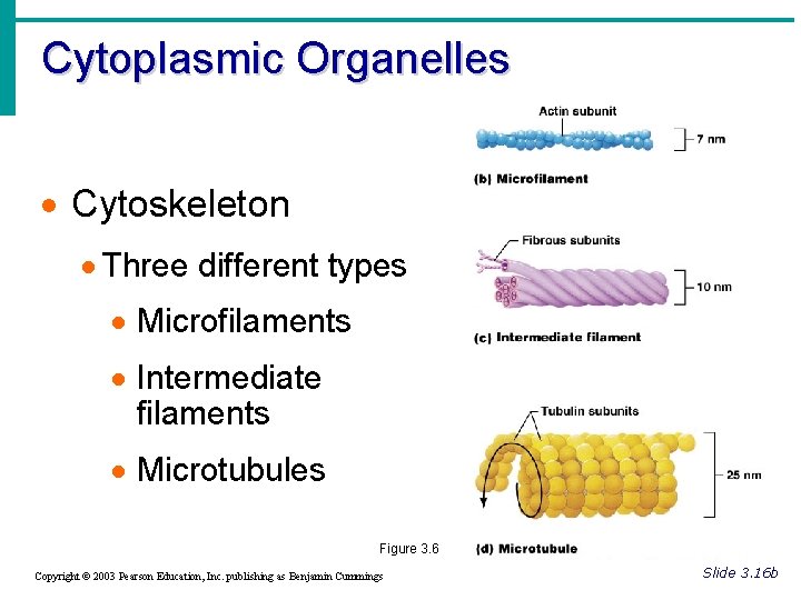
Cytoplasmic Organelles · Cytoskeleton · Network of protein structures that extend throughout the cytoplasm · Provides the cell with an internal framework Copyright © 2003 Pearson Education, Inc. publishing as Benjamin Cummings Slide 3. 16 a
Cytoplasmic Organelles · Cytoskeleton · Three different types · Microfilaments · Intermediate filaments · Microtubules Figure 3. 6 Copyright © 2003 Pearson Education, Inc. publishing as Benjamin Cummings Slide 3. 16 b
Anatomy Aug 26, 2015 • SAP 1 d: DESCRIBE HOW STRUCTURE AND FUNCTION ARE RELATED IN TERMS OF CELL AND TISSUE TYPE • WARM UP: Write and answer the following questions in your notebook. 1. What organelle produces ribosomes? 2. What organelle is responsible for cholesterol synthesis and fat metabolism? 3. What organelle digests bacteria and other harmful items that enter the cell? • CLASSWORK: Organelles graphic organizer; organelles analogies project • CLOSING: What analogy have you decided upon?
Cytoplasmic Organelles · Centrioles · Rod-shaped bodies made of microtubules · Direct formation of mitotic spindle during cell division Copyright © 2003 Pearson Education, Inc. publishing as Benjamin Cummings Slide 3. 17
Cellular Projections · Not found in all cells · Used for movement · Cilia moves materials across the cell surface · Flagellum propels the cell Copyright © 2003 Pearson Education, Inc. publishing as Benjamin Cummings Slide 3. 18
Anatomy Aug 31, 2015 • SAP 1 d: DESCRIBE HOW STRUCTURE AND FUNCTION ARE RELATED IN TERMS OF CELL AND TISSUE TYPE • WARM UP: Write and answer the following questions in your notebook. 1. Which organelle regulates what enters and leaves the cell? 2. Which organelle is responsible for packaging proteins to be sent out of cell? 3. What is the threadlike material in the nucleus that coils up into chromosomes prior to cell division? • CLASSWORK: Organelles review sheet; Notes on cell diversity. • CLOSING: Which organelle do you think is most important? Why
Anatomy Sept 1, 2015 • SAP 1 d: DESCRIBE HOW STRUCTURE AND FUNCTION ARE RELATED IN TERMS OF CELL AND TISSUE TYPE • WARM UP: look over your organelles sheets for quiz • CLASSWORK: Organelles Quiz; Notes/graphic organizer on cell diversity. • CLOSING: Why are cells so diverse?
• Nucleus • Lysosomes • Nucleolus • Peroxisomes • Chromatin • Plasma Membrane • Centrioles • Nuclear Envelope • Rough Endoplasmic Reticulum • Ribosomes • Smooth Endoplasmic Reticulum • Golgi Apparatus • Mitochondria • Microfilaments • Microtubules • Microvilli
Anatomy Sept 2, 2015 • SAP 1 d: DESCRIBE HOW STRUCTURE AND FUNCTION ARE RELATED IN TERMS OF CELL AND TISSUE TYPE • WARM UP: 1. 2. 3. 4. What are cells that bind other cells together? What cells function in reproduction? What cells fight diseases? What cells store nutrients? • CLASSWORK: Notes/graphic organizer on cell diversity; notes on cell transport • CLOSING: What is the difference between passive and active transport?
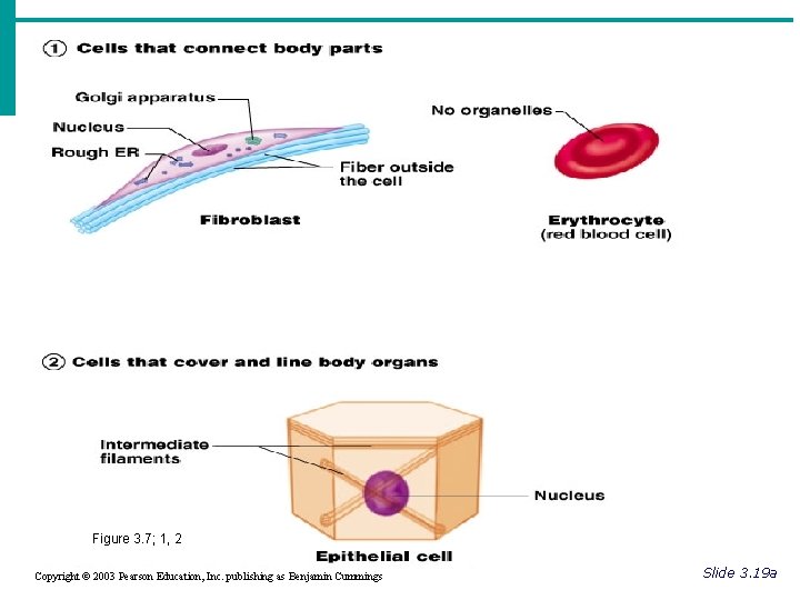
Cell Diversity Figure 3. 7; 1, 2 Copyright © 2003 Pearson Education, Inc. publishing as Benjamin Cummings Slide 3. 19 a
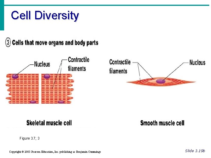
Cell Diversity Figure 3. 7; 3 Copyright © 2003 Pearson Education, Inc. publishing as Benjamin Cummings Slide 3. 19 b
Cell Diversity Figure 3. 7; 4, 5 Copyright © 2003 Pearson Education, Inc. publishing as Benjamin Cummings Slide 3. 19 c
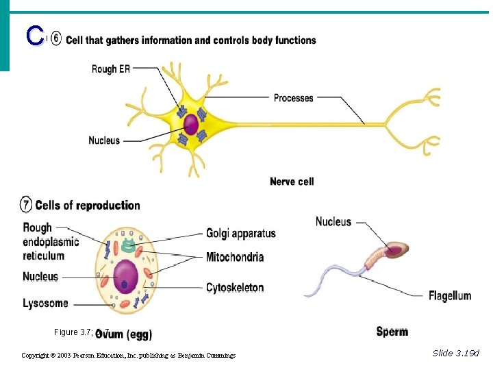
Cell Diversity Figure 3. 7; 6, 7 Copyright © 2003 Pearson Education, Inc. publishing as Benjamin Cummings Slide 3. 19 d
Cellular Physiology: Membrane Transport · Membrane Transport – movement of substance into and out of the cell · Transport is by two basic methods · Passive transport · No energy is required · Active transport · The cell must provide metabolic energy Copyright © 2003 Pearson Education, Inc. publishing as Benjamin Cummings Slide 3. 20
Solutions and Transport · Solution – homogeneous mixture of two or more components · Solvent – dissolving medium · Solutes – components in smaller quantities within a solution · Intracellular fluid – nucleoplasm and cytosol · Interstitial fluid – fluid on the exterior of the cell Copyright © 2003 Pearson Education, Inc. publishing as Benjamin Cummings Slide 3. 21
Selective Permeability · The plasma membrane allows some materials to pass while excluding others · This permeability includes movement into and out of the cell Copyright © 2003 Pearson Education, Inc. publishing as Benjamin Cummings Slide 3. 22
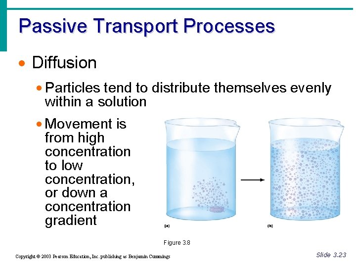
Passive Transport Processes · Diffusion · Particles tend to distribute themselves evenly within a solution · Movement is from high concentration to low concentration, or down a concentration gradient Figure 3. 8 Copyright © 2003 Pearson Education, Inc. publishing as Benjamin Cummings Slide 3. 23
Passive Transport Processes · Types of diffusion · Simple diffusion · Unassisted process · Solutes are lipid-soluble materials or small enough to pass through membrane pores Copyright © 2003 Pearson Education, Inc. publishing as Benjamin Cummings Slide 3. 24 a
Passive Transport Processes · Types of diffusion · Osmosis – simple diffusion of water · Highly polar water easily crosses the plasma membrane · Facilitated diffusion · Substances require a protein carrier for passive transport Copyright © 2003 Pearson Education, Inc. publishing as Benjamin Cummings Slide 3. 24 b
Diffusion through the Plasma Membrane Figure 3. 9 Copyright © 2003 Pearson Education, Inc. publishing as Benjamin Cummings Slide 3. 25
Passive Transport Processes · Filtration · Water and solutes are forced through a membrane by fluid, or hydrostatic pressure · A pressure gradient must exist · Solute-containing fluid is pushed from a high pressure area to a lower pressure area Copyright © 2003 Pearson Education, Inc. publishing as Benjamin Cummings Slide 3. 26
Active Transport Processes · Transport substances that are unable to pass by diffusion · too large · not lipid soluble · against a concentration gradient · Two common forms of active transport · Solute pumping · Bulk transport Copyright © 2003 Pearson Education, Inc. publishing as Benjamin Cummings Slide 3. 27
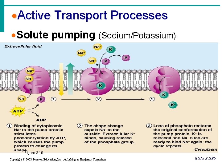
Active Transport Processes · Solute pumping (Sodium/Potassium) · Amino acids, some sugars and ions are transported by solute pumps · ATP energizes protein carriers, and in most cases, moves substances against concentration gradients Copyright © 2003 Pearson Education, Inc. publishing as Benjamin Cummings Slide 3. 28 a
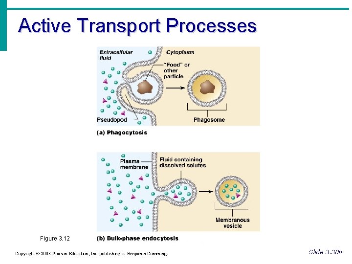
Active Transport Processes · Bulk transport · Exocytosis · Moves materials out of the cell · Material is carried in a membranous vesicle · Vesicle migrates to plasma membrane · Vesicle combines with plasma membrane · Material is emptied to the outside Copyright © 2003 Pearson Education, Inc. publishing as Benjamin Cummings Slide 3. 29 a
Active Transport Processes · Bulk transport · Endocytosis · Extracellular substances are engulfed by being enclosed in a membranous vescicle · Types of endocytosis · Phagocytosis – cell eating · Pinocytosis – cell drinking Copyright © 2003 Pearson Education, Inc. publishing as Benjamin Cummings Slide 3. 30 a
·Active Transport Processes ·Solute pumping (Sodium/Potassium) Figure 3. 10 Copyright © 2003 Pearson Education, Inc. publishing as Benjamin Cummings Slide 3. 28 b
Active Transport Processes Figure 3. 11 Copyright © 2003 Pearson Education, Inc. publishing as Benjamin Cummings Slide 3. 29 b
Active Transport Processes Figure 3. 12 Copyright © 2003 Pearson Education, Inc. publishing as Benjamin Cummings Slide 3. 30 b
Anatomy Sept 8, 2015 • SAP 1 e: DESCRIBE HOW CELLS GROW AND DIVIDE • WARM UP: 1. What are the 4 phases of mitosis? 2. What does the cell do during interphase? • CLASSWORK: Notes on Cell Cycle; • CLOSING: POPCORN
Cell Life Cycle · Cells have two major periods · Interphase · Cell grows · Cell carries on metabolic processes · Cell division · Cell replicates itself · Function is to produce more cells for growth and repair processes Copyright © 2003 Pearson Education, Inc. publishing as Benjamin Cummings Slide 3. 31
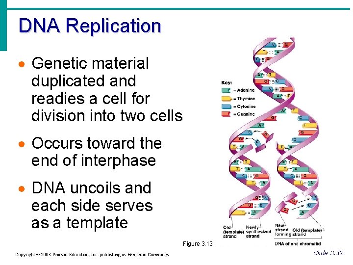
DNA Replication · Genetic material duplicated and readies a cell for division into two cells · Occurs toward the end of interphase · DNA uncoils and each side serves as a template Figure 3. 13 Copyright © 2003 Pearson Education, Inc. publishing as Benjamin Cummings Slide 3. 32
Events of Cell Division · Mitosis · Division of the nucleus · Results in the formation of two daughter nuclei · Cytokinesis · Division of the cytoplasm · Begins when mitosis is near completion · Results in the formation of two daughter cells Copyright © 2003 Pearson Education, Inc. publishing as Benjamin Cummings Slide 3. 33
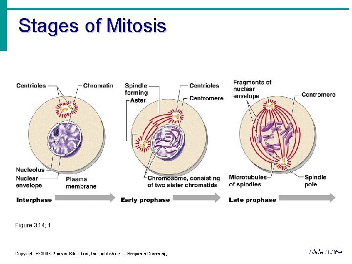
Stages of Mitosis · Interphase · No cell division occurs · The cell carries out normal metabolic activity and growth · Prophase · First part of cell division · Centromeres migrate to the poles Copyright © 2003 Pearson Education, Inc. publishing as Benjamin Cummings Slide 3. 34 a
Stages of Mitosis · Metaphase · Spindle from centromeres are attached to chromosomes that are aligned in the center of the cell Copyright © 2003 Pearson Education, Inc. publishing as Benjamin Cummings Slide 3. 34 b
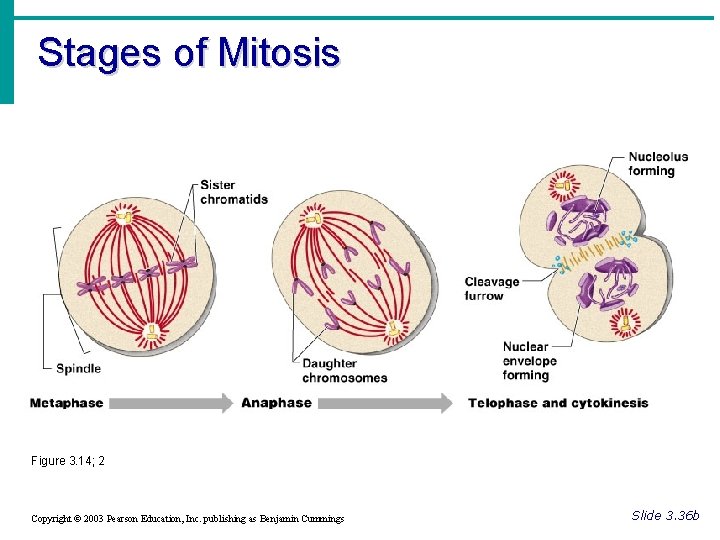
Stages of Mitosis · Anaphase · Daughter chromosomes are pulled toward the poles · The cell begins to elongate · Telophase · Daughter nuclei begin forming · A cleavage furrow (for cell division) begins to form Copyright © 2003 Pearson Education, Inc. publishing as Benjamin Cummings Slide 3. 35
Stages of Mitosis Figure 3. 14; 1 Copyright © 2003 Pearson Education, Inc. publishing as Benjamin Cummings Slide 3. 36 a
Stages of Mitosis Figure 3. 14; 2 Copyright © 2003 Pearson Education, Inc. publishing as Benjamin Cummings Slide 3. 36 b
Protein Synthesis · Gene – DNA segment that carries a blueprint for building one protein · Proteins have many functions · Building materials for cells · Act as enzymes (biological catalysts) · Proteins are made up of amino acids (building blocks of proteins) Copyright © 2003 Pearson Education, Inc. publishing as Benjamin Cummings Slide 3. 37
POPCORN
What is the 1 st phase of mitosis? • Prophase
What is the longest phase of mitosis? • Anaphase
What phase of mitosis do chromatids line up along the equator? • metaphase
When does the nuclear envelope disappear and the chromatin coil up to form chromosomes? • prophase
DNA segment that carries the blue print for building one protein? • GENE
What is the longest part of the cell cycle? • interphase
How many daughter cells are generated by each cell in mitosis? • 2
What would happen if the centromeres did not form spindle fibers? • The chromatids would not align and separate into the 2 daughter cells
What would happen if cytokinesis did not take place? • The 2 daughter cells would stay together.
What are the building blocks of proteins? • Amino Acids.
What are the building blocks of proteins? • Amino Acids.
Essentials of Human Anatomy & Physiology Seventh Edition Elaine N. Marieb Chapter 3 Cells and Tissues Slides 3. 38 – 3. 54 Lecture Slides in Power. Point by Jerry L. Cook Copyright © 2003 Pearson Education, Inc. publishing as Benjamin Cummings
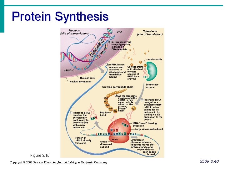
Protein Synthesis · Gene – DNA segment that carries a blueprint for building one protein · Proteins have many functions · Building materials for cells · Act as enzymes (biological catalysts) · RNA is essential for protein synthesis Copyright © 2003 Pearson Education, Inc. publishing as Benjamin Cummings Slide 3. 37
Role of RNA · Transfer RNA (t. RNA) · Transfers appropriate amino acids to the ribosome for building the protein · Ribosomal RNA (r. RNA) · Helps form the ribosomes where proteins are built · Messanger RN · Carries the instructions for building a protein from the nucleus to the ribosome Copyright © 2003 Pearson Education, Inc. publishing as Benjamin Cummings Slide 3. 38
Transcription and Translation · Transcription · Transfer of information from DNA’s base sequence to the complimentary base sequence of m. RNA · Translation · Base sequence of nucleic acid is translated to an amino acid sequence · Amino acids are the building blocks of proteins Copyright © 2003 Pearson Education, Inc. publishing as Benjamin Cummings Slide 3. 39
Protein Synthesis Figure 3. 15 Copyright © 2003 Pearson Education, Inc. publishing as Benjamin Cummings Slide 3. 40
Body Tissues · Cells are specialized for particular functions · Tissues · Groups of cells with similar structure and function · Four primary types · Epithelium · Connective tissue · Nervous tissue · Muscle Copyright © 2003 Pearson Education, Inc. publishing as Benjamin Cummings Slide 3. 41
Epithelial Tissues · Found in different areas · Body coverings · Body linings · Glandular tissue · Functions · Protection · Absorption · Filtration · Secretion Copyright © 2003 Pearson Education, Inc. publishing as Benjamin Cummings Slide 3. 42
Epithelium Characteristics · Cells fit closely together · Tissue layer always has one free surface · The lower surface is bound by a basement membrane · Avascular (have no blood supply) · Regenerate easily if well nourished Copyright © 2003 Pearson Education, Inc. publishing as Benjamin Cummings Slide 3. 43
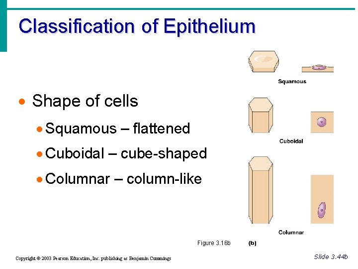
Classification of Epithelium · Number of cell layers · Simple – one layer · Stratified – more than one layer Figure 3. 16 a Copyright © 2003 Pearson Education, Inc. publishing as Benjamin Cummings Slide 3. 44 a
Classification of Epithelium · Shape of cells · Squamous – flattened · Cuboidal – cube-shaped · Columnar – column-like Figure 3. 16 b Copyright © 2003 Pearson Education, Inc. publishing as Benjamin Cummings Slide 3. 44 b
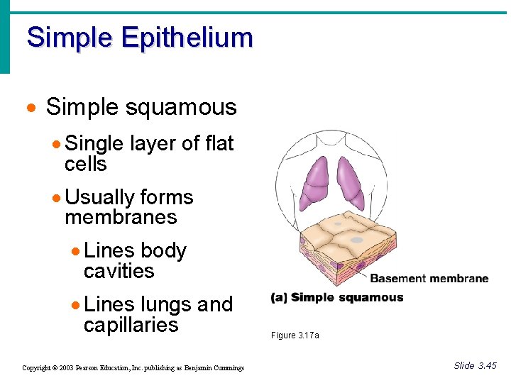
Simple Epithelium · Simple squamous · Single layer of flat cells · Usually forms membranes · Lines body cavities · Lines lungs and capillaries Copyright © 2003 Pearson Education, Inc. publishing as Benjamin Cummings Figure 3. 17 a Slide 3. 45
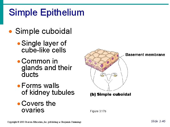
Simple Epithelium · Simple cuboidal · Single layer of cube-like cells · Common in glands and their ducts · Forms walls of kidney tubules · Covers the ovaries Copyright © 2003 Pearson Education, Inc. publishing as Benjamin Cummings Figure 3. 17 b Slide 3. 46
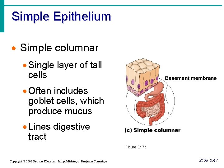
Simple Epithelium · Simple columnar · Single layer of tall cells · Often includes goblet cells, which produce mucus · Lines digestive tract Figure 3. 17 c Copyright © 2003 Pearson Education, Inc. publishing as Benjamin Cummings Slide 3. 47
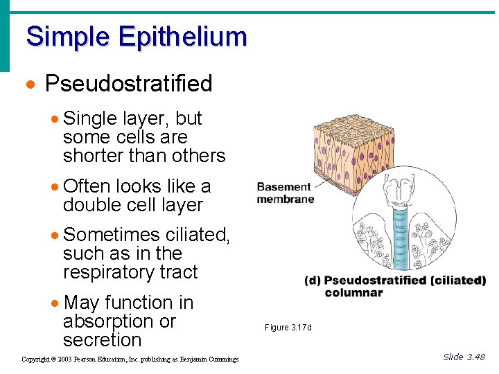
Simple Epithelium · Pseudostratified · Single layer, but some cells are shorter than others · Often looks like a double cell layer · Sometimes ciliated, such as in the respiratory tract · May function in absorption or secretion Copyright © 2003 Pearson Education, Inc. publishing as Benjamin Cummings Figure 3. 17 d Slide 3. 48
Stratified Epithelium · Stratified squamous · Cells at the free edge are flattened · Found as a protective covering where friction is common · Locations · Skin · Mouth · Esophagus Copyright © 2003 Pearson Education, Inc. publishing as Benjamin Cummings Figure 3. 17 e Slide 3. 49
Stratified Epithelium · Stratified cuboidal · Two layers of cuboidal cells · Stratified columnar · Surface cells are columnar, cells underneath vary in size and shape · Stratified cuboidal and columnar · Rare in human body · Found mainly in ducts of large glands Copyright © 2003 Pearson Education, Inc. publishing as Benjamin Cummings Slide 3. 50
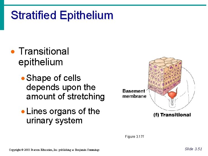
Stratified Epithelium · Transitional epithelium · Shape of cells depends upon the amount of stretching · Lines organs of the urinary system Figure 3. 17 f Copyright © 2003 Pearson Education, Inc. publishing as Benjamin Cummings Slide 3. 51
Glandular Epithelium · Gland – one or more cells that secretes a particular product · Two major gland types · Endocrine gland · Ductless · Secretions are hormones · Exocrine gland · Empty through ducts to the epithelial surface · Include sweat and oil glands Copyright © 2003 Pearson Education, Inc. publishing as Benjamin Cummings Slide 3. 52
Connective Tissue · Found everywhere in the body · Includes the most abundant and widely distributed tissues · Functions · Binds body tissues together · Supports the body · Provides protection Copyright © 2003 Pearson Education, Inc. publishing as Benjamin Cummings Slide 3. 53
Connective Tissue Characteristics · Variations in blood supply · Some tissue types are well vascularized · Some have poor blood supply or are avascular · Extracellular matrix · Non-living material that surrounds living cells Copyright © 2003 Pearson Education, Inc. publishing as Benjamin Cummings Slide 3. 54
Essentials of Human Anatomy & Physiology Seventh Edition Elaine N. Marieb Chapter 3 Cells and Tissues Slides 3. 55 – 3. 73 Lecture Slides in Power. Point by Jerry L. Cook Copyright © 2003 Pearson Education, Inc. publishing as Benjamin Cummings
Extracellular Matrix · Two main elements · Ground substance – mostly water along with adhesion proteins and polysaccharide molecules · Fibers · Produced by the cells · Three types · Collagen fibers · Elastic fibers · Reticular fibers Copyright © 2003 Pearson Education, Inc. publishing as Benjamin Cummings Slide 3. 55
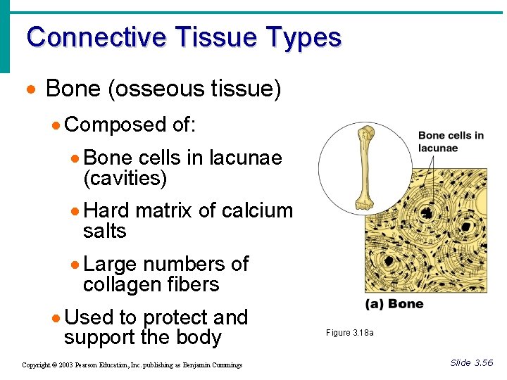
Connective Tissue Types · Bone (osseous tissue) · Composed of: · Bone cells in lacunae (cavities) · Hard matrix of calcium salts · Large numbers of collagen fibers · Used to protect and support the body Copyright © 2003 Pearson Education, Inc. publishing as Benjamin Cummings Figure 3. 18 a Slide 3. 56
Connective Tissue Types · Hyaline cartilage · Most common cartilage · Composed of: · Abundant collagen fibers · Rubbery matrix · Entire fetal skeleton is hyaline cartilage Copyright © 2003 Pearson Education, Inc. publishing as Benjamin Cummings Figure 3. 18 b Slide 3. 57
Connective Tissue Types · Elastic cartilage · Provides elasticity · Example: supports the external ear Copyright © 2003 Pearson Education, Inc. publishing as Benjamin Cummings Slide 3. 58 a
Connective Tissue Types · Fibrocartilage · Highly compressible · Example: forms cushion-like discs between vertebrae Figure 3. 18 c Copyright © 2003 Pearson Education, Inc. publishing as Benjamin Cummings Slide 3. 58 b
Connective Tissue Types · Dense connective tissue · Main matrix element is collagen fibers · Cells are fibroblasts · Examples · Tendon – attach muscle to bone · Ligaments – attach bone to bone Copyright © 2003 Pearson Education, Inc. publishing as Benjamin Cummings Figure 3. 18 d Slide 3. 59
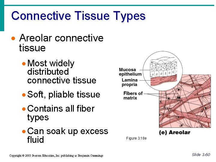
Connective Tissue Types · Areolar connective tissue · Most widely distributed connective tissue · Soft, pliable tissue · Contains all fiber types · Can soak up excess fluid Copyright © 2003 Pearson Education, Inc. publishing as Benjamin Cummings Figure 3. 18 e Slide 3. 60
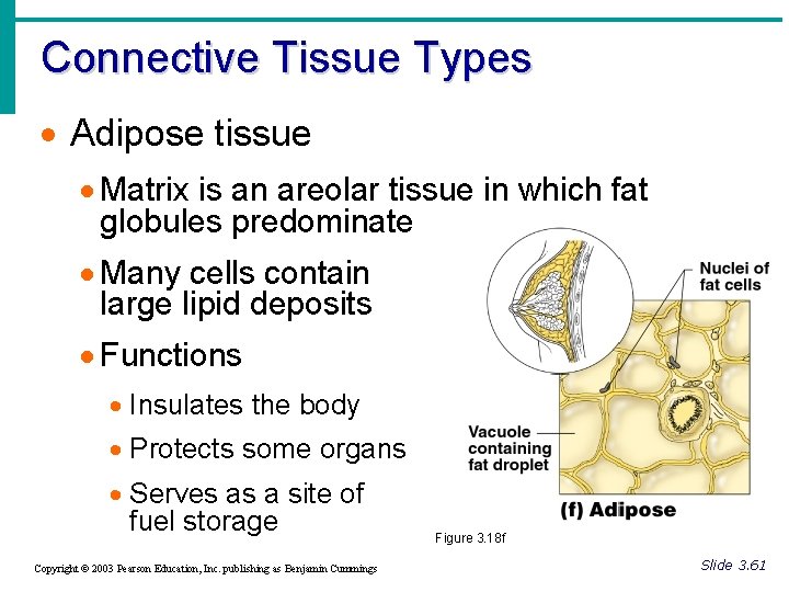
Connective Tissue Types · Adipose tissue · Matrix is an areolar tissue in which fat globules predominate · Many cells contain large lipid deposits · Functions · Insulates the body · Protects some organs · Serves as a site of fuel storage Copyright © 2003 Pearson Education, Inc. publishing as Benjamin Cummings Figure 3. 18 f Slide 3. 61
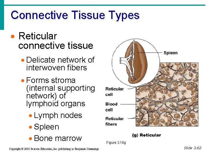
Connective Tissue Types · Reticular connective tissue · Delicate network of interwoven fibers · Forms stroma (internal supporting network) of lymphoid organs · Lymph nodes · Spleen · Bone marrow Copyright © 2003 Pearson Education, Inc. publishing as Benjamin Cummings Figure 3. 18 g Slide 3. 62
Connective Tissue Types · Blood cells surrounded by fluid matrix · Fibers are visible during clotting · Functions as the transport vehicle for materials Figure 3. 18 h Copyright © 2003 Pearson Education, Inc. publishing as Benjamin Cummings Slide 3. 63
Muscle Tissue · Function is to produce movement · Three types · Skeletal muscle · Cardiac muscle · Smooth muscle Copyright © 2003 Pearson Education, Inc. publishing as Benjamin Cummings Slide 3. 64
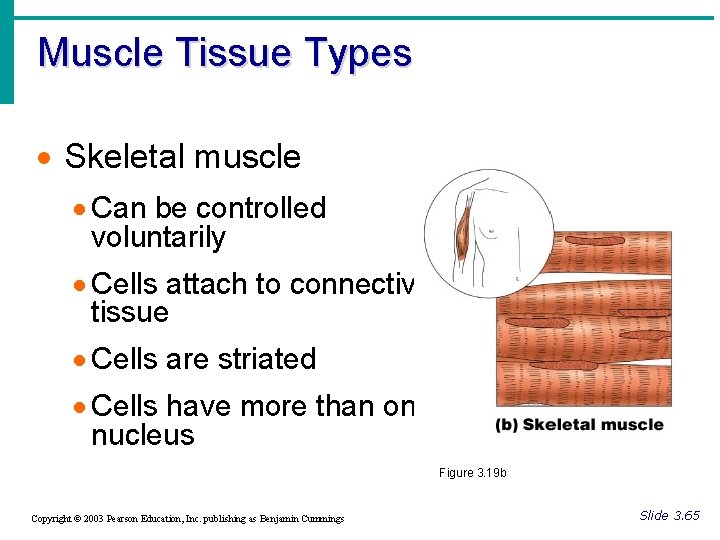
Muscle Tissue Types · Skeletal muscle · Can be controlled voluntarily · Cells attach to connective tissue · Cells are striated · Cells have more than one nucleus Figure 3. 19 b Copyright © 2003 Pearson Education, Inc. publishing as Benjamin Cummings Slide 3. 65
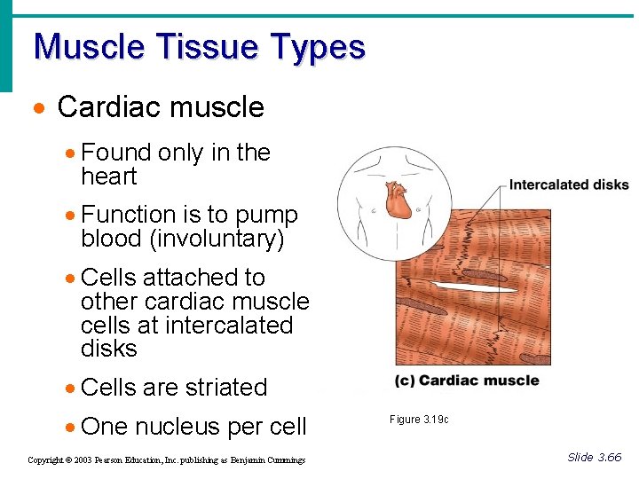
Muscle Tissue Types · Cardiac muscle · Found only in the heart · Function is to pump blood (involuntary) · Cells attached to other cardiac muscle cells at intercalated disks · Cells are striated · One nucleus per cell Copyright © 2003 Pearson Education, Inc. publishing as Benjamin Cummings Figure 3. 19 c Slide 3. 66
Muscle Tissue Types · Smooth muscle · Involuntary muscle · Surrounds hollow organs · Attached to other smooth muscle cells · No visible striations · One nucleus per cell Copyright © 2003 Pearson Education, Inc. publishing as Benjamin Cummings Figure 3. 19 a Slide 3. 67
Nervous Tissue · Neurons and nerve support cells · Function is to send impulses to other areas of the body · Irritability · Conductivity Copyright © 2003 Pearson Education, Inc. publishing as Benjamin Cummings Figure 3. 20 Slide 3. 68
Tissue Repair · Regeneration · Replacement of destroyed tissue by the same kind of cells · Fibrosis · Repair by dense fibrous connective tissue (scar tissue) · Determination of method · Type of tissue damaged · Severity of the injury Copyright © 2003 Pearson Education, Inc. publishing as Benjamin Cummings Slide 3. 69
Events in Tissue Repair · Capillaries become very permeable · Introduce clotting proteins · Wall off injured area · Formation of granulation tissue · Regeneration of surface epithelium Copyright © 2003 Pearson Education, Inc. publishing as Benjamin Cummings Slide 3. 70
Regeneration of Tissues · Tissues that regenerate easily · Epithelial tissue · Fibrous connective tissue and bone · Tissues that regenerate poorly · Skeletal muscle · Tissues that are replaced largely with scar tissue · Cardiac muscle · Nervous tissue within the brain and spinal cord Copyright © 2003 Pearson Education, Inc. publishing as Benjamin Cummings Slide 3. 71
Developmental Aspects of Tissue · Epithelial tissue arises from all three primary germ layers · Muscle and connective tissue arise from the mesoderm · Nervous tissue arises from the ectoderm · With old age there is a decrease in mass and viabililty in most tissues Copyright © 2003 Pearson Education, Inc. publishing as Benjamin Cummings Slide 3. 72