Essentials of Human Anatomy Physiology Elaine N Marieb

Essentials of Human Anatomy & Physiology Elaine N. Marieb Seventh Edition The Nervous System Modified by J. Kalinowski 1/2014 Copyright © 2003 Pearson Education, Inc. publishing as Benjamin Cummings

Functions of the Nervous System Sensory input – gathering information Uses sensory receptors to monitor changes (stimuli) occurring inside and outside the body Integration To process and interpret sensory input and decide if action is needed Copyright © 2003 Pearson Education, Inc. publishing as Benjamin Cummings Slide 7. 1 a

Functions of the Nervous System Slide 7. 1 b Motor output A response to integrated stimuli The response activates muscles or glands Copyright © 2003 Pearson Education, Inc. publishing as Benjamin Cummings

Functions of Neuroglia § § Myelin production Structural support Communication Environmental monitoring

Neuroglia can replicate but cannot conduct Supports neurons
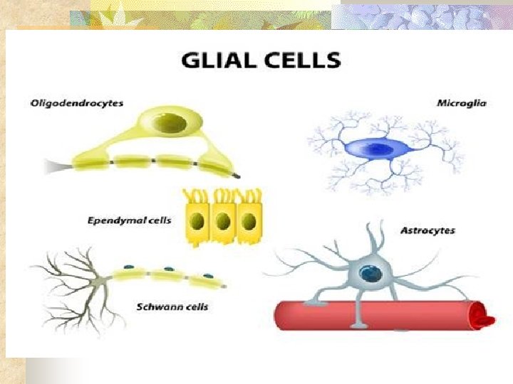
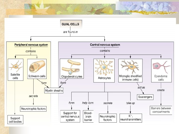

Neuroglial cells – write functions by the diagram • Microglia - scavenge and degrade dead cells, protecting the brain from invading microorganisms • Oligodendrocytes - form myelin sheaths around axons in the CNS • Schwann cells - form myelin sheaths around axons in the PNS

Neuroglial Cells • Ependymal cells - produce cerebrospinal fluid in brain and spinal cords • Astrocytes - provide nutrients to neurons, give synapses structural support, and block toxic substances

Nervous Tissue: Neurons = nerve cells Cells specialized to transmit messages – can conduct but cannot replicate Have 3 specialized characteristics Longevity: with nutrition, can live as long as you do Amitotic: unable to reproduce themselves (so cannot be replaced) High metabolic rate: require continuous oxygen & glucose (due to lots of activity) Copyright © 2003 Pearson Education, Inc. publishing as Benjamin Cummings Slide 7. 8

Neuroglia vs. Neurons Neuroglia divide. Neurons do not. Most brain tumors are “gliomas. ” Involve the neuroglia cells, not the neurons. As neuroglia grow out of control, they press on the neurons impairing their function
 of a neuron so important? It is")
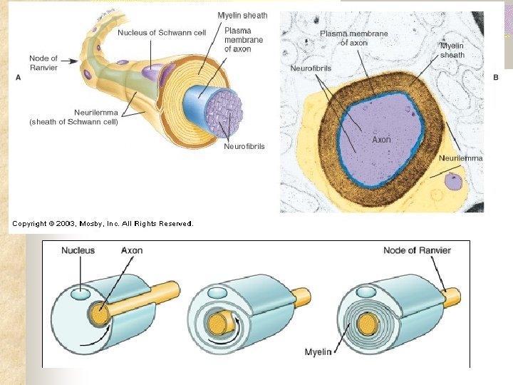
Neurilemma Why is the outer covering (neurilemma) of a neuron so important? It is the site of electrical signaling – also plays a crucial role in cell to cell interactions during development

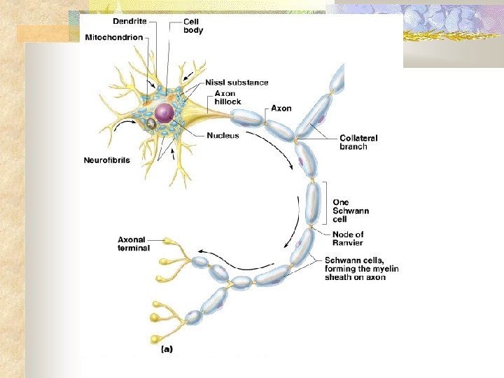
Major Regions of Neurons Cell body Contains the metabolic/biosynthetic center of the cell (location of the nucleus) Does not contain centrioles (reflects amitotic nature) but has the other organelles Copyright © 2003 Pearson Education, Inc. publishing as Benjamin Cummings Slide 7. 8

Neuron Anatomy Dendrites hundreds per cell – diffusely branched – close to cell body Receptive sites conduct impulses toward the cell body Immense surface area for reception Figure 7. 4 a Copyright © 2003 Pearson Education, Inc. publishing as Benjamin Cummings Slide 7. 10

Neuron Anatomy • Axon hillock: site of summation for incoming information where the "decision" to initiate or not initiate a nerve impulse takes place

Neuron Anatomy Axons Transmit impulses away from cell body Vary in length and diameter Larger diameter = faster conduction Figure 7. 4 a Copyright © 2003 Pearson Education, Inc. publishing as Benjamin Cummings Slide 7. 10

Neuron Anatomy Axons Axon terminals located at end of axon branches Figure 7. 4 a Copyright © 2003 Pearson Education, Inc. publishing as Benjamin Cummings Slide 7. 10

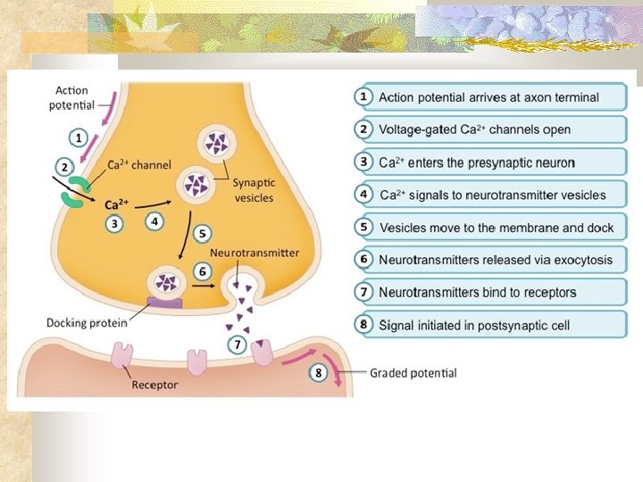
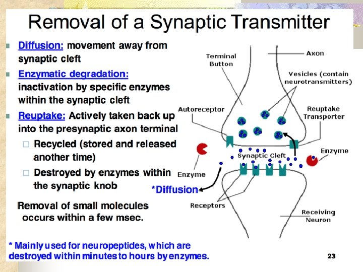
Axon terminals Contain vesicles with neurotransmitters – chemicals which transmit electrical impulses Axonal terminals are separated from the next neuron or effector by the Synaptic cleft Copyright © 2003 Pearson Education, Inc. publishing as Benjamin Cummings Slide 7. 11

Axon terminals Synaptic knob – site where action potential is converted into a chemical message Synapse – the entire junction between 2 nerves OR a nerve and another structure Copyright © 2003 Pearson Education, Inc. publishing as Benjamin Cummings Slide 7. 11

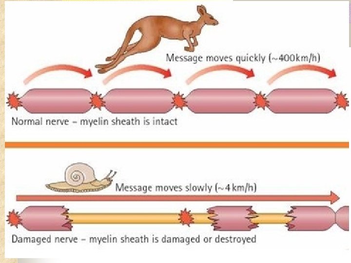
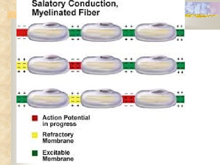
Myelin Sheath Function: Protects & insulates fibers Increases speed of transmission through a process known as Saltatory Conduction Figure 7. 5 Copyright © 2003 Pearson Education, Inc. publishing as Benjamin Cummings Slide 7. 12

End of Quiz #1 Material Study for quiz!!!
 Nerve fibers that carry information from sensory receptors")
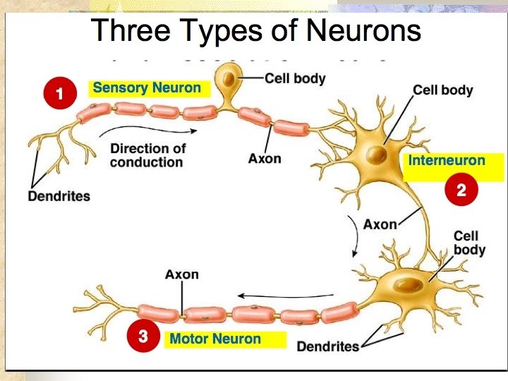
Functional Classification of Neurons Sensory (afferent) Nerve fibers that carry information from sensory receptors to the central nervous system (CNS) Ends of dendrites associated with specialized receptors Figure 7. 1 Copyright © 2003 Pearson Education, Inc. publishing as Benjamin Cummings
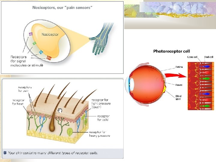
, thermoreceptors (heat & cold) • Proprioceptors:")
Sensory Receptors • Cutaneous receptors: pressure, nociceptors (pain), thermoreceptors (heat & cold) • Proprioceptors: muscles, tendons, organs: amount of stretch or tension • Specialized receptors in sense organs: photoreceptors (light), mechanoreceptors (hearing & equilibrium), chemoreceptors (smell & taste)
 division Nerve fibers that carry impulses from the central nervous")
Functional Classification Motor (efferent) division Nerve fibers that carry impulses from the central nervous system to muscles & glands Association or Interneurons Responsible for integration & reflex – connect motor & sensory neurons Make up over 99% of neurons Figure 7. 1 Copyright © 2003 Pearson Education, Inc. publishing as Benjamin Cummings Slide 7. 3 b

Reflex Activity Reflex: rapid predictable motor response to stimuli that the body is programmed to do Unlearned, unpremeditated, involuntary Withdrawal from pain Learned or acquired reflexes result from repetition or practice. Example: experienced driver drives a car Copyright © 2003 Pearson Education, Inc. publishing as Benjamin Cummings Slide 7. 58

Reflex Activity Two types: Autonomic: regulate the activity of smooth muscles, the heart, and glands Examples: salivary reflex, pupilary reflex, digestion, blood pressure Somatic reflexes: skeletal muscle reflexes Example: knee jerk reflex Copyright © 2003 Pearson Education, Inc. publishing as Benjamin Cummings Slide 7. 58

Reflex – define 5 elements of Know your diagram

Functional Properties of Neurons Two major functional properties of neurons resulting in electrochemical event Irritability - ability to respond to stimuli & convert it into a nerve impulse Conductivity – ability to transmit an impulse to other neurons, muscles, or glands Copyright © 2003 Pearson Education, Inc. publishing as Benjamin Cummings Slide 7. 17

Synapse – know the diagram Copyright © 2003 Pearson Education, Inc. publishing as Benjamin Cummings Slide 7. 11

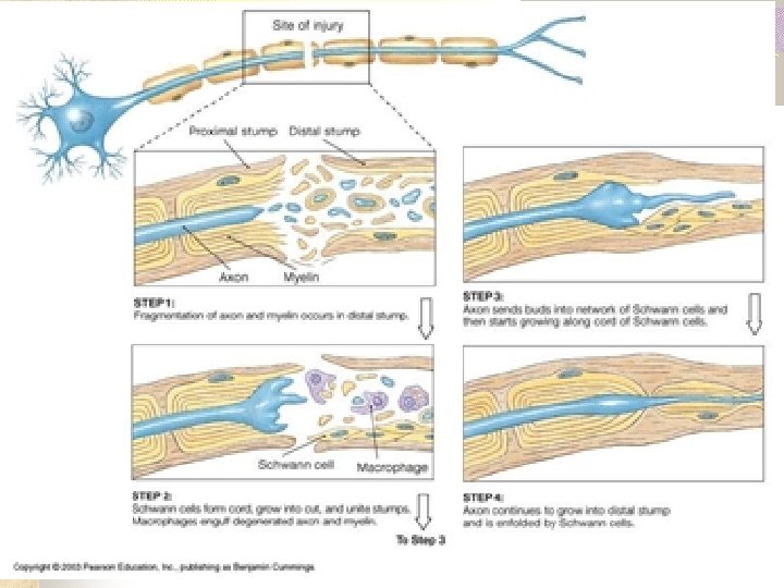
Regeneration • Mature neurons are incapable of mitosis. • PNS nerve axons can regenerate successfully if cell body is not destroyed • Uninjured cell body swells to prepare to synthesize proteins to support regeneration

Regeneration Axons regenerate at a rate of 1. 5 mm/day The greater the distance between severed nerve endings, the less chance of recovery. Axonal sprouts may grow into surrounding areas and form a mass called a neuroma. Surgical realignment can help. Retraining may be necessary once the connection is completed Copyright © 2003 Pearson Education, Inc. publishing as Benjamin Cummings Slide 7. 14 b

Neuroma Acoustic neuroma MRI

Regeneration PNS vs CNS In PNS axon regeneration, macrophages clean out the debris from the injury. Schwann cells will form a tunnel of neurolemma to guide severed nerve ending together. A growth factor is also released In CNS – No Schwann cells to do this. Slide Copyright © 2003 Pearson Education, Inc. publishing as Benjamin Cummings 7. 14 b

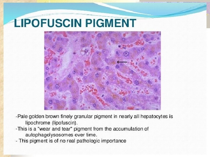
Age related changes Degenerative changes Loss of dendritic & synaptic connections Accumulation of lipofuscin

Age related changes Lead to: Loss of balance Insomnia Increased risk of depression, Alzheimer’s & Parkinson’s disease Fading memory Slowed responses & reflexes

End of Quiz #2 Material Don’t forget to complete your Quiz review for EC
- Slides: 46