Essentials of Human Anatomy Physiology Elaine N Marieb

Essentials of Human Anatomy & Physiology Elaine N. Marieb Seventh Edition Chapter 5 The Skeletal System Copyright © 2003 Pearson Education, Inc. publishing as Benjamin Cummings

The Skeletal System

Essential Question What is the purpose of the skeletal system?
 · Joints")
The Skeletal System · Parts of the skeletal system · Bones (skeleton) · Joints · Cartilages · Ligaments (bone to bone)(tendon=bone to muscle) Copyright © 2003 Pearson Education, Inc. publishing as Benjamin Cummings Slide 5. 1

The Skeletal System • Skeleton comes from a Greek word meaning dried up body. • Bone appears dead and dried up, but it is not! • Bone is living tissue • Newborn human has 350 bones • Adult human has 206 bones
 · Protection of soft organs")
Functions of Bones · Support of the body (framework) · Protection of soft organs · Serve as levers (with help from muscles) · Storage of minerals and fats (calcium) · Blood cell formation Copyright © 2003 Pearson Education, Inc. publishing as Benjamin Cummings Slide 5. 2

Bones of the Human Body · Two basic types of bone tissue · Compact bone · Dense/hard · Spongy bone · (Cancellous) · Many open spaces Figure 5. 2 b ·Decrease wt of bone/contain red bone Slide 5. 3 marrow Copyright © 2003 Pearson Education, Inc. publishing as Benjamin Cummings

Classification of Bones · Long bones · Typically longer than wide · Have a shaft with heads at both ends · Contain mostly compact bone · Found in legs and arms • Examples: Femur, humerus Copyright © 2003 Pearson Education, Inc. publishing as Benjamin Cummings Slide 5. 4 a

Classification of Bones · Short bones · Generally cube-shape and small · Contain mostly spongy bone · Found in wrist, ankles, and toes · Examples: Carpals, tarsals Copyright © 2003 Pearson Education, Inc. publishing as Benjamin Cummings Slide 5. 4 b

Classification of Bones on the Basis of Shape Figure 5. 1 Copyright © 2003 Pearson Education, Inc. publishing as Benjamin Cummings Slide 5. 4 c

Classification of Bones · Flat bones · Thin and flattened · Usually curved · Cover organs/provide surface for lg. muscle · Thin layers of compact bone around a layer of spongy bone · Examples: Skull, ribs, sternum Copyright © 2003 Pearson Education, Inc. publishing as Benjamin Cummings Slide 5. 5 a

Classification of Bones · Irregular bones · Irregular shape · Do not fit into other bone classification categories · Example: Vertebrae and hip

Gross Anatomy of a Bone · Diaphysis · Shaft · Composed of compact bone · Epiphysis · Ends of the bone · Composed mostly of spongy bone

Structure of a Long Bone · Periosteum · Outside covering of the diaphysis · Fibrous connective tissue membrane Serves as an attachment for muscle · Arteries · Supply bone cells with nutrients

Structure of a Long Bone · Articular cartilage · Covers the external surface of the epiphyses · Made of hyaline cartilage · Decreases friction at joint surfaces

Structure of a Long Bone · Medullary cavity · Cavity of the shaft · Contains yellow marrow (mostly fat) in adults · Contains red marrow (for blood cell formation) in infants

Changes in the Human Skeleton · In embryos, the skeleton is primarily hyaline cartilage · During development, much of this cartilage is replaced by bone · Cartilage remains in isolated areas · Bridge of the nose · Parts of ribs · Joints Copyright © 2003 Pearson Education, Inc. publishing as Benjamin Cummings Slide 5. 12

Bone Growth · Epiphyseal plates allow for growth of long bone during childhood · New cartilage is continuously formed · Older cartilage becomes ossified · Cartilage is broken down · Bone replaces cartilage Copyright © 2003 Pearson Education, Inc. publishing as Benjamin Cummings Slide

Bone Growth · Bones are remodeled and lengthened until growth stops · Grow longitudinally for height · Bones grow in width to support weight Copyright © 2003 Pearson Education, Inc. publishing as Benjamin Cummings Slide

Epiphyseal Disc • Refer to figure 8 -3 • Growth plate • The cartilage near the epiphyseal disc multiplies and eventually becomes ossified (turns to bone) • As long as new cartilage continues to form the bone continues to lengthen.
 • When the growth plate hardens and becomes ossified, growth")
Epiphyseal Disc (cont. ) • When the growth plate hardens and becomes ossified, growth stops • Hormones play a big part in this • Growth hormone stimulates growth • Sex hormones stop growth

Bone Width • Long after longitudinal bone growth has stopped, bones continue to grow in thickness and width. • Bones are continuously being reshaped

Types of Bone Cells · Osteocytes · Mature bone cells · Osteoblasts · Bone-forming cells · Osteoclasts · Bone-destroying cells · Break down bone matrix for remodeling and release of calcium · Bone remodeling is a process by both osteoblasts and osteoclasts Copyright © 2003 Pearson Education, Inc. publishing as Benjamin Cummings Slide 5. 15
 and osteoclasts (bone")
Bone Remodeling • A combined action of osteoblasts (bone forming cells) and osteoclasts (bone destroying cells) • Osteoblasts deposit bone on the external bone surface • Figure 8 -5 (like a brick layer) • Osteoclasts break down bone from the inside • Figure 8 -5 (like a sculptor)

Long Bone Formation and Growth Figure 5. 4 a Copyright © 2003 Pearson Education, Inc. publishing as Benjamin Cummings Slide

Bone Fractures · A break in a bone · Types of bone fractures · Closed (simple) fracture – break that does not penetrate the skin · Open (compound) fracture – broken bone penetrates through the skin · Bone fractures are treated by reduction and immobilization · Realignment of the bone Copyright © 2003 Pearson Education, Inc. publishing as Benjamin Cummings Slide 5. 16

Common Types of Fractures Table 5. 2 Copyright © 2003 Pearson Education, Inc. publishing as Benjamin Cummings Slide 5. 17

Handout #1 Injuries to bones

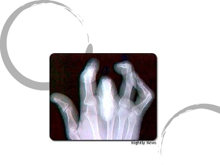
Dislocation of joint • Displacement of bones at the joint • Often caused by impact trauma to that joint • Can be more damaging and painful than a fracture • Damage to the joint capsule and surrounding ligaments and tendons often takes much longer to heal than bone tissue.

Dislocated Finger

In Groups of 4 • Locate the fracture/dislocation • Identify the type of fracture
 is formed · Break is splinted")
Repair of Bone Fractures · Hematoma (blood-filled swelling) is formed · Break is splinted by fibrocartilage to form a soft callus · Blood vessels grow into the hematoma · Fibrocartilage callus is replaced by a bony callus · Bony callus is remodeled to form a permanent patch Copyright © 2003 Pearson Education, Inc. publishing as Benjamin Cummings Slide 5. 18

Stages in the Healing of a Bone Fracture Figure 5. 5 Copyright © 2003 Pearson Education, Inc. publishing as Benjamin Cummings Slide 5. 19

Skeletal System • Divided into two divisions • Axial skeleton ~ bones of the cranium, face, vertebral column, and bony thorax. • Appendicular skeleton ~ includes the bones of the pelvic girdles, the upper extremities and lower extremities.
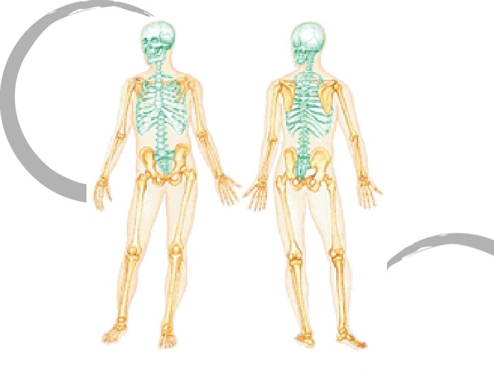

The Axial Skeleton · Forms the longitudinal part of the body · Divided into three parts · Skull · Vertebral column · Bony thorax Copyright © 2003 Pearson Education, Inc. publishing as Benjamin Cummings Slide

The Axial Skeleton Figure 5. 6 Copyright © 2003 Pearson Education, Inc. publishing as Benjamin Cummings Slide
 (18 names!) · Sits on top of the vertebral column")
The Skull (28 bones) (18 names!) · Sits on top of the vertebral column · Two sets of bones · Cranium (8 bones) · Facial bones (14 bones) · Bones are joined by sutures · Only the mandible is attached by a freely movable joint Copyright © 2003 Pearson Education, Inc. publishing as Benjamin Cummings Slide

The Cranium • Bony structure that encases and protects the brain. • 8 bones • Frontal Bone ~ forehead/upper part of the bony structure surrounding the eyes.

The Skull Figure 5. 7 Copyright © 2003 Pearson Education, Inc. publishing as Benjamin Cummings Slide

Bones of the Skull Figure 5. 11 Copyright © 2003 Pearson Education, Inc. publishing as Benjamin Cummings Slide 5. 22

Human Skull, Superior View Figure 5. 8 Copyright © 2003 Pearson Education, Inc. publishing as Benjamin Cummings Slide 5. 23

Human Skull, Inferior View Figure 5. 9 Copyright © 2003 Pearson Education, Inc. publishing as Benjamin Cummings Slide 5. 24
 ~ upper sides of the head and the")
The Cranium • Parietal Bone (2) ~ upper sides of the head and the roof of the cranial cavity (top of the head)

The Skull Figure 5. 7 Copyright © 2003 Pearson Education, Inc. publishing as Benjamin Cummings Slide

Bones of the Skull Figure 5. 11 Copyright © 2003 Pearson Education, Inc. publishing as Benjamin Cummings Slide 5. 22

Human Skull, Superior View Figure 5. 8 Copyright © 2003 Pearson Education, Inc. publishing as Benjamin Cummings Slide 5. 23
 ~ sides of the head, close to ears.")
The Cranium • Temporal Bones (2) ~ sides of the head, close to ears. • Commonly called the temples • Includes the external auditory meatus • Opening for the ear • Includes the zygomatic process • Part of the cheekbone
")
The Skull (lateral view)

The Skull
")
The Skull (superior view)

The Cranium • Occipital Bone ~ back and base of the cranium • Includes the foramen magnum • Foramen means hole • Large hole for the brainstem/spinal cord
")
The Skull (lateral view)

The Skull
")
The Skull (superior view)

The Cranium • Sphenoid Bone ~ forms sides of cranium and parts of orbits of the eyes • Butterfly shaped • Includes Sella Turcica (Turk’s Saddle) • Where pituitary gland sits
")
The Skull (lateral view)

The Skull
")
The Skull (superior view)

The Cranium • Ethmoid Bone ~ irregularly shaped bone located between the eye orbits • Major supporting bone of the nasal cavity
")
The Skull (lateral view)

The Skull
")
The Skull (superior view)

The Cranium • That’s it! No more cranium bones! • 206 bones - 8 bones = 198 bones to go…

Facial Bones • 14 bones • Most of these bones come in pairs • Only the mandible and vomer are single bones

Facial Bones • Mandible ~ the lower jaw bone • Carries the lower teeth • The anterior portion forms the chin • Only freely movable joint in the skull
")
The Skull (lateral view)

The Skull
")
The Skull (superior view)
 ~ Upper jaw • Two bones fused together •")
Facial Bones • Maxilla (2) ~ Upper jaw • Two bones fused together • Roof of the mouth • Also form parts of the nasal cavity and eye orbits
")
The Skull (lateral view)

The Skull
")
The Skull (superior view)
 ~ form the posterior part of the hard")
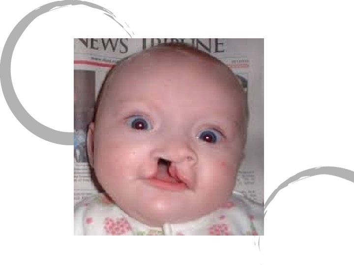
Facial Bones • Palantine Bones (2) ~ form the posterior part of the hard palate and the floor of the nasal cavity. • Failure of the palatine and/or maxillary bones to fuse causes a cleft palate.
")
The Skull (lateral view)

The Skull
")
The Skull (superior view)
 ~ the cheekbones • Also forms a")
The Facial Bones • Zygomatic Bones (2) ~ the cheekbones • Also forms a part of the orbits of the eyes
")
The Skull (lateral view)

The Skull
")
The Skull (superior view)
 ~ inner wall of")
Facial Bones • Other Facial Bones • Lacrimal Bones (2) ~ inner wall of eye sockets • Nasal Bones (2) ~ bridge of nose • Vomer ~ nasal septum • Inferior Nasal Conchae (2)
")
The Skull (lateral view)

The Skull
")
The Skull (superior view)

Facial Bones • That’s it! No more facial bones! • 198 bones - 14 bones = 184 bones to go…

Paranasal Sinuses · Functions of paranasal sinuses · Air filled cavities · Lighten the skull · Give resonance and amplification to voice Figure 5. 10 Copyright © 2003 Pearson Education, Inc. publishing as Benjamin Cummings Slide

The Fetal Skull · The fetal skull is large compared to the infants total body length Figure 5. 13 Copyright © 2003 Pearson Education, Inc. publishing as Benjamin Cummings Slide

The Fetal Skull · Fontanelles – fibrous membranes connecting the cranial bones · Allow the brain to grow · Convert to bone within 24 months after birth Figure 5. 13 Copyright © 2003 Pearson Education, Inc. publishing as Benjamin Cummings Slide

The Hyoid Bone · U shaped · Found in the upper neck · The only bone that does not articulate with another bone · Serves as a moveable base for the tongue Figure 5. 12 Copyright © 2003 Pearson Education, Inc. publishing as Benjamin Cummings Slide 5. 26

Hyoid Bone

Middle Ear • • • 3 Tiny bones ~ transmit vibrations All derived from Latin words Malleus (hammer) Incus (anvil) Stapes (stirrup) • Smallest bone in the body

Middle Ear and Hyoid Bones • That’s it! • 184 bones - 4 bones = 180 bones to go…

The Vertebral Column • The backbone or spine • Consists of 26 bones called vertebrae
 ·")
The Vertebral Column · Vertebrae separated by intervertebral discs (act as shock absorbers) · The spine has a normal curvature · Each vertebrae is given a name according to its location Copyright © 2003 Pearson Education, Inc. publishing as Benjamin Cummings Figure 5. 14 Slide 5. 28

Vertebral column • C 1 -C 7 ~ in the neck region • 7 cervical vertebrae • T 1 -T 12 ~ located in the chest region • 12 thoracic vertebrae • L 1 -L 5 ~ located in the lower back • 5 lumbar vertebrae

Vertebral column • Sacrum ~ curved bone of the lower back (posterior wall of the pelvis) • fused sacral vertebrae • 5 vertebrae at birth • Coccyx ~ the tailbone • 4 vertebrae at birth

Vertebral Column • The vertebrae become larger as the vertebral column descends…. . WHY? • Vertebral foramen ~ opening for spinal cord. • What is the opening for the spinal cord in the skull called?

Vertebral Column • 180 bones - 26 vertebral column bones = 154 bones to go!
 · The chest region · Forms a cage to")
The Bony Thorax (Thoracic Cage) · The chest region · Forms a cage to protect major organs · Composed of sternum, ribs and thoracic vertebrae. Copyright © 2003 Pearson Education, Inc. publishing as Benjamin Cummings Figure 5. 19 a Slide

Thoracic Cage • Sternum ~ breastbone. • Dagger-shaped bone located along the midline of the anterior chest.

Thoracic Cage • Ribs ~ 12 pairs of ribs attach posteriorly to the thoracic vertebrae • True ribs ~ first 7 pair • False ribs ~ last 5 pairs

That is it for the axial skeleton! • 154 bones - 24 ribs -1 sternum = 129 bones to go!!!
 · Pectoral (shoulder) girdle · Pelvic girdle Copyright")
The Appendicular Skeleton · Limbs (appendages) · Pectoral (shoulder) girdle · Pelvic girdle Copyright © 2003 Pearson Education, Inc. publishing as Benjamin Cummings Slide

The Appendicular Skeleton Figure 5. 6 c Copyright © 2003 Pearson Education, Inc. publishing as Benjamin Cummings Slide
 Girdle · Composed of two bones · Clavicle – collarbone ·")
The Pectoral (Shoulder) Girdle · Composed of two bones · Clavicle – collarbone · Scapula – shoulder blade · These bones allow the upper limb to have exceptionally free movement Copyright © 2003 Pearson Education, Inc. publishing as Benjamin Cummings Slide 5. 33

Bones of the Shoulder Girdle Figure 5. 20 a, b Copyright © 2003 Pearson Education, Inc. publishing as Benjamin Cummings Slide

Bones of the Upper Limb · The arm is formed by a single bone · Humerus · Head of humerus allows for rotation Copyright © 2003 Pearson Education, Inc. publishing as Benjamin Cummings Figure 5. 21 a, b Slide

Bones of the Upper Limb • The forearm has two bones • Ulna • Radius Figure 5. 21 c Copyright © 2003 Pearson Education, Inc. publishing as Benjamin Cummings Slide

Radius • Radius ~ locate on the lateral or thumb side when the palm of the hand is facing forward.

Ulna • Ulna~ the longer of the two forearm bones. • Located on the medial or little finger side of the forearm.

Bones of the Upper Limb · The hand · Carpals – wrist · Metacarpals – palm · Phalanges – fingers Figure 5. 22 Copyright © 2003 Pearson Education, Inc. publishing as Benjamin Cummings Slide 5. 36
 ·")
Bones of the Pelvic Girdle · Composed of two coxal bones (hip bones) · Composed of three pair of fused bones · Ilium · Ischium · Pubis · The total weight of the upper body rests on the pelvis · Protects several organs · Reproductive organs · Urinary bladder · Part of the large intestine Copyright © 2003 Pearson Education, Inc. publishing as Benjamin Cummings Slide 5. 37

The Pelvis Figure 5. 23 a Copyright © 2003 Pearson Education, Inc. publishing as Benjamin Cummings Slide

Gender Differences of the Pelvis Figure 5. 23 c Copyright © 2003 Pearson Education, Inc. publishing as Benjamin Cummings Slide 5. 39

Bones of the Lower Limbs · The thigh has one bone · Femur – thigh bone Figure 5. 35 a, b Copyright © 2003 Pearson Education, Inc. publishing as Benjamin Cummings Slide

Bones of the Lower Limb • Patella ~ knee cap • Triangular bone located within a tendon that passes over the knee.

Bones of the Lower Limbs · The leg has two bones · Tibia ~ shin bone · larger · Fibula · Long and thin Figure 5. 35 c Copyright © 2003 Pearson Education, Inc. publishing as Benjamin Cummings Slide
– ankle · Metatarsals")
Bones of the Lower Limbs · The foot · Tarsal (7)– ankle · Metatarsals (5)– sole/instep · Phalanges (14) – toes Copyright © 2003 Pearson Education, Inc. publishing as Benjamin Cummings Figure 5. 25 Slide 5. 41

Joints · Articulations of bones · Functions of joints · Hold bones together · Provide flexibility · Ways joints are classified · By their function · By their structure Copyright © 2003 Pearson Education, Inc. publishing as Benjamin Cummings Slide 5. 43

Functional Classification of Joints · Synarthroses – immovable joints · Amphiarthroses – slightly moveable joints · Diarthroses – freely moveable joints Copyright © 2003 Pearson Education, Inc. publishing as Benjamin Cummings Slide 5. 44

Structural Classification of Joints · Fibrous joints · Generally immovable · Cartilaginous joints · Immovable or slightly moveable · Synovial joints · Freely moveable Copyright © 2003 Pearson Education, Inc. publishing as Benjamin Cummings Slide 5. 45

Fibrous Joints · Bones united by fibrous tissue – synarthrosis or largely immovable. Figure 5. 27 d, e Copyright © 2003 Pearson Education, Inc. publishing as Benjamin Cummings Slide 5. 46

Cartilaginous Joints – mostly amphiarthrosis · Bones connected by cartilage · Examples · Pubic symphysis · Intervertebral joints Figure 5. 27 b, c Copyright © 2003 Pearson Education, Inc. publishing as Benjamin Cummings Slide 5. 47

Synovial Joints · Articulating bones are separated by a joint cavity · Synovial fluid is found in the joint cavity · Reinforced by ligaments Figure 5. 27 f–h Copyright © 2003 Pearson Education, Inc. publishing as Benjamin Cummings Slide 5. 48

The Synovial Joint Figure 5. 28 Copyright © 2003 Pearson Education, Inc. publishing as Benjamin Cummings Slide 5. 51

6 Types of Synovial Joints • Hinge joint • Movement is like two boards joined together by a hinge • Movement in one direction • Elbow, knees, fingers

Types of Synovial Joints • Ball and Socket Joint • When ball-shaped end of one bone fits into the cup-shaped socket of another • Bones can move in many directions • Shoulder, hip

Types of Synovial Joints • Pivot Joint • Allows for rotation around the length of a bone. • Allows only for rotation • Head (side to side “no” action) • Forearm joints (palms) supination/pronation

Types of Synovial Joints • Saddle Joint • When the surfaces of both articulation bones are saddle-shaped • Concave/convex • Thumb • Wide range of motion

Types of Synovial Joints • Gliding Joint • Interaction of flat surfaces of articulating bones • Limited but complex movement • Wrist, ankle

Types of Synovial Joints • Condyloid Joint • Oval-shaped articular surface of one bone fits into the oval-shaped depression of another • Mandible, knuckles

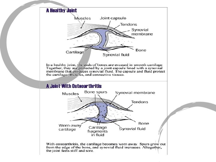
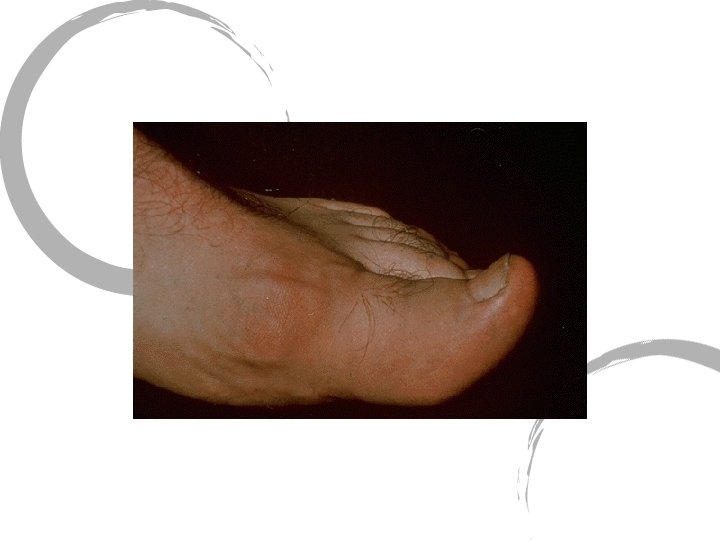
Inflammatory Conditions Associated with Joints · Bursitis – inflammation of a bursa usually caused by a blow or friction · Tendonitis – inflammation of tendon sheaths · Arthritis – inflammatory or degenerative diseases of joints · Over 100 different types · The most widespread crippling disease in the United States Copyright © 2003 Pearson Education, Inc. publishing as Benjamin Cummings Slide 5. 53

Clinical Forms of Arthritis · Osteoarthritis · Most common chronic arthritis · Probably related to normal aging processes · Rheumatoid arthritis · An autoimmune disease – the immune system attacks the joints · Symptoms begin with bilateral inflammation of certain joints · Often leads to deformities Copyright © 2003 Pearson Education, Inc. publishing as Benjamin Cummings Slide

Clinical Forms of Arthritis · Gouty Arthritis · Inflammation of joints is caused by a deposition of urate crystals from the blood · Can usually be controlled with diet · Red meat and wine are high in uric acid Copyright © 2003 Pearson Education, Inc. publishing as Benjamin Cummings Slide
- Slides: 139