Essentials of Human Anatomy Physiology Elaine N Marieb

Essentials of Human Anatomy & Physiology Elaine N. Marieb Seventh Edition BIOL 2402 The Cardiovascular System Copyright © 2003 Pearson Education, Inc. publishing as Benjamin Cummings

The Cardiovascular System A closed system of the heart and blood vessels The heart pumps blood Blood vessels allow blood to circulate to all parts of the body The function of the cardiovascular system is to deliver oxygen and nutrients and to remove carbon dioxide and other waste products Copyright © 2003 Pearson Education, Inc. publishing as Benjamin Cummings Slide 11. 1

The Cardiovascular System Cells make the exchange of nutrients & wastes only with the fluid in their immediate vicinity. Changing & refreshing these fluids is necessary to prevent buildup of wastes and to replenish the nutrient supply The lymphatic system aids with this process. Copyright © 2003 Pearson Education, Inc. publishing as Benjamin Cummings Slide 11. 1

The Heart Location Mediastinum - between the lungs Posterosuperior base points toward right shoulder beneath 2 nd rib Anteroinferior apex directed toward left hip, rests on diaphragm at top of 6 th rib About the size of your fist Weighs less than a pound: 250 -350 grams Copyright © 2003 Pearson Education, Inc. publishing as Benjamin Cummings Slide 11. 2 a

Exterior surface of heart

Exterior surface of heart

Sulci

The Heart Figure 11. 1 Copyright © 2003 Pearson Education, Inc. publishing as Benjamin Cummings Slide 11. 2 b

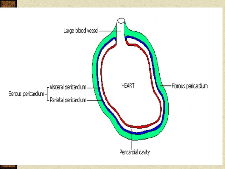
The Heart: Double layered membranous sac Fibrous Pericardium - outer layer Superficial layer of dense irregular CT Functions: Protect heart Secure heart to mediastinum Copyright © 2003 Pearson Education, Inc. publishing as Benjamin Cummings Slide 11. 3

The Heart: Coverings Serous Pericardium – a double serous membrane Visceral pericardium/epicardium · Next to heart Parietal pericardium · Outside layer Serous fluid (reduce friction) fills the pericardial cavity: space between layers of pericardium Copyright © 2003 Pearson Education, Inc. publishing as Benjamin Cummings Slide 11. 3
")
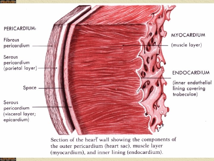
The Heart: Heart Wall Epicardium/visceral pericardium · Friction reducing delicate layer (simple squamous epithelium) Myocardium · Middle layer · Mostly cardiac muscle with fibrous CT support · Actual contracting tissue layer Endocardium · Inner layer · Endothelium (simple squamous epithelium) lining reduces friction and continues into BV Copyright © 2003 Pearson Education, Inc. publishing as Benjamin Cummings

The Heart: Four Chambers Right Atrium Receives blood from body through superior & inferior vena cavae & coronary sinus Thin walled – does not need great pumping power Auricle – pouch like structure that increases atrial volume Pectinate muscles: anterior wall of right atrium Interatrial septum Copyright © 2003 Pearson Education, Inc. publishing as Benjamin Cummings Slide 11. 6

The Heart: Four Chambers Left Atrium Receives blood from lungs through right & left pulmonary veins Thin walled – does not need great pumping power Auricle – pouch like structure that increases atrial volume No pectinate muscles Copyright © 2003 Pearson Education, Inc. publishing as Benjamin Cummings Slide 11. 6

The Heart: Chambers Four chambers Ventricles - discharging chambers · Right ventricle – goes to lungs through pulmonary arteries · Left ventricle – goes to body through aorta · Thick muscular walls – need pumping power ·Left ventricle pumps to entire body Interventricular Septum Copyright © 2003 Pearson Education, Inc. publishing as Benjamin Cummings Slide 11. 6

The Heart: Septum: divides heart into right and left halves Superior vena cava: returns blood to heart from head, shoulders, and arms Inferior vena cava: returns blood to heart from rest of body Copyright © 2003 Pearson Education, Inc. publishing as Benjamin Cummings Slide 11. 6

External Heart Anatomy Copyright © 2003 Pearson Education, Inc. publishing as Benjamin Cummings Figure 11. 2 a Slide 11. 5

External Heart Anatomy Pectinate muscles Chordae Tendinae Trabeculae carnae Papillary Muscle Copyright © 2003 Pearson Education, Inc. publishing as Benjamin Cummings Figure 11. 2 a Slide 11. 5

The Heart: Pulmonary trunk: BV out of right ventricle to pulmonary arteries Pulmonary arteries: to lungs Pulmonary veins: back to left side of heart Aorta: Largest Artery in body Copyright © 2003 Pearson Education, Inc. publishing as Benjamin Cummings Slide 11. 6
 Allow blood to flow in only one direction")
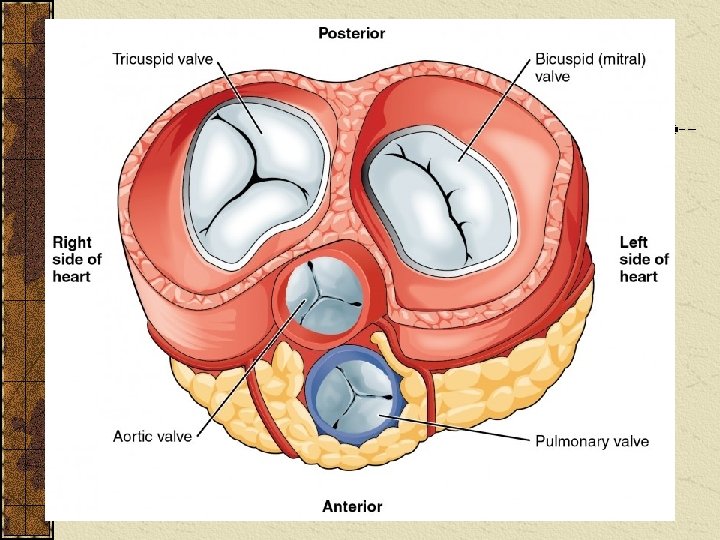
The Heart: Valves (notes page 5) Allow blood to flow in only one direction Four valves Atrioventricular valves – between atria and ventricles · Papillary muscles: attached to chordae tendineae to help prevent valves from allowing backflow · Bicuspid valve (left) – aka. Mitral valve · Tricuspid valve (right) Copyright © 2003 Pearson Education, Inc. publishing as Benjamin Cummings Slide 11. 8
 ·Aortic")
The Heart: Valves Semilunar valves between ventricle and artery ·Pulmonary semilunar valve (right) ·Aortic semilunar valve (left) Copyright © 2003 Pearson Education, Inc. publishing as Benjamin Cummings Slide 11. 8

The Heart: Valves Know how valves open & close to prevent backflow of blood which is important in keeping oxygenated & deoxygenated blood from mixing and gets them to the right structures. Copyright © 2003 Pearson Education, Inc. publishing as Benjamin Cummings Slide 11. 9

Operation of Heart Valves Figure 11. 4 Copyright © 2003 Pearson Education, Inc. publishing as Benjamin Cummings Slide 11. 10

Heart Sounds Heart sounds - result from closing of valves 1. Lub - AV valve closure – first sound 2. Dup - semilunar valve closure – second sound

Coronary Circulation: See Lab Portfolio for specifics blood contained in the heart does not nourish the heart right & left coronary arteries and their major branches supply blood to heart cardiac veins empty into coronary sinus which drains into right atrium left ventricle works hardest so needs most blood Slide Copyright © 2003 Pearson Education, Inc. publishing as Benjamin Cummings 11. 12
- Slides: 27