ESOPHAGUS Dr Bushra AlTarawneh MD NORMAL HISTOLOGY OF

ESOPHAGUS Dr. Bushra Al-Tarawneh, MD

NORMAL HISTOLOGY OF ESOPHAGUS

OBSTRUCTIVE DISEASES - - Mechanical Obstruction: Stenosis most often is due to inflammation and scarring, which may be caused by chronic gastroesophageal reflux, irradiation, or caustic injury. Absence, or agenesis, of the esophagus is extremely rare. Atresia, in which a thin, noncanalized cord replaces a segment of esophagus. Functional Obstruction: Achalasia is characterized by the triad of incomplete LES relaxation, increased LES tone, and esophageal aperistalsis. Caused by Degenerative changes in neural innervation. Can be primary or secondary.

Esophageal Varices - Portal hypertension induces development of collateral channel that enlarge the subepithelial and submucosal venous plexi within the distal esophagus. - Develop in 90% of cirrhotic patients, most commonly in association with alcoholic liver disease. Worldwide, hepatic schistosomiasis is the second most common cause of varices. - Varices often are asymptomatic, but their rupture can lead to massive hematemesis and death. Lacerations: -The most common esophageal lacerations are Mallory. Weiss tears, which are often associated with severe retching or vomiting. - By contrast, Boerhaave syndrome, characterized by transmural esophageal tears and mediastinitis, occurs rarely and is a catastrophic event.

ESOPHAGITIS 1 - Chemical and Infectious Esophagitis Ø Medicinal pills may lodge and dissolve in the esophagus, rather than passing into the stomach intact, resulting in a condition termed pill-induced esophagitis. Ø Esophagitis due to chemical injury generally causes only self-limited pain, particularly odynophagia (pain with swallowing). Hemorrhage, stricture, or perforation may occur in severe cases. Ø Iatrogenic esophageal injury may be caused by cytotoxic chemotherapy, radiation therapy, or graft-versus-host disease. Ø Candidiasis is characterized by adherent, graywhite pseudomembranes composed of densely matted fungal hyphae and inflammatory cells covering the esophageal mucosa.

Herpesviruses typically cause punched-out ulcers and histopathologic analysis demonstrates nuclear viral inclusions within a rim of degenerating epithelial cells at the ulcer edge). CMV causes shallower ulcerations and characteristic nuclear and cytoplasmic inclusions within capillary endothelium and stromal cells.
 GERD is most common in adults older")
REFLUX ESOPHAGITIS AND GASTROESOPHAGEAL REFLUX DISEASE (GERD) GERD is most common in adults older than 40 years of age but also occurs in infants and children. The most frequently reported symptoms are heartburn and dysphagia. Reflux of gastric contents into the lower esophagus is the most frequent cause of esophagitis. The stratified squamous epithelium of the esophagus is resistant to abrasion from foods but is sensitive to acid. Constant LES tone prevents reflux of acidic gastric contents. Conditions that decrease LES tone or increase abdominal pressure contribute to GERD and include alcohol and tobacco use, obesity, central nervous system depressants, pregnancy, hiatal hernia delayed gastric emptying, and increased gastric volume. In many cases, no definitive cause is identified.

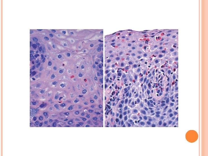
Simple hyperemia, evident to the endoscopist as redness. Microscopically: Eosinophils are recruited into the squamous mucosa, followed by neutrophils. Basal zone hyperplasia elongation of lamina propria papillae. Treatment with proton pump inhibitors reduces gastric acidity and typically provides symptomatic relief. Complications include esophageal ulceration, hematemesis, melena, stricture development, and Barrett esophagus. Hiatal hernia is characterized by separation of the diaphragmatic crura and protrusion of the stomach into the thorax through the resulting gap.

EOSINOPHILIC ESOPHAGITIS The cardinal histologic feature is epithelial infiltration by large numbers of eosinophils. Failure of high-dose proton pump inhibitor treatment and the absence of acid reflux, are also typical. A majority of persons with eosinophilic esophagitis are atopic, and many have atopic dermatitis, allergic rhinitis, asthma, or modest peripheral eosinophilia. Treatments include dietary restrictions to prevent exposure to food allergens, and topical or systemic corticosteroids.

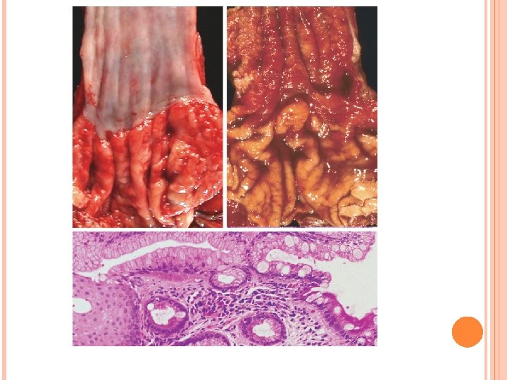
BARRETT ESOPHAGUS Barrett esophagus is a complication of chronic GERD that is characterized by intestinal metaplasia within the esophageal squamous mucosa. The incidence of Barrett esophagus is 10% of persons with symptomatic GERD. White males are affected most often and typically present between 40 and 60 years of age. Clinical Features Diagnosis of Barrett esophagus requires endoscopy and biopsy, usually prompted by GERD symptoms. Although the vast majority of esophageal adenocarcinomas are associated with Barrett esophagus, it should be noted that most persons with Barrett esophagus do not develop esophageal cancer.

Barrett esophagus is recognized endoscopically as tongues or patches of red, velvety mucosa extending upward from the gastroesophageal junction. Histologically : documented gastric or intestinal metaplasia for diagnosis of Barrett esophagus. Goblet cells, which have distinct mucous vacuoles that stain pale blue by H&E and impart the shape of a wine goblet to the remaining cytoplasm. Dysplasia is classified as low-grade or high-grade on the basis of morphologic criteria. Intramucosal carcinoma is characterized by invasion of neoplastic epithelial cells into the lamina propria.

ESOPHAGEAL TUMORS 1 - Esophageal adenocarcinoma typically arises in a background of Barrett esophagus and long-standing GERD. Risk of adenocarcinoma is tobacco use, obesity, and previous radiation therapy. Conversely, reduced adenocarcinoma risk is associated with diets rich in fresh fruits and vegetables. Esophageal adenocarcinoma occurs most frequently in whites and shows a strong gender bias, being seven times more common in men than in women. Esophageal adenocarcinoma usually occurs in the distal third of the esophagus and may invade the adjacent gastric cardia. While early lesions may appear as flat or raised patches in otherwise intact mucosa, tumors may form large exophytic masses, infiltrate diffusely, or ulcerate and invade deeply. It manifests with pain or difficulty in swallowing, progressive weight loss, chest pain, or vomiting.
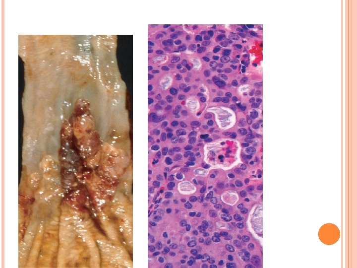

2 - squamous cell carcinoma of the esophagus It occurs in adults older than 45 years of age and affects males four times more frequently than females. Risk factors include alcohol and tobacco use, poverty, caustic esophageal injury, achalasia, Plummer-Vinson syndrome, frequent consumption of very hot beverages, and previous radiation therapy to the mediastinum. In contrast to the distal location of most adenocarcinomas, half of squamous cell carcinomas occur in the middle third of the esophagus. Clinical manifestations of squamous cell carcinoma of the esophagus begin insidiously and include dysphagia, odynophagia (pain on swallowing), and obstruction. Early lesions appear as small, gray-white plaquelike thickenings. Over months to years they grow into tumor masses that may be polypoid and protrude into and obstruct the lumen. Other tumors are either ulcerated or diffusely infiltrative lesions.

SQUAMOUS CELL CARCINOMA COMPOSED OF NESTS OF MALIGNANT CELLS THAT PARTIALLY RECAPITULATE THE STRATIFIED ORGANIZATION OF SQUAMOUS EPITHELIUM.
- Slides: 17