Esophagus Anatomy Physiology Corrosive stricture Perforation of Esophagus


 most common site of perforation during endoscopy is at")
 Always occurs on the")
 The MC cause is chest injury by a")
 Caustic Injury Drug Induced eg. tetracycline, KCL,")


- Slides: 49
Esophagus: Anatomy, Physiology, Corrosive stricture & Perforation of Esophagus Dr Amit Gupta Associate Professor Dept. of Surgery
Anatomy The primitive foregut forms during the fourth week of gestation by a longitudinal folding and incorporation of the dorsal part of the yolk sac into the embryo. 34 th day: The distal esophagus elongates first, followed by the proximal. 6 th week: Mesenchymal circular muscle coat develops Three to nine weeks later, longitudinal musculature appears.
Seventh to eighth week: Esophageal lumen is almost filled with cells from the proliferated esophageal epithelium. During the 4 th month, the muscularis mucosa appears
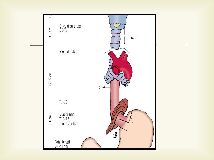
narrowest tube of the gastrointestinal tract Midline structure anterior to the spine and posterior to the trachea Length: ranges from 21 cm-34 cm (27 cm average).
Classical anatomy divides the esophagus into three parts: Cervical Thoracic Abdominal
Function divides the esophagus according to its differing forms of motility into the following three zones : Upper esophageal sphincter (UES) Esophageal body Lower esophageal sphincter (LES)
Arterial Supply
Venous Supply
Innervation
Lymphatic drainage
Histology It lacks a serosal coating The four layers are: mucosa Submucosa muscularis tunica adventitia
Corrosive stricture
Stricture formation, which usually develops between 3 and 8 weeks after the initial injury but sometimes requires a much longer period for evolution
Etiology Alkaline caustics, acid or acidlike corrosives, and household bleaches. Hydrochloric, sulfuric, nitric, and phosphoric acids are contained in automobile battery acids.
Age 75% of injuries involving children younger than 5 years and a much lower, secondary peak occurring in 20 -30
Type of caustic related to injury Acid – Generally less severe injury Alkaline • Liquefactive necrosis • Sodium hydroxide – Coagulative necrosis – Coagulum lessen tissue penetration • Very hazardous • 30% causes full thickness necrosis
The severity of esophageal and gastric damage resulting from a caustic ingestion depends on Corrosive properties Concentration of the agent Quantity swallowed
Pathogenesis Corrosive enter to stomach ‐> reflex pyloric spasm Limit passage of corrosive to duodenum Regurgitation of corrosive against a closed cricopharyngeus ‐> damage to esophagus and Stomach 3‐ 5 mins ‐> gastric atonia ‐> opening of pylorus
Goal of emergency management Limit and treat the immediately life-threatening consequences Control subsequent stricture formation
Endoscopic findings Zargar et al GIE 1991; Orringer 1993
Early management Resuscitation Upper airway – Assessment of severity of damage – Secure the airway Fiberoptic intubation Tracheostomy
Contraindication Emetics OG or NG Neutralization Alkali ---try Milk Acid---- do not try anything
Surgery is warranted if evidence of Perforation of the esophagus or stomach Mediastinitis Peritonitis exists
Treatment Corticosteroids to modify the inflammatory response to the burn injury Antibiotics to control secondary bacterial infection Esophagoscopy within 12 -24 hrs
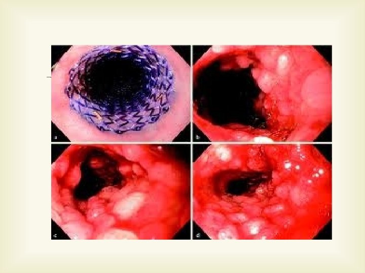
Bougienage Esophageal stents Colon interposition Forearm tube Free jejunal flap
Perforation of Esophagus
Introduction Grand Admiral of Holland died of spontaneous rupture of the esophagus in 1724 J. R. Meyer of Berlin was the first to recognize this disease prior to death Barrett made the first early diagnosis and performed the first surgical repair in 1946
Anatomy Esophagus lacks serosa More likely to rupture Site of rupture: More commonly on left side Due to instrumentation: distal esophagus Spontaneous: posterolateral esophagus Tears are usually longitudinal
Etiology Iatrogenic Instrumentation (MC cause) most common site of perforation during endoscopy is at the cricopharyngeus Surgical injury
Boerhaave Syndrome (barogenic perforation, postemetic perforation, spontaneous esophageal rupture) Always occurs on the left side of the distal third of esophagus Most tears occur along the longitudinal axis (0. 6 to 8. 9 cm) long The mucosal tear is often longer than the muscle tear, which is important to repair the esophageal wall completely
Trauma (8% to 15. 3%) The MC cause is chest injury by a steering wheel in a traffic accident The incidence of esophageal perforation by penetrating injuries is 11% to 17% Perforation is more common in the cervical than thoracic esophagus The overall mortality rate remains high (15% to 40%).
Tumor Foreign Body ( 7 -14%) Caustic Injury Drug Induced eg. tetracycline, KCL, quinidine, NSAID’s Infection Other Causes eg. Barrett ulcer and ulcerative esophagitis with Zollinger-Ellison syndrome
Pathophysiology Air, Saliva, and Gastric contents released mediastinitis pneumomediastinum empyema can progress to sepsis, shock, resp failure
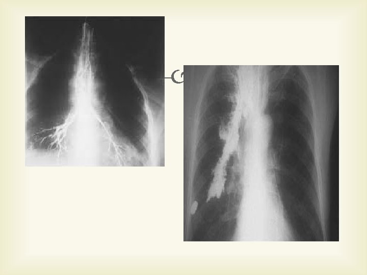
Diagnosis Chest X ray Chest radiographs appear normal in the early phase Emphysema becomes manifestated by 1 hour after the perforation Pleural effusion is detected several hours after the perforation Pneumomediastinum is present in 60% of cases. Perforation of the mid-thoracic esophagus is associated with right-sided pleural effusion and perforation of the distal thoracic esophagus is associated with left-sided pleural effusion
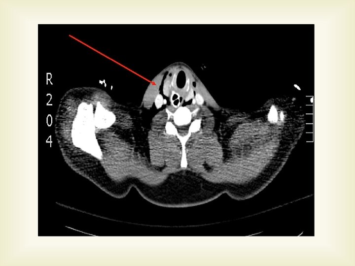
Esophagography The detection rate is 60% for cervical perforation and 90% for surgically confirmed perforations. Computerized tomography (CT) Endoscopy Diagnostic thoracentesis
Treatment The goal of treatment is to: Prevent further contamination Eliminate infection produced by contamination Restore the integrity and continuity of the GIT Restore and maintain adequate nutrition
There are two major types of treatment Surgical Nonsurgical
Surgical treatment Primary closure Reinforced closure Resection Drainage alone T-tube drainage Exclusion and diversion Intraluminal stents
Primary repair of Esophagus
The principles of surgical treatment are : Debridement of all infected and necrotic tissue Secure closure of the perforation Correction or elimination of distal obstruction Drainage of contaminated and infected areas An enteral nutrition route, such as a jejunostomy, should be added for nutritional support to any surgical method
Choice of Treatment Surgical Non Surgical Patient selection according to strict criteria is necessary to make such comparisons Indications for nonsurgical treatment are limited.
Survival depends on rapid diagnosis and surgery Within 24 hours of rupture: 70 -75% survival Within 25 -48 hours: 35 -50% survival Beyond 48 hours: 10% survival
Conclusion Diagnosis & treatment of esophageal perforation remains a challenge to surgeons Early diagnosis and treatment are important to prevent morbidity and mortality Optimal treatment consists of complete repair with tissue reinforcement and elimination of distal obstruction Esophagectomy should be performed in patients with cancer or extensive necrosis of the esophagus Nonsurgical treatment may be used in carefully selected patients
Thank you