ERYTHROCYTESPART 3 MS NELSON LVT BLOOD COLLECTION PCV

ERYTHROCYTES-PART 3 MS. NELSON, LVT

BLOOD COLLECTION, PCV, TOTAL PROTEINS & BLOOD FILMS Let’s put it together!

BLOOD COLLECTION Determine what tests are needed Determine the needles you will need and the type of tubes you will use Preferred blood source should almost always be fresh blood, not dated. Use the appropriate needle that your patient can tolerate Choose the best size syringe to best match the amount of blood you will need to collect.

NEEDLE & SYRINGE VARIETIES

VACUTAINER COLLECTION SETS

BLOOD COLLECTION The amount of blood collected from an animal depends on the amount of tests or blood needed, as well as the size of the animal Enough blood should be taken to run the required tests three times.

WHEN COLLECTING WHOLE BLOOD… Choose the correct container with the proper anticoagulant to prevent clotting Once the blood is collected, gently mix the blood with a gentle rocking motion Shaking the sample vigorously can cause hemolysis of the cells

HOW DO I KNOW WHICH TUBE TO USE? Each tube contains chemicals that prevent or delay the clotting process of blood. Depending on the test, you will choose the appropriate collection tube.

RED TOPPED TUBE Contains no anticoagulant Used for serum or clotted whole blood
 TUBE/SERUM SEPARATOR Contains no anticoagulant. Has a yellowish ‘plug’ of clot")
TIGER TOPPED (STRIPED) TUBE/SERUM SEPARATOR Contains no anticoagulant. Has a yellowish ‘plug’ of clot activation gel that separates serum from plasma when spun. Used for serum samples.

WAIT, WHAT’S THE DIFFERENCE BETWEEN PLASMA & SERUM? ? Plasma is the fluid portion of whole blood, in which the cells are suspended. It is composed of approx. 90% water and 10% dissolved constituents (vitamins, hormones, enzymes, lipids, etc. ) Serum is plasma from which fibrinogen is removed. During the clotting process, the soluble fibrinogen in plasma is converted to an insoluble fibrin clot matrix.

LAVENDER TOPPED TUBE Contains the anticoagulant EDTA or Ethlenediamine tetracetic acid Used for whole blood samples or plasma samples Used for complete blood counts because it does not alter cellular morphology. HOWEVER, an excess of anticoagulant in a sample may cause cells to shrink and invalidate cell counts done on automated analyzers.

GREY TOPPED TUBE Contains the anticoagulant Sodium Fluoride. Best for glucose preservation Interferes with many other tests performed on serum

BLUE TOPPED TUBE Contains the anticoagulant Sodium Citrate. Commonly used for clotting times. Sodium Citrate interferes with Sodium assays and many common serum tests.

GREEN TOPPED TUBE Contains the anticoagulant Heparin. Can be used for most tests that require plasma blood samples Should never be used for differential blood film analysis, because the anticoagulant interferes with the staining of WBCs
")
LET’S MOVE ON TO PACKED CELL VOLUME (PCV)

PCV In a CBC, we determine the number of RBC’s in several different ways. The quickest and easiest is called the microhematocrit/hematocrit, also referred to as the packed cell volume (PCV) The PCV will tell you if the animal is anemic or dehydrated.

PCV Normal PCV Values Canine: 37 -55% Feline: 30 -45% Equine: 32 -52% Bovine: 24 -46%
 tube, and then")
PCV, CONTINUED Whole blood is collected in an anticoagulant (usually EDTA) tube, and then placed in a capillary tube Microhematocrit tubes should be filled to the designated line, with one end plugged with clay sealant.

PCV, CONTINUED… Blood sample should be spun in a hematocrit centrifuge for 2 -5 minutes Lay the tube in the centrifuge with the plugged end facing the outside of the centrifuge. Making sure that a balancing tube is placed opposite or have another sample across from yours Cells are heavier than plasma, and are compacted at the end of the tube that has the clay plug.
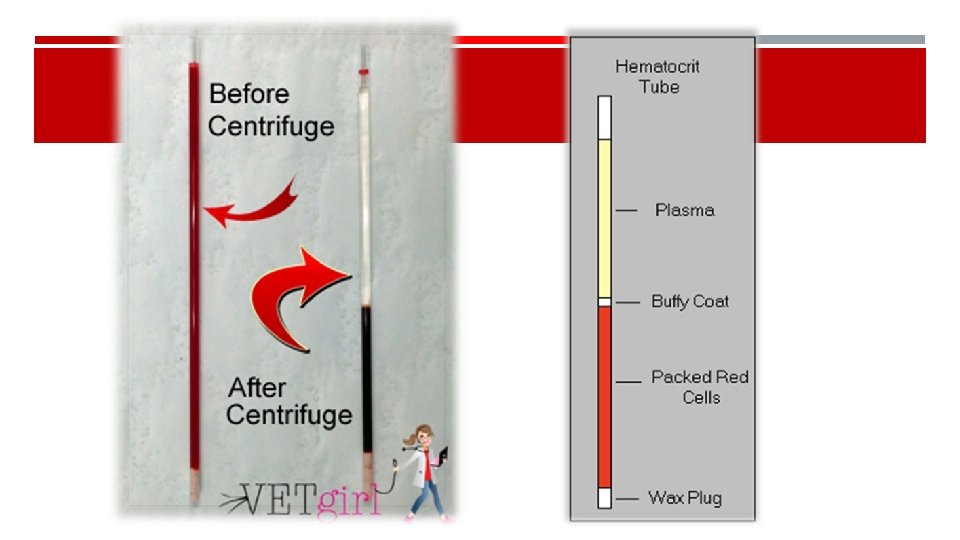

PACK CELL VOLUME EXPLAINED Increased Pack Cell Volume = Dehydrated Ex: From Heat Exhaustion Decreased Pack Cell Volume = Anemic

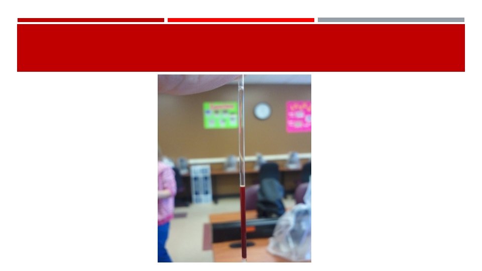
PCV, CONTINUED… Reading your PCV

PLASMA EVALUATION Plasma color and transparency may be helpful in determining a diagnosis and should be recorded in your findings Normal color plasma is clear or a pale straw/yellow color Cloudy plasma = lipemic Reddish tinge = hemolyzed Yellow = icteric (possible liver disease)

CONCENTRATION OF TOTAL PROTEIN/TOTAL SOLIDS Plasma protein concentrations estimated by refractometry is an important component of the CBC in all species Plasma used to determine the TP/TS is collected by breaking the hematocrit tube just above the buffy coat/plasma interface.

The plasma is allowed to flow onto the refractometer. (blow gently through the open end of the hematocrit tube with the broken end of the tube over the prism of your refractometer) Hold the refractometer up to the light and record your findings Make sure to wipe your refractometer after each use.


BLOOD FILMS Blood films are used to perform the differential WBC count, estimate platelet numbers, and evaluate the morphological features of WBCs, RBCs and platelets Wedge smears are prepared by placing a small drop of blood on a clean glass microscope slide.

BLOOD FILM

STAINING A SLIDE Always stain using the lightest to darkest stain Remember which side of your slide is the top Rinse off from back side of slide May heat fix to speed up process

WE FOCUS ON THE MONOLAYER. . DO NOT VENTURE ANYWHERE ELSE

PERFORMING THE DIFFERENTIAL COUNT This is where the different white blood cells are tallied separately. This can be done by a blood counting machine, or by hand To manually count the different cells, first you must make a perfect slide and stain Using a cell counter, you will tally a total of 100 cells This will make it easy to turn the numbers into a percent

Time to go To the lab!
- Slides: 34