Erythrocyte Morphology Normal Morphology Size abnormalities Shape abnormalities

Erythrocyte Morphology Normal Morphology Size abnormalities Shape abnormalities Color abnormalities RBC Inclusions

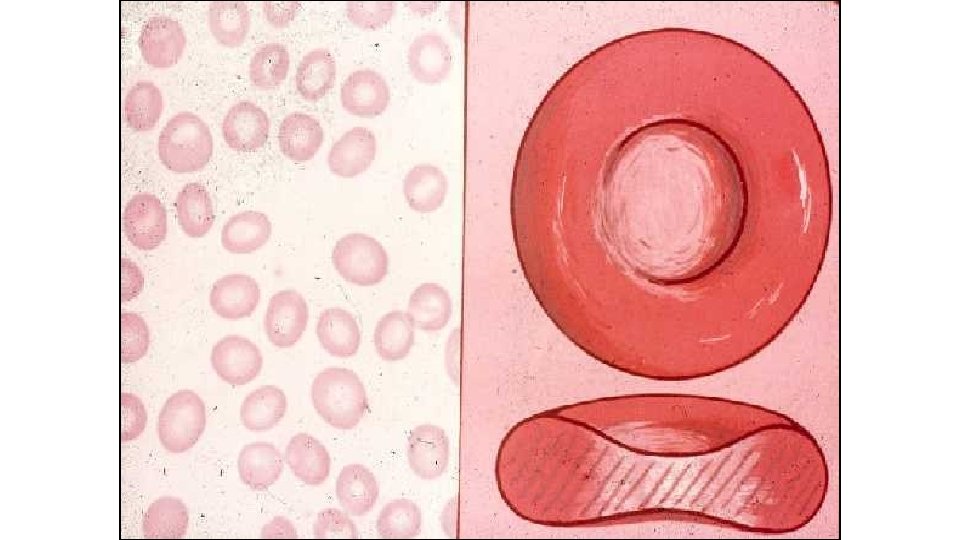
Normal Erythrocyte Morphology • Most common of blood cells on a blood smear • Biconcave disc • No nuclei in mammal RBC’s • Nuclei present normally in bird and reptile blood • Normal canine RBC’s have a central pallor (lightness) to them

Erythrocytes on a Needle

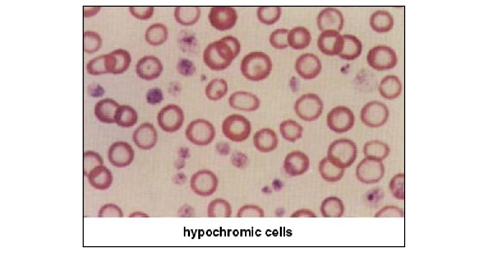
RBC Size & Pigment • Normocytic • Macrocytic • Microcytic • Normochromic • Hypochromic • No such thing as “Hyperchromic” • Why?

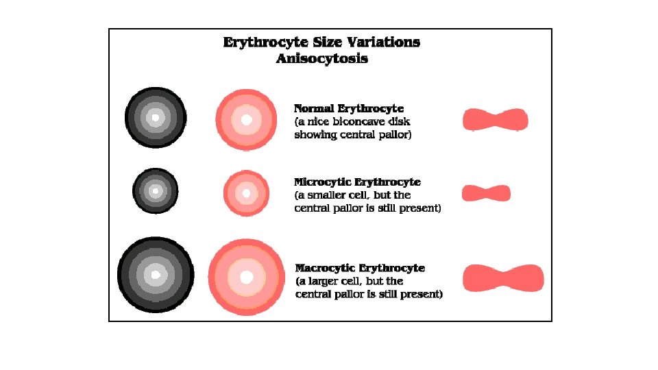
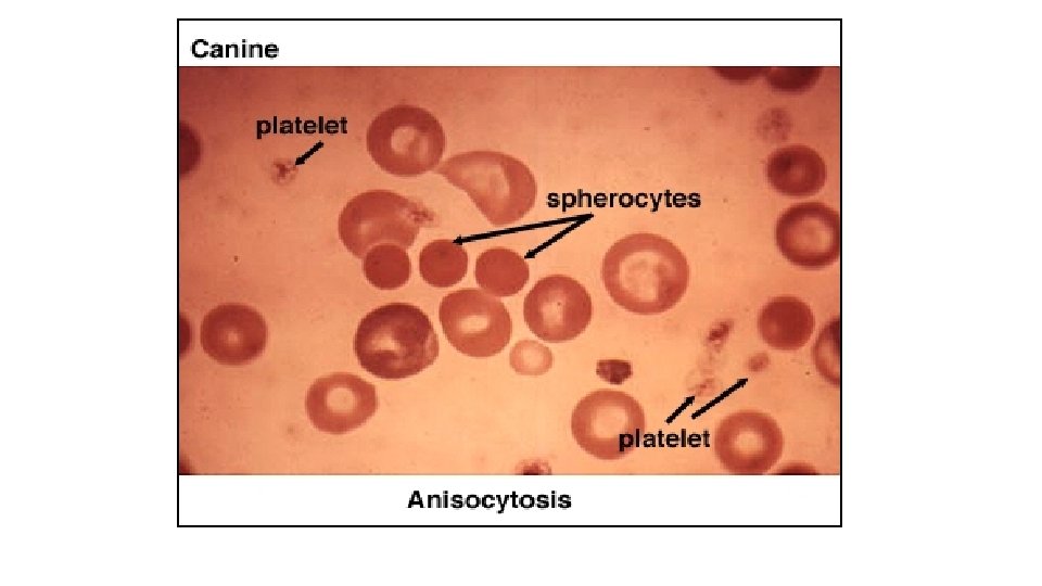
Erythrocyte Size Abnormalities • Record abnormal findings under “General Comments” on the hemogram • Anisocytosis • Macrocytes • Microcytes • Normocytic
 Schistocytes Acanthocytes Crenation (Echinocytes) Keratocytes Spherocytes Target Cells (Codocytes)")
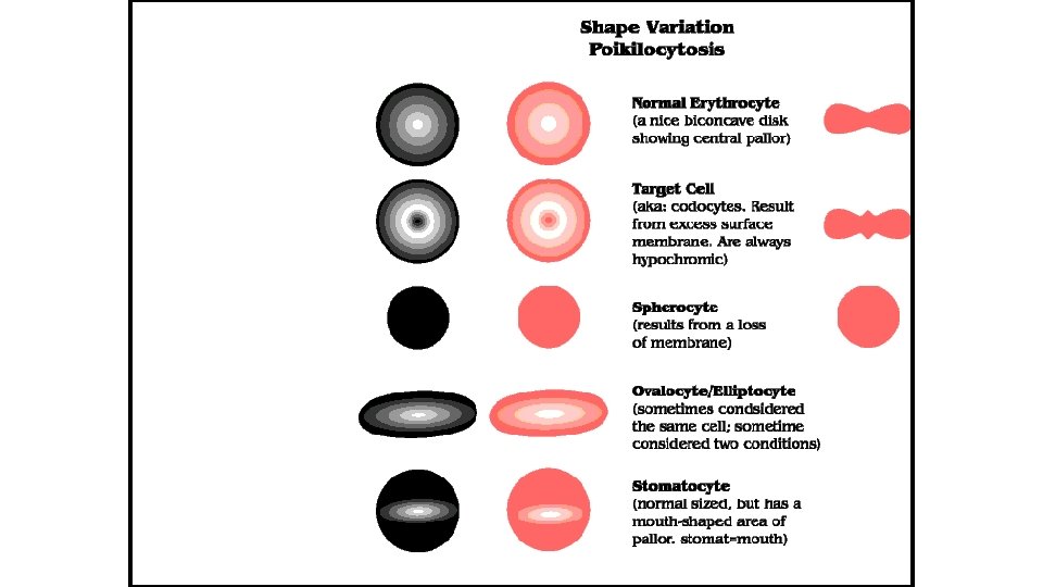
Erythrocyte Shape Abnormalities (Poikilocytosis) Schistocytes Acanthocytes Crenation (Echinocytes) Keratocytes Spherocytes Target Cells (Codocytes)

Erythrocyte Shape Abnormalities • Poikilocytosis – general term for shape abnormalities
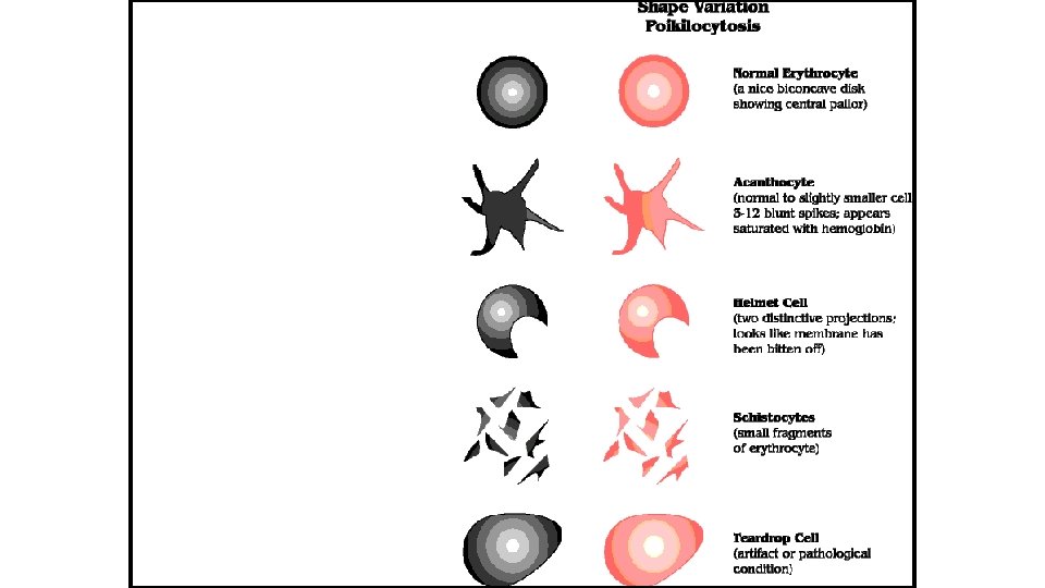

Schistocytes • RBC fragments
 • Unevenly distributed surface projections of different lengths")
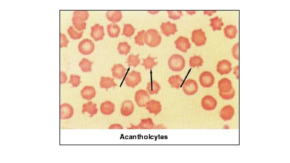
Acanthocytes (Spur cells) • Unevenly distributed surface projections of different lengths
 – spiculated (pointed) cells with short, evenly spaced surface")
Crenation • Echinocytes (Burr cells) – spiculated (pointed) cells with short, evenly spaced surface projections • Artifact? – slow drying of blood films • Feline blood
")
Crenation (Echinocytes)

Keratocytes • Helmet cells – contain a vacuole?

Spherocytes • Darkly staining RBC’s with no central pallor Canine only? • Autoimmune hemolytic anemia
 • RBCs with central rounded area of hemoglobin surrounded by clear")
Target Cells (Codocytes) • RBCs with central rounded area of hemoglobin surrounded by clear zone • A few in normal blood? • Associated with anemias

Target Cells

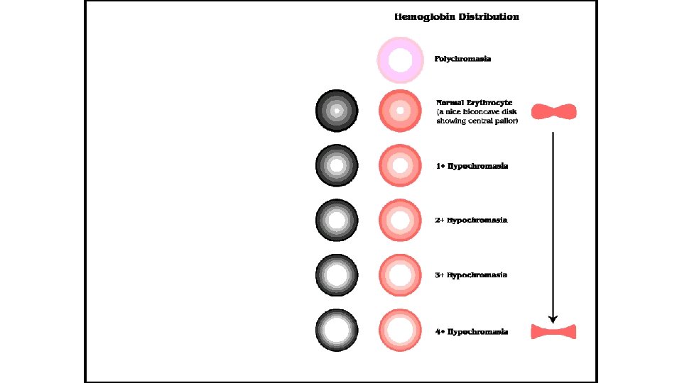
Erythrocyte Color Abnormalities • Normochromic • Polychromasia – polychromatophilic RBCs • Blue tint to cytoplasm, due to presence of organelles remaining in cytoplasm (young cells) • Hypochromic – decreased staining due to insufficient hemoglobin in cells Hyperchromic (no such thing!)
")
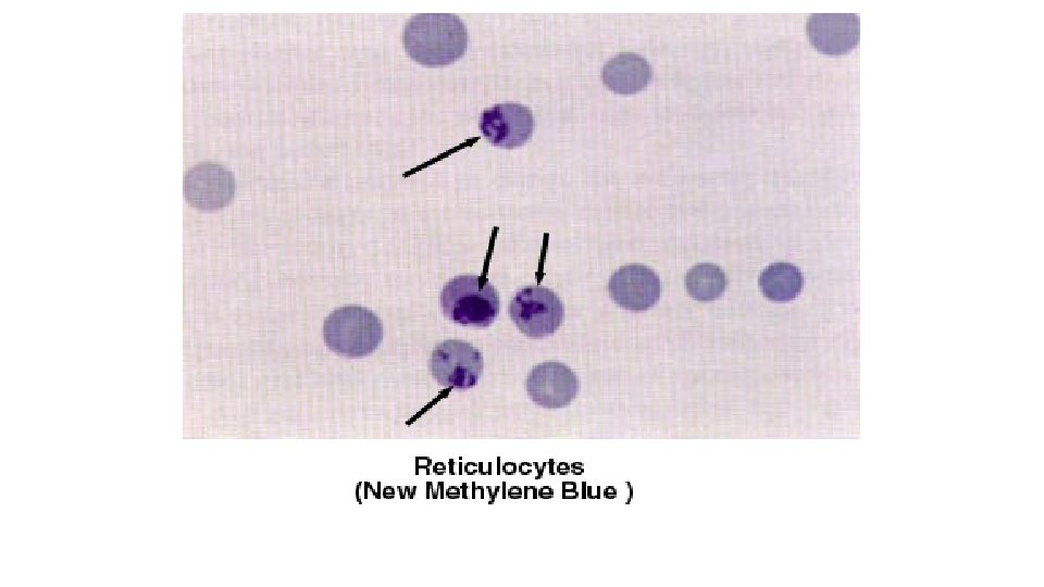
RBC Inclusions Reticulocytes Basophilic Stippling Howell-Jolly Bodies Heinz Bodies Nucleated RBCs (NRBCs)
 • As cell matures they are")
Reticulocytes • Immature RBCs that contain organelles (ribosomes) • As cell matures they are lost • Account for diffuse bluegray stain with Wright’s stain • 1% in normal circulation • Special stains needed usually

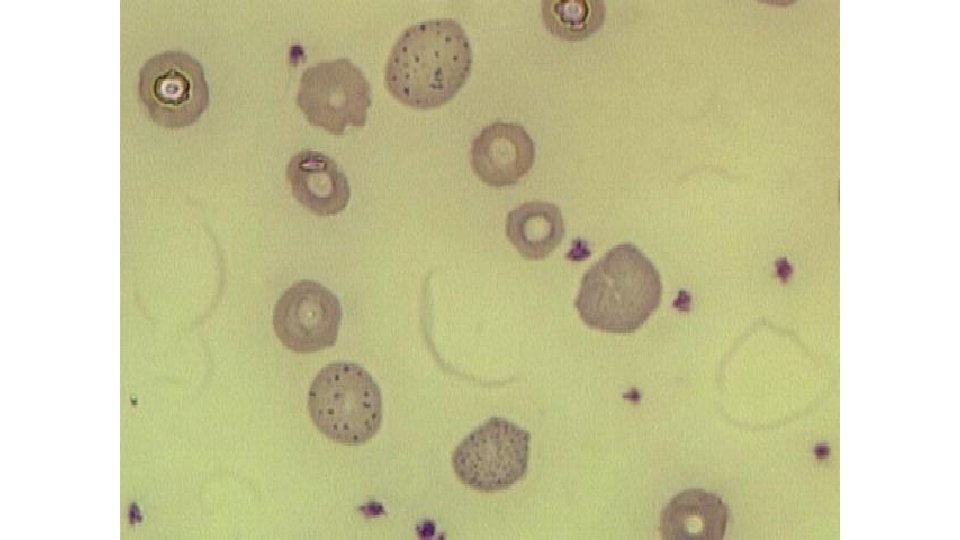
Basophilic Stippling • Presence of small, darkblue bodies all over RBC • Represents residual RNA • Common in immature RBCs

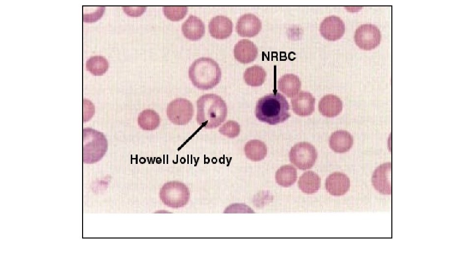
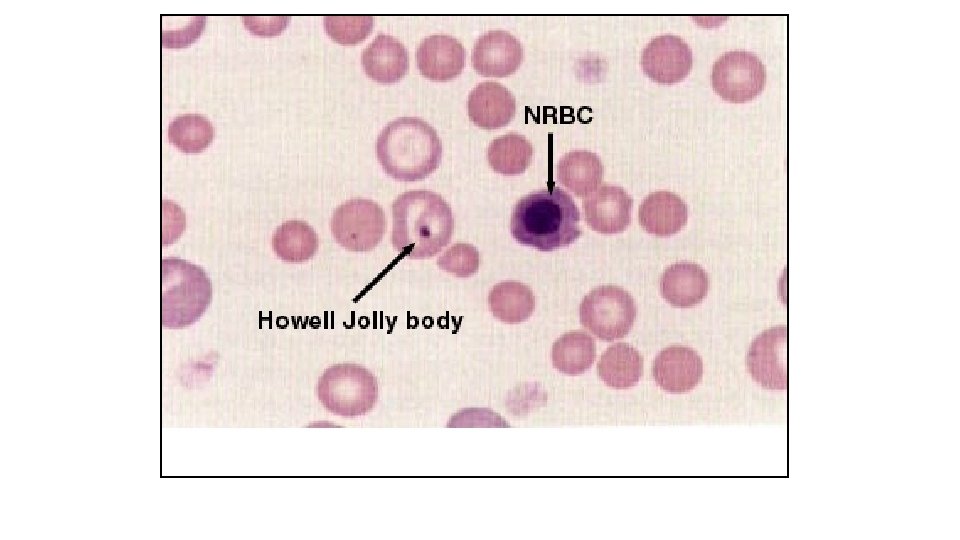
Howell-Jolly Bodies • Basophilic nuclear remnants • Seen in young RBCs in response to anemia • Phagocytes in spleen remove these remnants

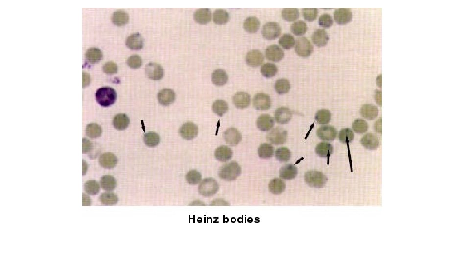
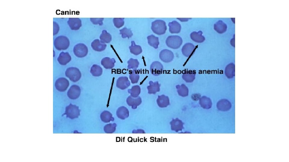
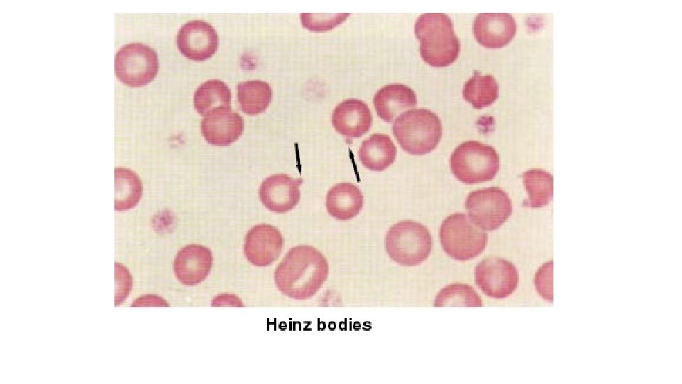
Heinz Bodies • Rounded structures representing denatured hemoglobin • Pale or blue area, depending on stain • Cause: certain oxidants or drugs • Normal cats may have them

Heinz Bodies
 • Also called metarubricytes • Represent early release of immature cells")
Nucleated RBCs (NRBCs) • Also called metarubricytes • Represent early release of immature cells during anemia • If > than 5 per 100 WBC’s • Correct WBC Count • Normal in bird & reptile blood

NRBC’s – How Many Do You See?
- Slides: 39