ERRORS IN THE DETECTION AND IDENTIFICATION OF HEMOGLOBIN

ERRORS IN THE DETECTION AND IDENTIFICATION OF HEMOGLOBIN VARIENTS Peter J. Howanitz MD Professor and Vice Chair Department of Pathology SUNY Downstate, Brooklyn NY, USA (Peter. Howanitz@downstate. edu)

GOALS AND OBJECTIVES • Describe Measurements Of Hemoglobins • Introduce Role of HPLC • Case Studies • New Finding--Only A 1 C Detects Variant • Questions And Answers

REASONS FOR HEMOGLOBIN ID AND QUANTIFICATION • Newborn Screening • Prenatal Screening • Follow-up Newborn Screening • Diagnosis Cause of Microcytosis • Anemia, Polycythemia, Chronic Hemolysis • Hemoglobinopathy Blood Replacement • Unexplained A 1 c Results

WHY USE HPLC? • Advantages – Throughput 11 Specimens/hour, 24 Hr Cal. – Analytic Sensitivity @ Low Concentrations – Improved Precision – Better Separation – Less Referrals For ID • Disadvantages – More Complex→ Higher Skill Level – Co-elution Of Hemoglobins

Hemoglobin Electrophoresis Patterns

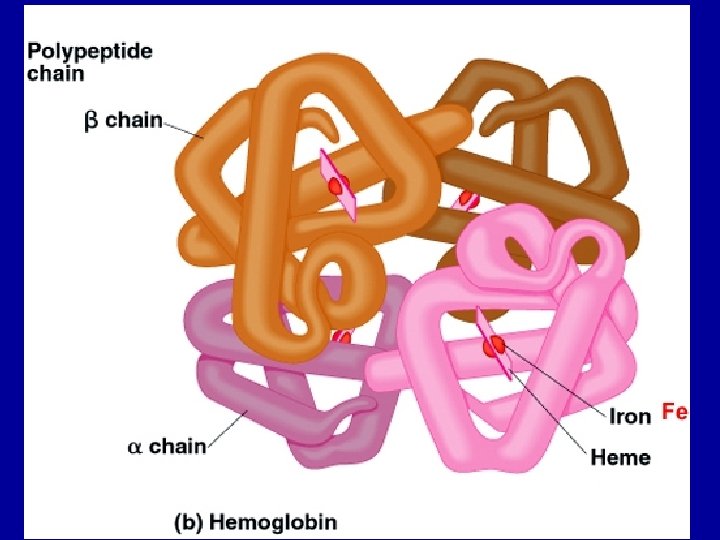
STRUCTURE HEMOGLOBINS Hemoglobin A Globin Chain α 2 β 2 A 2 α 2 δ 2 F α 2 γ 2 Adult Level A >95% 2 -3% F< 2. 0%
")
COMMON HEMOGLOBIN POINT MUTATIONS • Alpha Chain Variants – G Philadelphia (α 68 Asn→Lys) – – S (β 6 Glu→Val) C (β 6 Glu→Lys) E (β 26 Glu→Lys) D Los Angeles (β 22 Glu→Gln) • Beta Chain Variants • Delta Chain Variants -- A 2’ (δ 16 Gly→Arg)

INTERPRETATION OF HPLC RESULTS • • Hemoglobin Retention Time Variant Hemoglobin Percentage* A 2 Percentage* Number of Variants* CBC Indices* Transfusion History Age Clinical Course* • * Changed By Thalassemia
 RETENTION TIME (MIN) F")
BIO-RAD VARIANT WINDOWS PEAK NAME RETENTION PEAK NAME TIME (MIN) RETENTION TIME (MIN) F Window P 2 Window P 3 Window 0. 98 -1. 20 1. 24 -1. 40 -1. 90 3. 30 -3. 90 -4. 30 -4. 90 A 2 Window D Window S Window A 0 Window 1. 90 -3. 10 C Window 4. 90 -5. 30
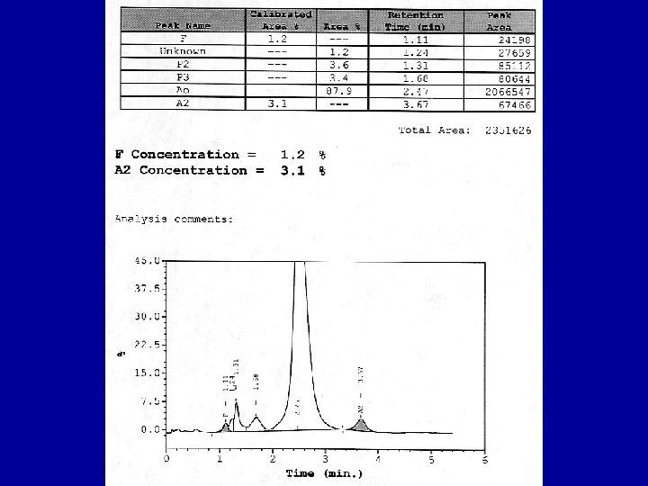
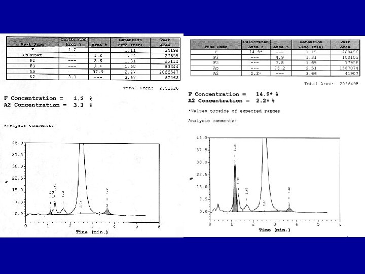
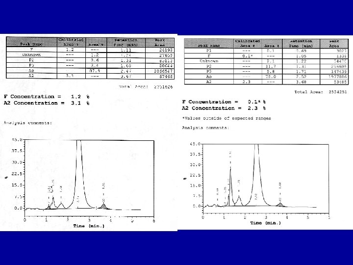
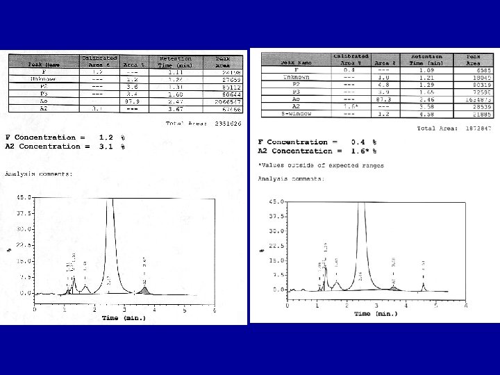
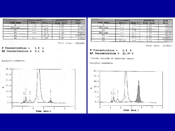
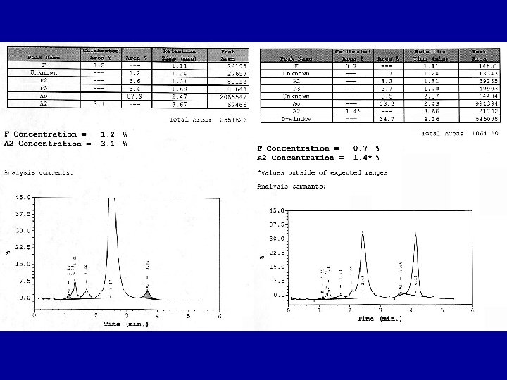
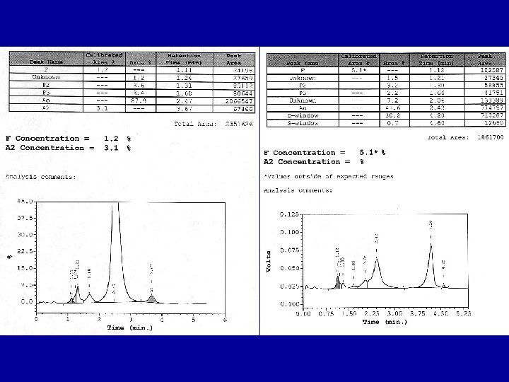
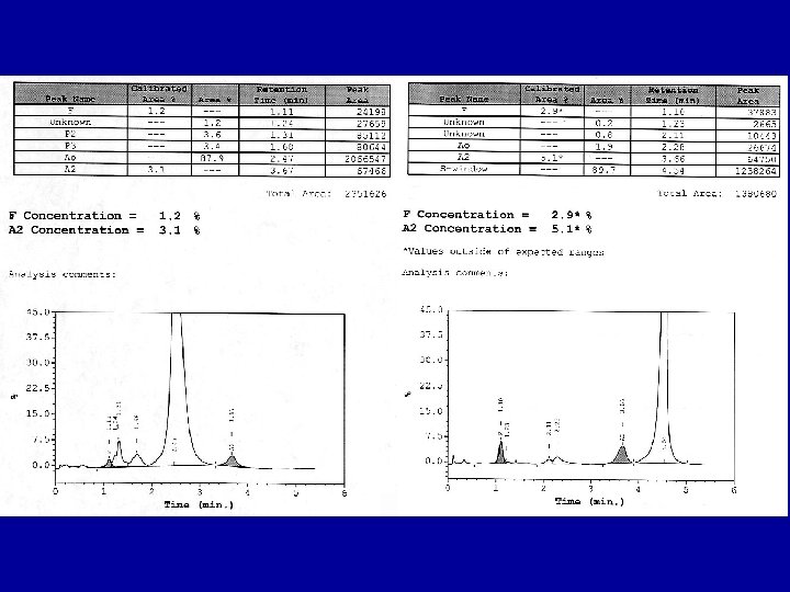
 A% A 2% VARIANT EXAMPLE 1 (25")
INTREPRATION OF RESULTS # Abnormal Peaks (%) A% A 2% VARIANT EXAMPLE 1 (25 -40) 50 -60 3. 5 -4. 5 β-Chain AS, AC 2 (25, 1. 0) 70 -80 1. 5 -2. 2* α-Chain AG-Phil 2 (50, 45) 0 3 40 -50 (12, 20, 14) 3. 5 -4. 5 2. 0* 2 β-Chain SC 1 α-, 1 β-, ASG 1αβPhilly Chain

INTREPRATION OF RESULTS • Hemoglobin F – >2 -80% Babies – 90 -100% Homozygous Hereditary Persistence Fetal Hemoglobin, β 0, δβ 0 -Thal – 15 -40% Heterozygous HPFH – 10 -25% SS, Hydroxyurea Treated – 3 -10% Homozygous Hemoglobinopathies, Anemias, Leukemias, Malignancies, – < 5% β-Thal, Lepore

INTREPRATION OF RESULTS • Hemoglobin A – Increased P 2 -? Diabetes (↑A 1 C>7%) – Increased P 3 -(>P 2) Old Specimen – Inverse of Other Hemoglobins – Focus on Abnormal Hemoglobins

HEMOGLOBIN A 2’ • Elutes in S Window • Δ 16 Gly→Arg • Characteristic Low A 2 Percentage (1. 0 -2. 5%) • Most Common In Blacks (2%) • CBC Normal • Little Consequence, Except β-Thal (add A 2)

INTREPRATION OF RESULTS • Hemoglobin A 2 – Increased • 4. 0 -7. 0% Β-Thalassemia, Sβ+ Thal • 3. 5 -4. 5% Hb AS, AC, SS, CC • 6. 5 -14. 0% Hb Lepore • 25 -30% Hb E – Decreased • 1. 3 -1. 7% Iron Deficiency, Sideroblastic, Aplastic Anemias • 1. 5 -2. 3% δ Chain Variant (A 2’), α Chain Variant

HEMOGLOBIN E • Found in SE Asia, β 26 Glu→Lys • Most Common Hemoglobinopathy Worldwide • Complicated by Iron Def, Thalassemia, A 2 • Elution Trait (Hb AE) – Asymtomatic, No CBC Abnormalities • Disease (Hb EE) – – Mild Anemia, Target Cells, ↓RBC Survival ↓Osmotic Fragility +Beta Thal = Severe, As Homozygous β-Thal +Alpha Thal=↓Hb E

HEMOGLOBIN D D Window On Bio-Rad Variant Β 121 Glu→Gln Found In India (D-Punjab/D-Los Angeles) Most Common D In U. S. Blacks (< 0. 02%) Trait Asymtomatic, No Anemia, Normal CBC Disease Asymtomatic, No Anemia/ Hemolysis D Los-Angeles. S = Symptoms of Sickle Cell Disease

HEMOGLOBIN G PHILADELPHIA • Elutes In D-Window • α 68 Asn→Lys of Hb A and A 2 • Heterozygote-CBC Normal – Most Common α Chain Variant In Blacks, Italians (25%), Chinese – Associated With α-Thal (30%, 45%G) • Association With S or C Common (Double Heterozygote)
 β 6 Glu→Val Common In Blacks; Other Populations")
HEMOGLOBIN S S Trait (Hemoglobin AS) β 6 Glu→Val Common In Blacks; Other Populations Asymptomatic, Blood Sickles in Vitro Protective Against Malaria S Disease (Hemoglobin SS) Severe Symptoms, Sickling in Vivo Hydroxy Urea Treatment→Induces F Crises→Bone Pain, Hemolysis, Stroke, etc Similar Symptoms Other Double Heterozygotes (SC)


HEMOGLOBIN C • Prevalent in West Africa, 3% U. S Blacks • Trait (Hb AC) β 6 Glu→Lys – No Symptoms or Anemia, – Hypochromia, Up to 40% Target Cells • Disease (Hb CC) – Mild Hemolytic Anemia, Spenomegly – Rod Shaped Crystals in RBCs – Normochromic, Normocytic Anemia, – 40 -90% Target Cells

MORE RARE VARIANTS?

BIORAD TURBO A 1 C-CHROMATOGRAM

BIO-RAD A 1 C-AS CHROMATOGRAM

BIO-RAD A 1 C AC CHROMATOGRAM

BIO-RAD UNKNOWN VARIANT A 1 C CHROMATOGRAM TYPE 1

BIO-RAD UNKNOWN VARIANT A 1 C CHROMATOGRAM TYPE 2

HEMOGLOBIN A 1 C CHROMATOGRAPHS CONTROL PATIENT 1 PATIENT 2 A 1 C HPLC results of a control specimen and the patients’ specimens. Note the variant eluting at 0. 872 & 0. 853 minutes in chromatograms of patient 1 and patient 2 depicted by an arrow.

HEMOGLOBIN IDENTIFICATION CHROMATOGRAMS CONTROL PATIENT 1 PATIENT 2 Hemoglobin HPLC results of a control specimen and the patients’ specimens. A hemoglobin variant is not identified in either chromatogram.

HEMOGLOBIN IDENTIFICATION CAPILLARY ELECTROPHORETOGRAMS CONTROL PATIENT 1 PATIENT 2 Capillary electrophoresis of a control specimen and the patients’ specimens. A hemoglobin variant is not identified in either electrophoretogram

HEMOGLOBIN ELECTROPHORESIS ALKALINE GEL ACID GEL Hemoglobin electrophoresis on alkaline and acid gel. The patient’s specimen migrates as S on alkaline gel, and a split A band on acid gel, identified as an arrow. Electrophoresis of the specimen from the second patient was identical to the first (not shown). Controls for C, S, F and A are the top two specimens in either gel.

GENETIC ANALYSIS OF VARIANT • DNA Sequence Analysis – Alpha-2 Substitution – Codon 95 CCG To CTG, Pro To Leu • Hemoglobin G-Georgia – Compatible With Other Lab Findings

HEMOGLOBIN G-GEORGIA • Five Cases In Literature • Found In Blacks & Portuguese • Increased 02 Affinity, Decreased Heme Interaction • No CBC Abnormalities • Double Heterozygote With S & C

CONCLUSIONS • HPLC Valuable Laboratory Technique • Discussed Common Variants • Interpreted Chromatograms–Case Studies • New-Hemoglobin G-Georgia Noted • Important To ID A 1 c Variants • Questions?
- Slides: 42