Epithelial cell abnormality in Pap smear in women


Epithelial cell abnormality in Pap smear in women with postmenopausal bleeding By Ghada Ahmed Asker Assistant Lecturer in Pathology Department

Introduction Cancer of the uterine cervix is the fourth most common cancer worldwide in females, and the seventh most common cancer overall (Ferlay et al. , 2013).

In Egypt cervical cancer ranks as the 13 th most frequent cancer among women and the 10 th most frequent cancer among women between 15 and 44 years of age (ICO Information Centre on HPV and Cancer, 2016).

Cancer cervix is a preventable and curable disease due to effective screening methods. Uterine cervix is ideal for screening due to easy accessibility of the cervix for inspection, palpation and exfoliative cytology (Gupta et al. , 2013).

Cervical smears in the screening programme help the early detection of cervical carcinoma and its precursors as an easy, costless, accessible, and reliable manner.

The study was conducted in the Cytology Section of Department of Pathology, Sohag University Hospital. Fifty peri and postmenopausal women were included in this study.

The cervical smears were taken in the Outpatient Clinic of Gynecology and Obstetrics Department.

Each patient were subjected to accurate history taking regarding parity, contraceptive use, menstrual history, and duration of marital life and the status (healthy or unhealthy) of per-vaginal examination of the patients.

The included fifty cases; 2 slides of Pap smears were prepared for each case; one was taken by spatula and the other was taken by brush representing the ectocervix and endocervix respectively. The age of the patients ranged from 38 to 70 years.

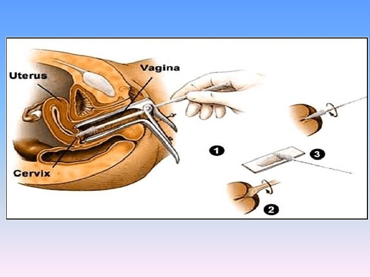
The specimens were collected as follows; firstly the speculum was inserted without lubricant. Any mucous, discharge or blood was removed with dry cotton. A wooden spatula was used for sampling ectocervix which is rotated 360 degrees.


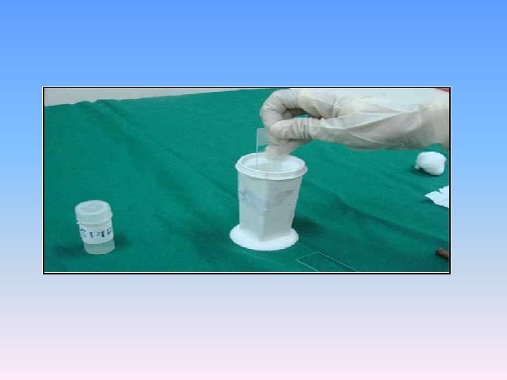
For sampling endocervix, the brush was rotated only 90 degrees as the circumferential bristles were in contact with the entire surface the moment the brush was inserted. The samples were applied to the slides which were encoded and immediately fixed in 95% ethanol and stained by the standard Pap stain.

Types of screening: • Conventional Pap: samples are smeared directly onto a slide after collection. • Liquid based cytology: sample is put in a bottle of preservative for transport to the laboratory, where it is then smeared on the slide.
 ____________________ Specimen type")
• • The 2014 Bethesda System (Nayar and, Wilbur, 2015) ____________________ Specimen type Indicate conventional smear (Pap smear) vs. liquid based preparation vs. other Specimen adequacy General categorization (optional) Negative for intraepithelial lesion or malignancy (NILM) Other Epithelial cell abnormality

Interpretation/Result I. NILM Organisms • Non neoplastic cellular variation • Reactive cellular changes associated with: • Glandular cells status post-hysterectomy II. Other • Endometrial cells in a woman >40 years of age (specify if negative for squamous intraepithelial lesion)
 Ø of")
III. Epithelial cell abnormalities A. Squamous cell • Atypical squamous cells (ASC) Ø of undetermined significance (ASC-US) Ø cannot exclude HSIL (ASC-H) • Low-grade squamous intraepithelial lesion (LSIL) Ø (Encompassing: HPV/mild dysplasia/CIN 1) • High-grade squamous intraepithelial lesion (HSIL) Ø (Encompassing: moderate and severe dysplasia, carcinoma in situ, CIN and CIN 3) A. with features suspicious for invasion (if invasion is suspected) • Squamous cell carcinoma
 Ø")
B. Glandular cell • Atypical Ø Endocervical cells (NOS or specify in comments) Ø Endometrial cells (NOS or specify in comments) Ø Glandular cells (NOS or specify in comments) • Atypical Ø endocervical cells, favor neoplastic Ø glandular cells, favor neoplastic • Endocervical adenocarcinoma in situ (AIS) • Adenocarcinoma Other malignant neoplasms (specify)
 and women 21 (42%) were postmenopausal. Bleeding was the main")
Twenty nine premenopausal (58%) and women 21 (42%) were postmenopausal. Bleeding was the main complaint in 92% patient and the remaining 8% complained from vaginal discharge and itching.

Classification of the studied cases according to TBS 2014 Diagnosis Number of patient Percentage (%) 39 78 Atrophy with no other finding 6 12 Atrophy with follicular cervicitis 3 6 NILM Atrophy with infection 6 Suggestive of Trichomonas 3 2 HSV 1 2 Bacterial vaginosis 1 22 Non specific 11 12 Atrophy with metaplastic changes 6 16 Normal smear 8 ASCUS 2 4 ASC-H 2 4 HSIL 3 6 SCC 1 2 Insufficient smear 3 6
: Atrophic changes in postmenopausal women showing parabasal cells (oval cells")
Papanicolaouea stained smear (Spatula): Atrophic changes in postmenopausal women showing parabasal cells (oval cells with dense cytoplasm)
: Immature squamous metaplastic cells showing high N/C ratio, prominent nucleoi")
Papanicolaouea stained smear (Spatula): Immature squamous metaplastic cells showing high N/C ratio, prominent nucleoi and finely ganular chomatin
: Mature squamous metaplastic cells (cells lying singly cytoplasm showing dense")
Papanicoaouea stained smear (Spatula): Mature squamous metaplastic cells (cells lying singly cytoplasm showing dense outer zone, “ectoplasm, ” and lighter inner zone “endoplasm”)
: HSV infection showing multinucleation, margination of chromatin and moulding")
Papanicoaouea stained smear (Spatula): HSV infection showing multinucleation, margination of chromatin and moulding
: Follicular cervicitis showing groups of mature lymphocytes")
Papanicoaouea stained smear (Spatula): Follicular cervicitis showing groups of mature lymphocytes
: Bacterial vaginosis showing cocci replacing the lactobacilli")
Papanicoaouea stained smear (Spatula): Bacterial vaginosis showing cocci replacing the lactobacilli
: Bacterial vaginosis showing clue cell (bacteria adhere to squamous cells)")
Papanicoaouea stained smear (Spatula): Bacterial vaginosis showing clue cell (bacteria adhere to squamous cells)
: Tight clusters of neutrophils representing BB shot suggesting infection with")
Papanicoaouea stained smear (Spatula): Tight clusters of neutrophils representing BB shot suggesting infection with Trichomonas vaginalis
: Reparative changes")
Papanicoaouea stained smear (Spatula): Reparative changes
: ASCUS showing mild nuclear pleomorphism, moulding, and occasional hyperchromatism and")
Papanicolaouea stained smear (Spatula): ASCUS showing mild nuclear pleomorphism, moulding, and occasional hyperchromatism and slight increase in N/C ratio.
: ASC-H showing moderate nuclear pleomorphism, hyperchromatism, and occasional irregularities in")
Papanicolaouea stained smear (Spatula): ASC-H showing moderate nuclear pleomorphism, hyperchromatism, and occasional irregularities in the nuclear contour and crowdening.
: HSIL showing crowded parabasal like cells, increase N/C ratio, nuclear")
Papanicolaouea stained smear (Spatula): HSIL showing crowded parabasal like cells, increase N/C ratio, nuclear hyperchromatism, coarse and unevenly distributed chromatin and irregular contour
: SCC with cell nest formation")
Papanicolaouea stained smear (Spatula): SCC with cell nest formation
: SCC with central keratinization")
Papanicolaouea stained smear (Spatula): SCC with central keratinization
: SCC showing a tadpole cell and fiber cells, arrow: tadpole")
Papanicolaouea stained smear (Spatula): SCC showing a tadpole cell and fiber cells, arrow: tadpole cell
: Tumour giant cell beside group of malignant cells")
Papanicolaouea stained smear (Spatula): Tumour giant cell beside group of malignant cells

Conclusion This study highlight the importance of using cervical smears in the screening programme of early detection of cervical carcinoma and its precursors as an easy, costless, accessible, and reliable manner in the Outpatient Clinic of Gynecology and Obstetrics Department.

And any premenopausal woman should have cervical screening if she complaint from bleeding, vaginal discharge or itching.

Thank you

BB shot a small pellet fired from an air rifle or BB gun. B B pellet, shot a solid missile discharged from a firearm; "the shot buzzed past his ear"
- Slides: 42