
EpilepsyAssociated Neuroplasticity in the Dentate Gyrus Bret N
Epilepsy-Associated Neuroplasticity in the Dentate Gyrus Bret N. Smith, Ph. D. Epilepsy Center Department of Physiology University of Kentucky
Disclosure No financial relationships to disclose
Educational Need = To better understand the relationship between epileptogenesis and reactive synaptic reorganization in the brain.
Objectives Ø Describe models of TLE and PTE; Ø Identify biological similarities and/or differences between models Ø Construct cellular hypotheses to study epileptogenesis; Ø Demonstrate experimentally a set of functional changes in brain circuitry involved in epileptic seizure generation; Ø Identify effects of m. TOR inhibition on reactive synaptic reorganization Ø Construct a working model of local-circuit neural plasticity associated with epileptogenesis.
Expected Outcome • Prompt/reinforce a “translational” mindset regarding development of new therapies for refractory epilepsy, including an appreciation of how mechanistic analyses might influence clinical outcomes.
Acknowledgements Laura Haselhorst Jennifer Rios-Pilier Corwin Butler Kathleen Schoch, Ph. D Robert F Hunt III, Ph. D Deepak Bhaskaran, MD, Ph. D Jeffrey Boychuk, Ph. D Tim Kubal , MD Heather Shibley, MD Ron Winokur, MD Eva Bach, Ph. D Katalin Cs Halmos, MPH Dan Liu Stephen Scheff, Ph. D Kathryn Saatman, Ph. D Epilepsy Foundation NINDS Do. D
Temporal Lobe Epilepsy Models Premise: To study epilepsy preclinically, we need a model that is “epileptic” SEIZURE GENERATION (the clinical manifestation of an abnormal and excessive excitation of a population of cortical neurons) vs EPILEPSY (a tendency toward recurrent seizures unprovoked by systemic or neurologic insults)
Characteristics of epilepsy models In rodents, chemical or electrical induction of status epilepticus (SE) or traumatic brain injury (TBI) leads to TLE. Characteristics in common with patients (from resected tissue): • Insult (e. g. , kindling, SE, TBI) followed by a “silent period”, then spontaneous seizures; • Cellular correlates include selective cell loss, axon sprouting, increased local excitability. Injury Latency SE, TBI Epileptogenesis Epilepsy permanent structure/function change
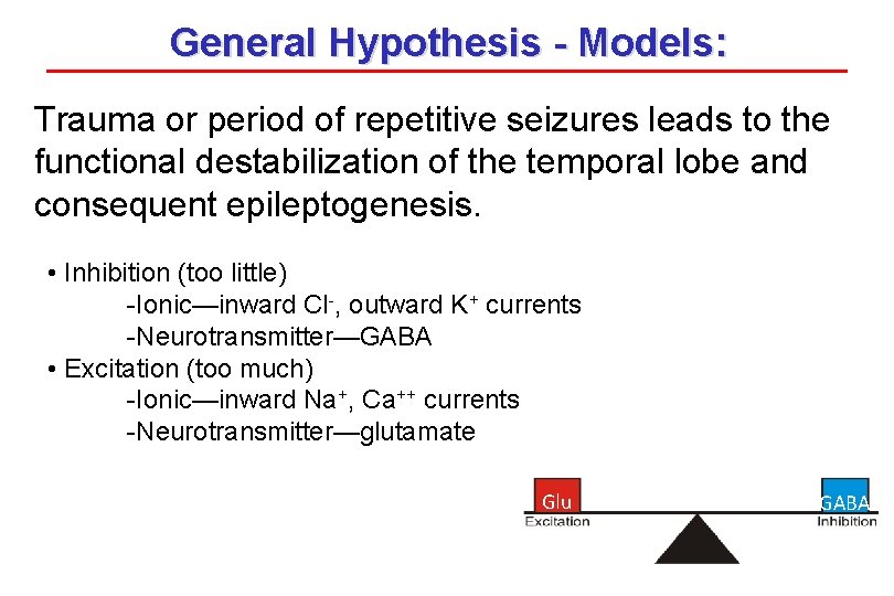
General Hypothesis - Models: Trauma or period of repetitive seizures leads to the functional destabilization of the temporal lobe and consequent epileptogenesis. • Inhibition (too little) -Ionic—inward Cl-, outward K+ currents -Neurotransmitter—GABA • Excitation (too much) -Ionic—inward Na+, Ca++ currents -Neurotransmitter—glutamate Glu GABA
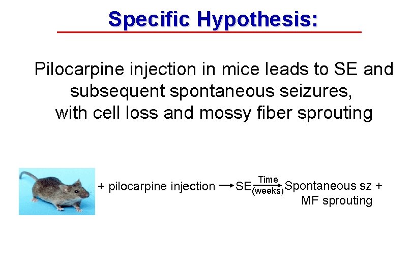
Specific Hypothesis: Pilocarpine injection in mice leads to SE and subsequent spontaneous seizures, with cell loss and mossy fiber sprouting + pilocarpine injection Time SE(weeks) Spontaneous sz + MF sprouting
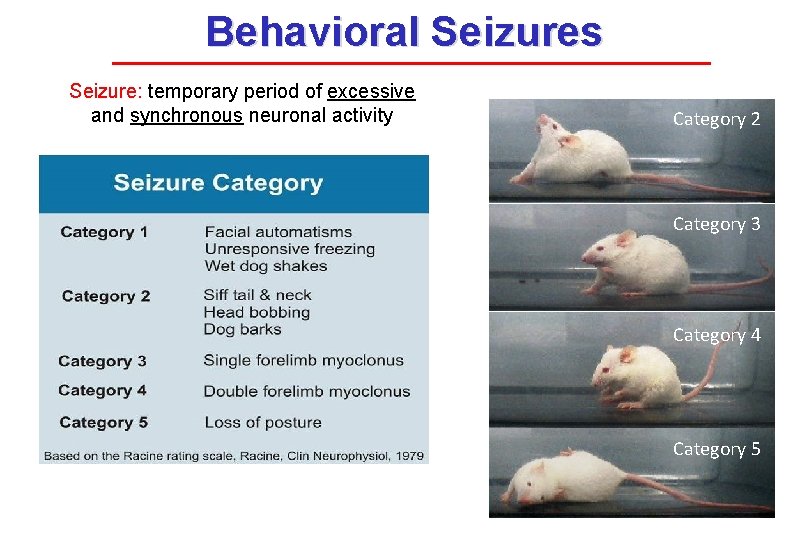
Behavioral Seizures Seizure: temporary period of excessive and synchronous neuronal activity Category 2 Category 3 Category 4 Category 5
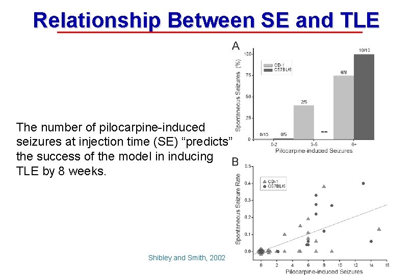
Relationship Between SE and TLE The number of pilocarpine-induced seizures at injection time (SE) “predicts” the success of the model in inducing TLE by 8 weeks. Shibley and Smith, 2002
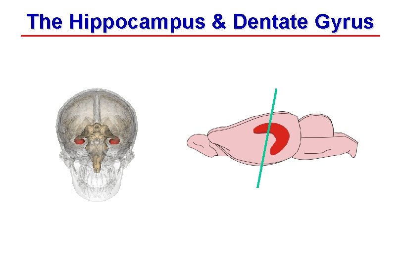
The Hippocampus & Dentate Gyrus Mouse Database Center for Life Sciences (DCLS)
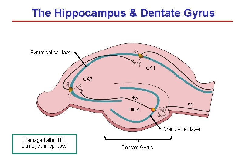
The Hippocampus & Dentate Gyrus Pyramidal cell layer CA 1 CA 3 MF Hilus PP Granule cell layer Damaged after TBI Damaged in epilepsy Dentate Gyrus
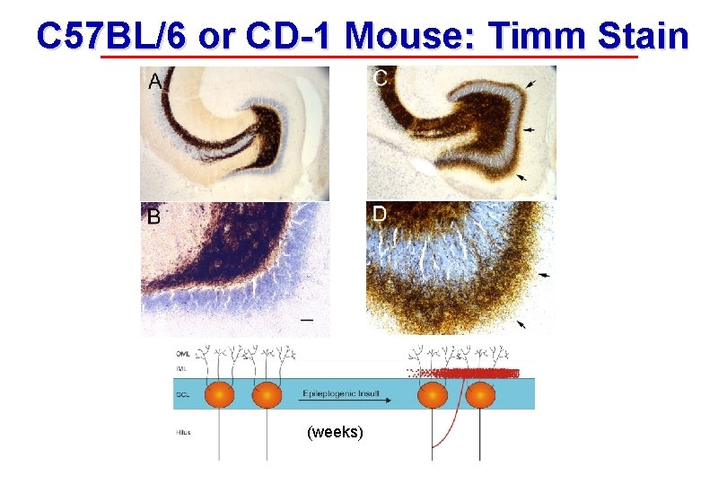
C 57 BL/6 or CD-1 Mouse: Timm Stain (weeks)
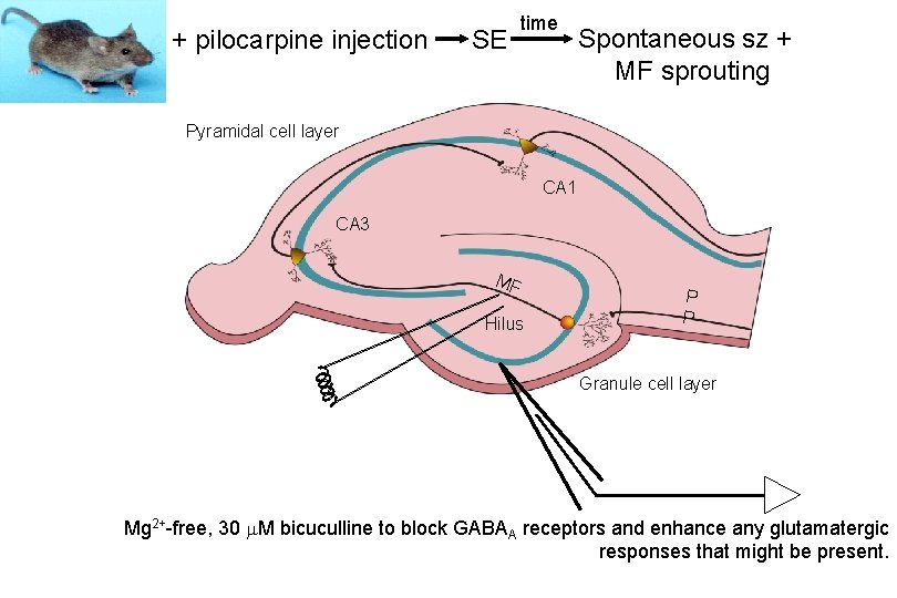
+ pilocarpine injection SE time Spontaneous sz + MF sprouting Pyramidal cell layer CA 1 CA 3 MF Hilus P P Granule cell layer Mg 2+-free, 30 m. M bicuculline to block GABAA receptors and enhance any glutamatergic responses that might be present.
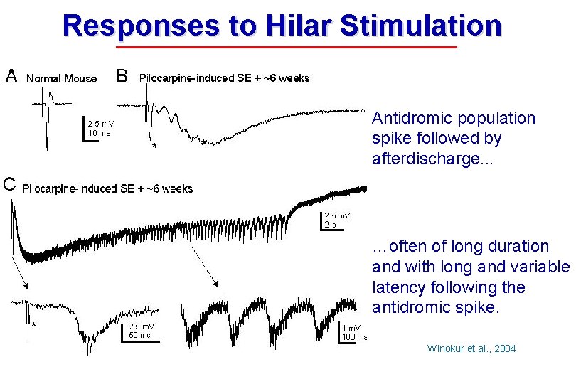
Responses to Hilar Stimulation Antidromic population spike followed by afterdischarge. . . …often of long duration and with long and variable latency following the antidromic spike. Adapted from Winokur et al. , 2004
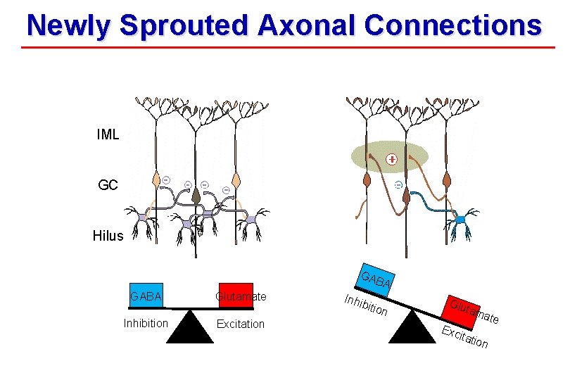
Newly Sprouted Axonal Connections IML GC Hilus GAB A GABA Inhibition Glutamate Excitation Inhi bitio n Glu ta Exc mat itati on e
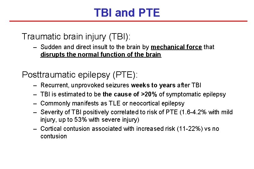
TBI and PTE Traumatic brain injury (TBI): – Sudden and direct insult to the brain by mechanical force that disrupts the normal function of the brain Posttraumatic epilepsy (PTE): – – Recurrent, unprovoked seizures weeks to years after TBI is estimated to be the cause of >20% of symptomatic epilepsy Commonly manifests as TLE or neocortical epilepsy Severity of TBI positively correlated to risk of PTE (1. 6 -4. 2% with mild injury, up to 53% with severe injury) – Cortical contusion associated with increased risk (11 -22%) vs no contusion
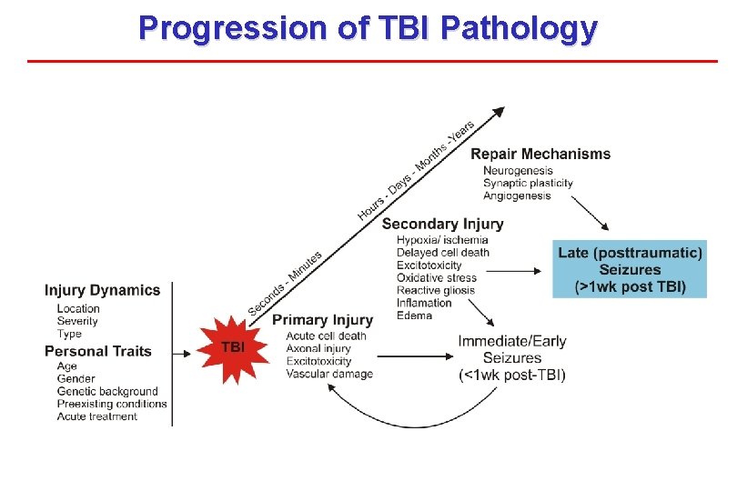
Progression of TBI Pathology
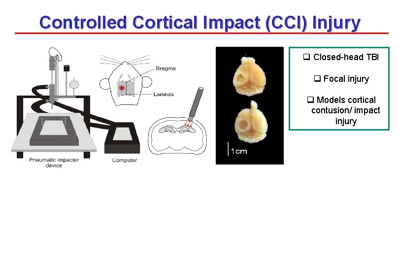
Controlled Cortical Impact (CCI) Injury q Closed-head TBI q Focal injury q Models cortical contusion/ impact injury
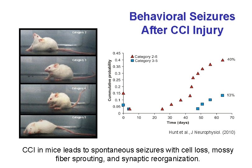
Category 2 Behavioral Seizures After CCI Injury Category 3 Category 4 Category 5 Hunt et al. , J Neurophysiol. (2010) CCI in mice leads to spontaneous seizures with cell loss, mossy fiber sprouting, and synaptic reorganization.
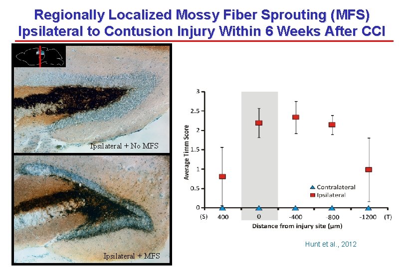
Regionally Localized Mossy Fiber Sprouting (MFS) Ipsilateral to Contusion Injury Within 6 Weeks After CCI Ipsilateral + No MFS Hunt et al. , 2012 Ipsilateral + MFS
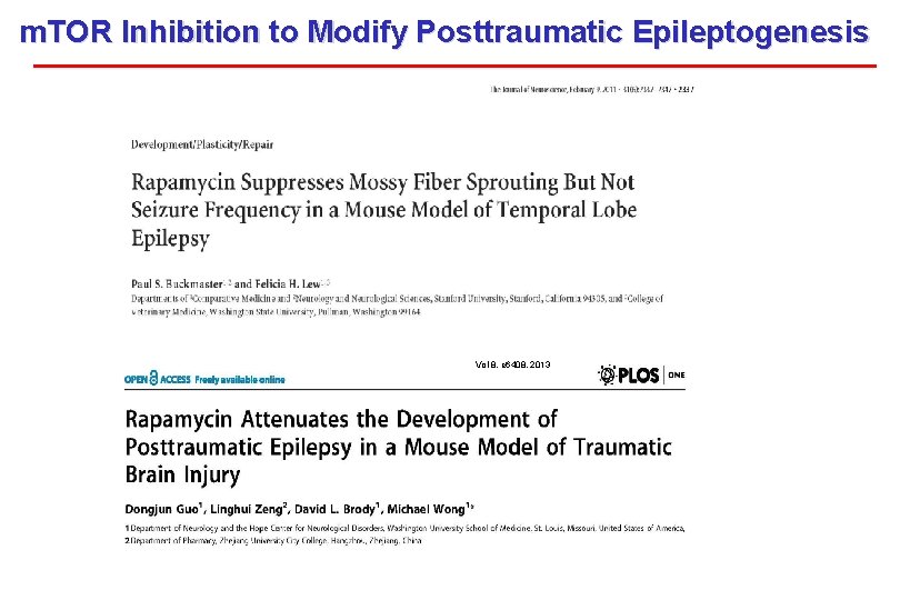
m. TOR Inhibition to Modify Posttraumatic Epileptogenesis Vol 8, e 6408, 2013
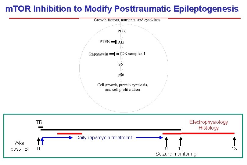
m. TOR Inhibition to Modify Posttraumatic Epileptogenesis Electrophysiology Histology TBI Wks post-TBI Daily rapamycin treatment 0 8 10 Seizure monitoring 13
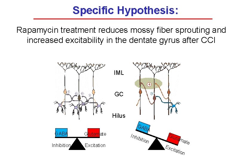
Specific Hypothesis: Rapamycin treatment reduces mossy fiber sprouting and increased excitability in the dentate gyrus after CCI IML GC Hilus GABA Inhibition Glutamate Excitation A Inhi bitio n Glu tam Exc itati ate on
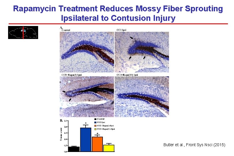
Rapamycin Treatment Reduces Mossy Fiber Sprouting Ipsilateral to Contusion Injury Butler et al. , Front Sys Nsci (2015)
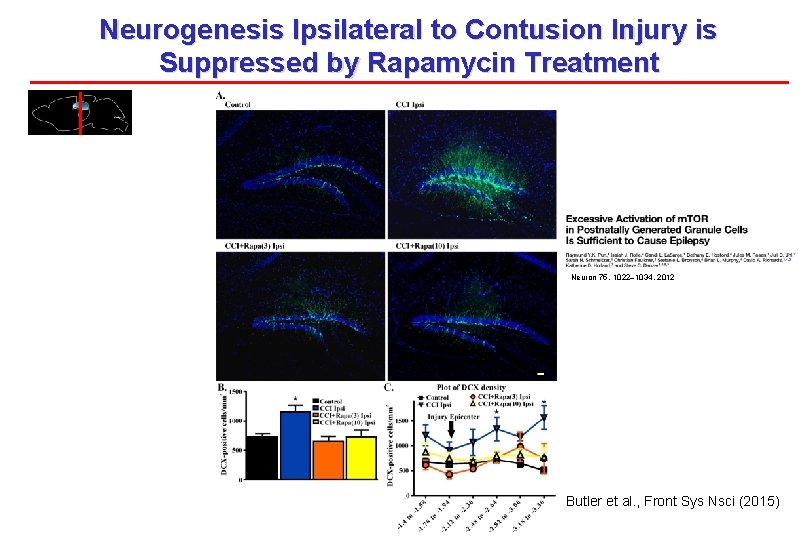
Neurogenesis Ipsilateral to Contusion Injury is Suppressed by Rapamycin Treatment Neuron 75, 1022– 1034, 2012 Butler et al. , Front Sys Nsci (2015)
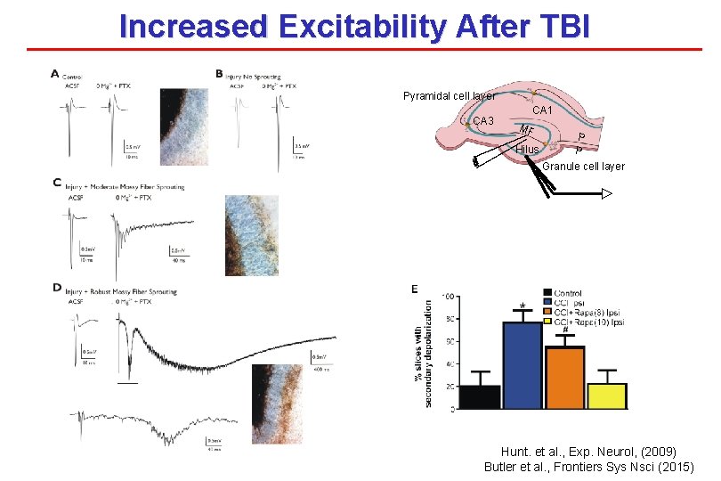
Increased Excitability After TBI Pyramidal cell layer CA 3 CA 1 MF Hilus P P Granule cell layer Hunt. et al. , Exp. Neurol, (2009) Butler et al. , Frontiers Sys Nsci (2015)
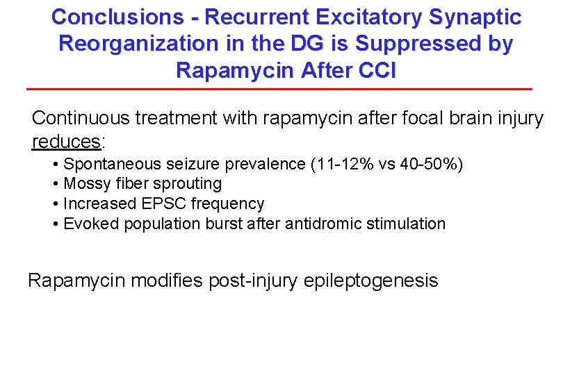
Conclusions - Recurrent Excitatory Synaptic Reorganization in the DG is Suppressed by Rapamycin After CCI Continuous treatment with rapamycin after focal brain injury reduces: • Spontaneous seizure prevalence (11 -12% vs 40 -50%) • Mossy fiber sprouting • Increased EPSC frequency • Evoked population burst after antidromic stimulation Rapamycin modifies post-injury epileptogenesis
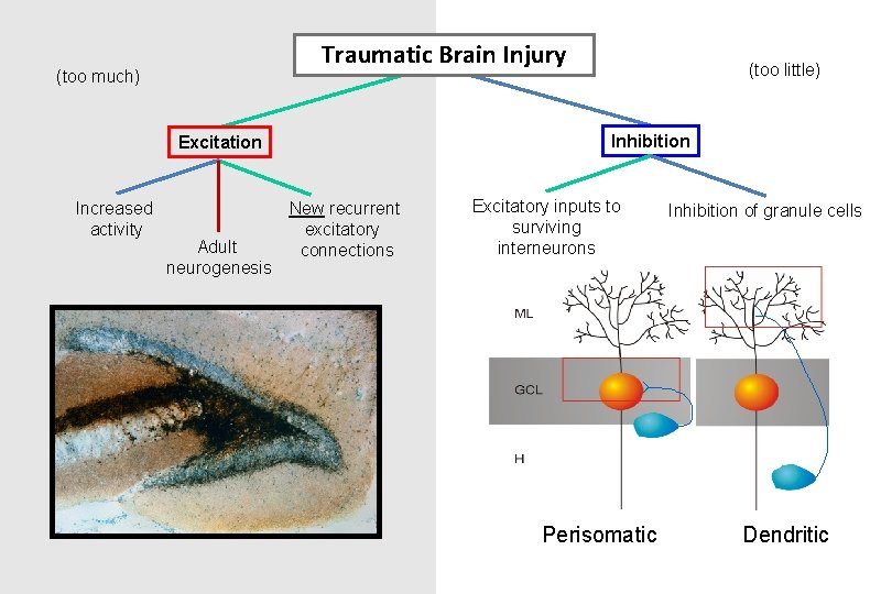
Traumatic Brain Injury (too much) Inhibition Excitation Increased activity Adult neurogenesis (too little) New recurrent excitatory connections Excitatory inputs to surviving interneurons Perisomatic Inhibition of granule cells Dendritic
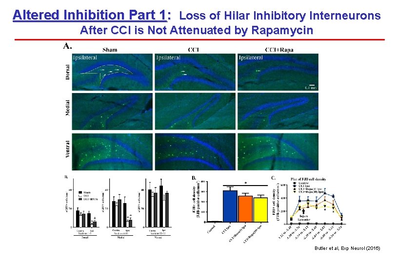
Altered Inhibition Part 1: Loss of Hilar Inhibitory Interneurons After CCI is Not Attenuated by Rapamycin Butler et al, Exp Neurol (2016)
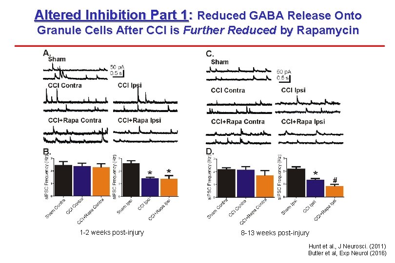
Altered Inhibition Part 1: Reduced GABA Release Onto Granule Cells After CCI is Further Reduced by Rapamycin 1 -2 weeks post-injury 8 -13 weeks post-injury Hunt et al. , J Neurosci. (2011) Butler et al, Exp Neurol (2016)
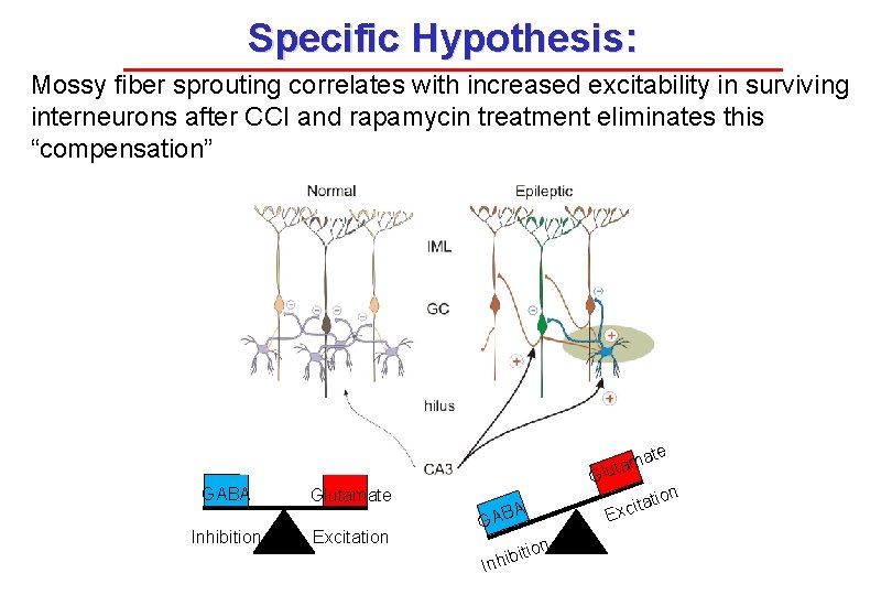
Specific Hypothesis: Mossy fiber sprouting correlates with increased excitability in surviving interneurons after CCI and rapamycin treatment eliminates this “compensation” ate m a t lu GABA Glutamate Inhibition Excitation G A GAB tion bi Inhi tion ita Exc
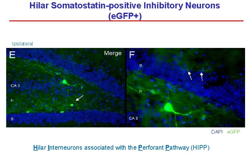
Hilar Somatostatin-positive Inhibitory Neurons (e. GFP+) Ipsilateral DAPI e. GFP Hilar Interneurons associated with the Perforant Pathway (HIPP)
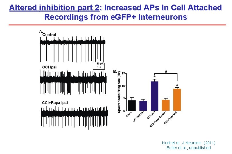
Altered inhibition part 2: Increased APs In Cell Attached Recordings from e. GFP+ Interneurons Hunt et al. , J Neurosci. (2011) Butler et al. , unpublished
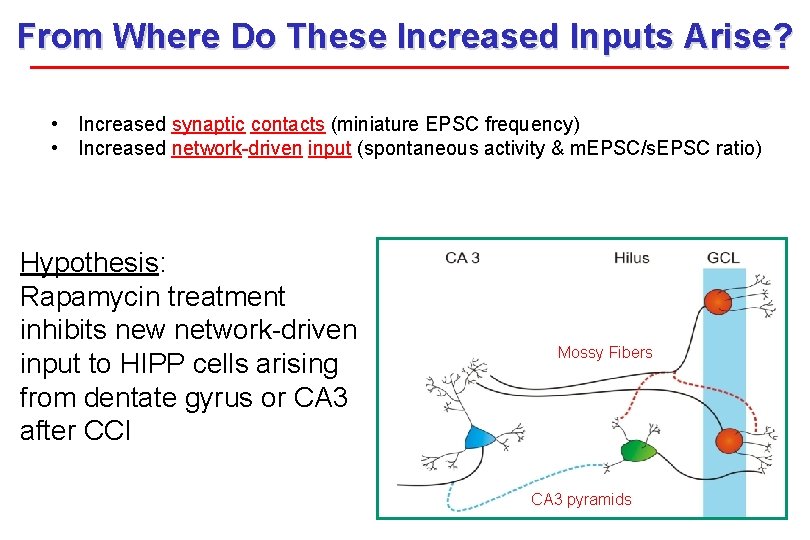
From Where Do These Increased Inputs Arise? • Increased synaptic contacts (miniature EPSC frequency) • Increased network-driven input (spontaneous activity & m. EPSC/s. EPSC ratio) Hypothesis: Rapamycin treatment inhibits new network-driven input to HIPP cells arising from dentate gyrus or CA 3 after CCI Mossy Fibers CA 3 pyramids
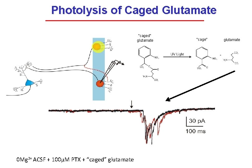
Photolysis of Caged Glutamate “caged” glutamate 0 Mg 2+ ACSF + 100μM PTX + “caged” glutamate “cage” glutamate
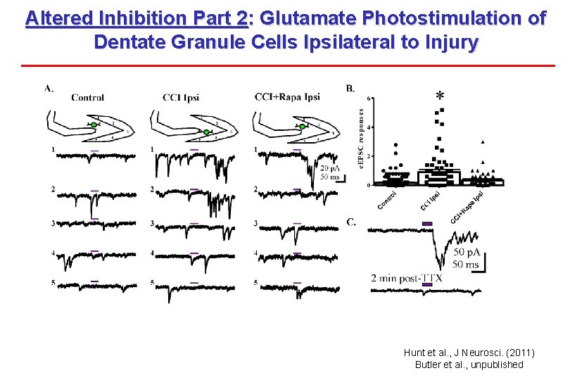
Altered Inhibition Part 2: Glutamate Photostimulation of Dentate Granule Cells Ipsilateral to Injury Hunt et al. , J Neurosci. (2011) Butler et al. , unpublished
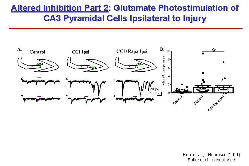
Altered Inhibition Part 2: Glutamate Photostimulation of CA 3 Pyramidal Cells Ipsilateral to Injury Hunt et al. , J Neurosci. (2011) Butler et al. , unpublished
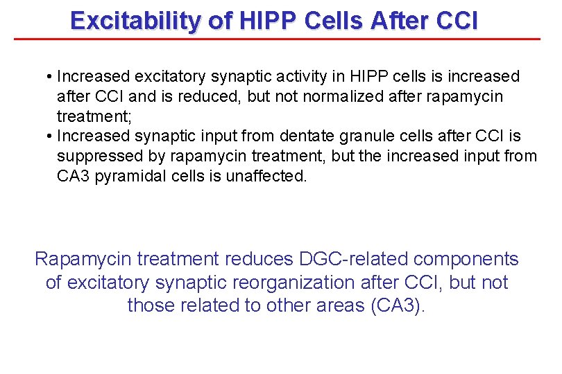
Excitability of HIPP Cells After CCI • Increased excitatory synaptic activity in HIPP cells is increased after CCI and is reduced, but normalized after rapamycin treatment; • Increased synaptic input from dentate granule cells after CCI is suppressed by rapamycin treatment, but the increased input from CA 3 pyramidal cells is unaffected. Rapamycin treatment reduces DGC-related components of excitatory synaptic reorganization after CCI, but not those related to other areas (CA 3).
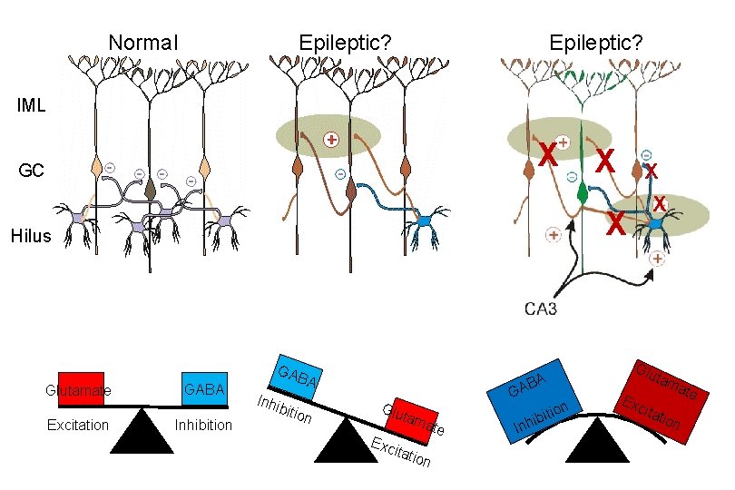
Normal Epileptic? IML X GC Excitation X X X Hilus Glutamate X GABA Inhibition GAB A Inhi bitio n A Glu tam ate Exc itati on B GA i Inh on biti Glu ta Exc ma te itat i on
Does synaptic reorganization cause TLE? “Those guys are out of breath and they need more oxygen. So they’re bending over because it helps them breathe if they’re closer to the grass. . . …’cause there’s oxygen in the grass” --John Madden
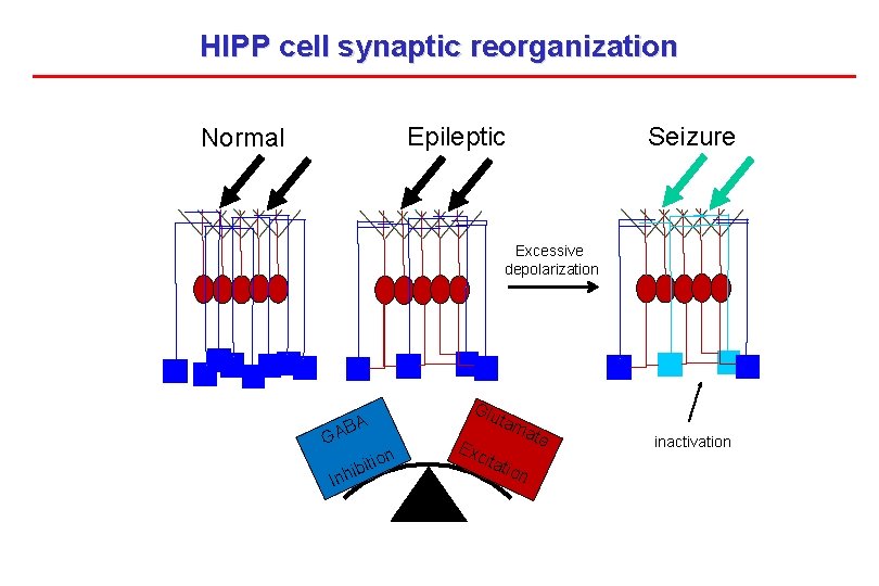
HIPP cell synaptic reorganization Seizure Epileptic Normal Excessive depolarization Glu A AB G I bit nhi ion tam Exc itat ate ion inactivation
- Slides: 44