Epilepsy Dr Omar Alrawashdeh Classification Generalized seizures Focal


 convulsive")


- Slides: 42
Epilepsy Dr Omar Alrawashdeh
Classification • Generalized seizures • Focal or partial seizures: 1. Focal seizures without loss of consciousness 2. Focal seizures with loss of consciousness 3. Focal seizures with secondary generalisation
Focal seizures presentation • Cognitive symptoms: Seizures can start with aura in the form of automatism (hand or oral) and it can be reactive (drinking from a cup in the hand) or perseverative ( continuation of complex act). This form is usually localised to the temporal lobe This can be followed by staring, fearful or sad expression. This is usually localised to the amygdala or anterior cingulate Gelastic or dacrystic auomatisms ( hypothalamic hamartomas)
Focal seizures presentation • Mesial temporal lobe epilepsy Typically begins with aura consisting of epigastric rising, fear, staring, oroalimantary automatism. The most common cause is hippocampal sclerosis
Focal seizures Neocortical temporal cortex epilepsy Auditory hallucination, vertigo, language disturbances, musicogenic seizures
Focal seizures • Frontal seziures bizarre motor automatims, hypermotor activity, salivation, spitting • Cingulate gyrus focus may causes expression of a particular affect such as laughter • Orbitofrontal cortex: complex motor automatism and olfactory hallucinations. • Clonic, or Tonic clonic movement in primary motor area according to the motor homunculus involved. • An example of partial seizures that affect the primary motor cortex is Jacksonian march
Focal seizures • Fencers posture: asymmetric bilateral movement of the upper limbs: supplementary motor cortex. This can be bilateral without loss of consciousness • Unilateral forced gaze and head deviation (versive seizures): premotor cortex and forntal eyes field. Arm and facial seizures, with laryngeal movement, tachycardia, : contraateral insula
Tonic clonic seizures • convulsive activity typically lasts < 1 minute. • Mood changes can precede seizures by days • Immediately pretonic-clonic phase: a few myoclonic jerks or brief clonic seizure activity; occasionally begins with forced eye and head deviation • Tonic phase: contracture of the axial musculature with upward eye deviation, pupillary dilation, and forced expiration of air {epileptic cry}; usually involves some decerebrate posturing. Tongue and jaw muscle tonus causes perioral injury, typically lateral tongue biting. Frequently, patient becomes cyanotic, tachycardic, and hypertensive
Tonic clonic seziures • Clonic phase: starts as low-amplitude, highfrequency (~ 8 Hz) convulsive movements of the extremities > the thorax and abdomen that progresses to high-amplitude, lowfrequency (~ 4 Hz) movements. Development of atonia breaks the seizure and causes incontinence
Tonic clonic seizures • Postictal phase: patient is poorly responsive and hypotonic; confusion and memory impairment may last a few minutes to hours, occasionally followed by psychiatric changes (depression, psychosis, anxiety, irritability) that can persist for about a day (a) Postictal phase involves generalized fatigue, soreness, and migrainous headaches
Tonic sezires • diffuse contraction of the axial musculature, sometimes involving the proximal limbs or entire limbs; It can be in the form of startle response that can follow a stimulus of any type
Atonic seizures • Sudden loss of consciousness and muscle tone especially of the head. This can results in a drop attacks • These are usually preceded by myoclonic jerks • EEG may demonstrate polyspikes and waves during the ictal period and followed by generalised slowing of the central area
Myoclonic seizures • Shock like movement of one muscle or a group of muscles. • Irregular and can be singular or repetitive. • It affects the eyelid, facial muscles, upper limbs ad lower limbs • EEG: Polyspikes and waves
Epilepsy syndromes Generalised epilpesy syndromes • Infantile spasms • Lennox – Gastaut syndrome • Juvenile myoclonic epilepsy • Absence epilepsy
Epilepsy syndromes • • • Focal epilepsy syndromes Landau Kleffner syndrome AD nocturnal frontal lobe epilpesy Benign occipital epilepsies Benign rolandic epilepsy Rasmussen syndrome
West syndrome • Seizures: classically manifest as clusters of symmetric extensor and/ or flexor movements, followed by tonic contractions lasting < 10 seconds; seizures most commonly occur after awakening (i) Abdominal flexions are often mistaken for colic (b) Autism (c) Progressive mental retardation (d) Microcephaly
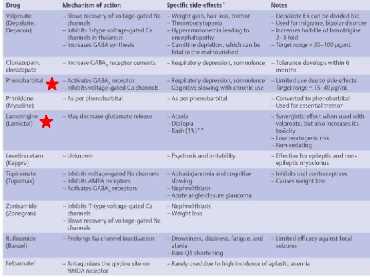
West Syndrome • In 75 % of cases there is an underlying structural lesion which can be idiopathic or acquired cerebral injury , intracranial hemorrhage or infection, or genetic abnormality such as Down syndrome • EEG: Hypsarrhythmia • Treatment: ethosuximide or Na valproate
West Syndrome • EEG: Hypsarrhythmias
Lennocx- Gastaut Syndrome • Symptoms: onset between 3– 5 years of age with • Multiple seizure types 1. Tonic seizures (90%). 2. Atypical absence seizures (65%) that involve repetitive facial motions followed by loss of tone in head and face. 3. Myoclonic seizures (20%)
Lennox – Gastaut Syndrome • Interictal EEG: exhibit 2– 2. 5 -Hz slow spike-and -wave complexes with frontotemporal predominance; paroxysmal fast activity > 10 Hz is apparent while sleeping • Ictal EEG: Atypical absence seizures exhibit 2– 2. 5 -Hz spike-and-wave similar to interictal pattern
Lennox – Gastaut Syndrome
Lennox – Gastaut Syndrome • Pathophysiology: 60% of cases are related to structural brain abnormalities; 20% of cases develop from infantile spasms • medications (usually fail) Ketogenic diet or Vagal nerve stimulator; corpus callosotomy
Juvenile Myoclonic epilepsy • Symptoms: seizures involve bilateral but asymmetric flexor movements of the upper extremities or rarely of the lower extremities that develop after awakening • usually no loss of consciousness • Other types of seizures commonly exist such as absence and tonic clonic • Photic stimulation provokes discharges
Juvenile Myoclonic epilepsy • interictal EEG demonstrates bursts of bilateral, symmetric 3. 5– 6 -Hz spike-and-wave and polyspike discharges; ictal EEG exhibits diffuse polyspike activity followed by 1– 3 -Hz slow waves • Pathophysiology: genetic, but usually with complex inheritance pattern • Treatment: valproate > lamotrigine, levetiracetam, topiramate, zonisamide
Absence epilpsy • Symptoms: 5– 10 -second long unresponsive staring spells ± rhythmic facial movements or picking behaviors; does not have an aura or postictal state • Childhood-onset form is usually self-limited, whereas the juvenile-onset form is more likely to persist into adulthood • autosomal dominant inheritance • slow-wave discharges on EEG may relate to cyclic activity of T-type calcium channels and repolarizing potassium currents in the reticular thalamic nucleus
Absence epilpsy • Occur on a daily basis in the childhood-onset form, more infrequently in the juvenile-onset form • 50% also have generalized tonic-clonic seizures, more commonly in the juvenile-onset form; these are infrequent and can be well controlled with medication • Diagnostic testing: EEG demonstrates bilateral spike-and-wave complexes at 3 Hz that are reliably activated by hyperventilation.
treatment • Treatment: 1. ethosuximide, valproate, acetazolamide 2. Avoid carbamazepine, phenytoin, and gabapentin, which have all been found to increase seizure frequency and may induce absence status epilepticus
Absence epilepsy
Landau-Kleffner syndrome • receptive aphasia progressing to global aphasia over a period of several days a. • Progression of aphasia is not related to frequency of seizures and is fluctuant • Auditory agnosia may occur with involvement of the non-dominant hemisphere • Clinical seizures (70%) usually occur during sleep and may be focal or generalized motor or hemi-clonic status epilepticus
Landau-Kleffner syndrome • Behavioral changes: hyperactivity, aggression • Transient motor impairments • Treatment : 1. Glucocorticoids often improve seizures and aphasia. 2. Antiepileptic drugs control seizures but have an unclear effect on aphasia 3. In severe cases, subpial transection to isolate seizure focus or surgical resection of seizure focus • Prognosis: seizures remit by age 15; aphasia may persist into early adulthood before improving
Benign occipital epilepsy • Pathophysiology: unknown; frequently occur in patients with migraine, but not associated with structural lesions • Symptoms: seizures involve unformed visual hallucinations, visual illusions (micropsia, macropsia, metamorphopsia), vision loss (either complete or hemianopic), vomiting and drooling, and/ or forced blinking and forced gaze deviation; some patients will have seizures followed by a loss of consciousness lasting several hours • Seizures usually develop after a migraine headache, and migraine headaches typically develop in patients who lose consciousness after the seizure once they regain consciousness • (
Benign occipital epilepsy • Seizures usually occur during sleep onset • Diagnostic testing: interictal EEG demonstrates unilateral or bilateral high-amplitude occipital spike-and-wave discharges at 1. 5– 2. 5 Hz that are inhibited by eye opening; ictal EEG is similar except that the spike-and-wave discharges are at higher frequencies • Epileptiform activity is reliably induced by photic stimulation
Benign rolandic epilepsy • Symptoms: perioral paresthesias followed by twitching of face and mouth with speech arrest and drooling but without impairment of consciousness. • convulsions may develop in an upper extremity or generalize, usually in patients < 5 years of age • Diagnostic testing: EEG demonstrates typical high -amplitude, diphasic spike with prominent aftergoing slow wave, maximum in the centrotemporal regions
Benign rolandic epilepsy • Treatment: only if the seizures are frequent; carbamazepine controls seizures in 65% (5) Prognosis: spontaneous resolution of seizures by age 14; withdrawal of antiepileptic medication after 2 years leaves 80% seizurefree
Rasmussen syndrome • focal motor seizures that spread to contiguous muscle groups; 60% develop focal status epilepticus {epilepsia partialis continua} before the disease remits after 2– 10 years • inflammatory degeneration of the cortex and supratentorial subcortical structures inflammatory degeneration • It does not involve the contralateral hemisphere or posterior fossa structures (
Rasmussen syndrome • EEG demonstrates continuous focal spike discharges that spread to contiguous areas of cortex and eventually to mirror foci on the contralateral hemisphere • Neuroimaging: may be normal initially, but hemispheric atrophy and ipsilateral hydrocephalus ex vacuo develop within 6 months • Treatment: refractory to antiepileptic medications; limited benefit from immunosuppressive agents; early hemispherectomy can be curative
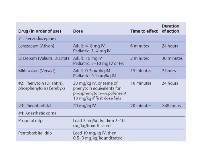
Antiepileptic drugs • Predictive factors for successful seizure remission 1. A single type of seizure 2. Had no seizures for > 2 years 3. A normal neurological exam and IQ 4. A normalized EEG on antiepileptic treatment
Epilpesy treatment • Management of medication failure 1. If maximal doses of an antiepileptic drug (AED) fail to control the seizures, there is only a 10% likelihood of developing control over seizures with a second AED 2. There is < 5% likelihood of developing control over seizures with a third AED or by using multiple AEDs after failing a second AED 3. 25% of patients with chronic refractory seizures are eventually found to have the wrong diagnosis
AED prophylaxis • Prophylactic AED use in patients with another neurological disorder, but without seizures, is usually not recommended 1. Brain tumor: prophylaxis does not seem to reduce development of seizures in patients with primary or metastatic tumors 2. Stroke: not indicated for ischemic stroke, although AEDs are routinely used short-term in subarachnoid hemorrhage patients because of the fear of increased intracranial pressure that accompanies seizures 3. Severe head trauma : (prolonged loss of consciousness, amnesia, depressed skull fracture, contusion, or hematoma) can be treated with acute prophylaxis limited to 1 week