Epidermis Dermis Lectures Objectives Describe the layers of

Epidermis & Dermis

Lectures Objectives • Describe the layers of the epidermis and development of the integumentary system. • Describe structure of the dermis, compare the structure and distribution of hair follicles, nails, sebaceous and sweat glands. • Explain bases of skin color.

Introduction • The organs of the integumentary system include the skin and its accessory structures including hair, nails, and glands, as well as blood vessels, muscles and nerves • Dermatology is the medical specialty for the diagnosis and treatment of disorders of the integumentary system.
 covers the body and is")
Structure of the Skin • The skin (cutaneous membrane) covers the body and is the largest organ of the body by surface area and weight • Its area is about 2 square meters (22 square feet) and weighs 4. 5‐ 5 kg (10‐ 11 lb), about 16% of body weight • It is 0. 5 – 4 mm thick, thinnest on the eyelids, thickest on the heels; the average thickness is 1 – 2 mm
 – – Epidermis proper Hair follicles")
Skin Layers • Epidermis and derivatives – (ectoderm) – – Epidermis proper Hair follicles Nails Glands • Sebaceous • Sweat (eccrine & apocrine) • Mammary • Dermis – Corium (mesoderm) – Papillary layer – loose CT – Reticular layer – dense Ct • Subcutaneous (hypodermis) subcutis (mesoderm) – Areolar CT – Fatty components

Development of the Integumentary System

Functions of Skin • Major functions of skin – Protection – Thermoregulation – Excretion – Sensory reception – communication – Immunoprotection – there are more lymphocytes in the skin than in circulation

Functions of Skin • Problems of burn victims exemplify the importance of skin. • Patients with severe burns has – Loss of sensation to temperature – Pain and pressure – Ionic balance drastically off – Fluid loss – Wildly fluctuating body temperature (5 -8 degrees) – Bacterial and fungal infections increase

Epidermis • Most superficial layer of skin • Cornified stratified squamous epithelium

Epidermis • Tow major types of skin based on thickness of epidermis – Thick skin on non hairy surfaces (epithelium up to 1. 0 mm) – Thin skin on most hairy surfaces of the body (epithelium generally 0. 1 mm) Thick vs thin does not reflect true thickness of skin including dermis • Skin on the back called thin but actually is thickest when dermal layer is included
 –")
Cells of Epidermis • Four cell types in the epidermis – Keratinocytes (squames) – production of keratin and water barrier. More than 90% of cells in epidermis – Melanocytes – pigment production less than 5% of cells in epidermis – Langerhans cells – immune response, 1 -3% cells in epidermis – Merkel cells – sensory reception and local endocrine control, 1 -3% cells of epidermis
 – Change shape (flatten) – Lose organelles –")
Epidermis • Differentiation of keratinocytes (cytomorphosis) – Change shape (flatten) – Lose organelles – Form fibrous proteins – Become dehydrated – Produce products that thicken plasma membranes
 of the Epidermis • • • Stratum corneum Stratum lucidum Stratum granulosum")
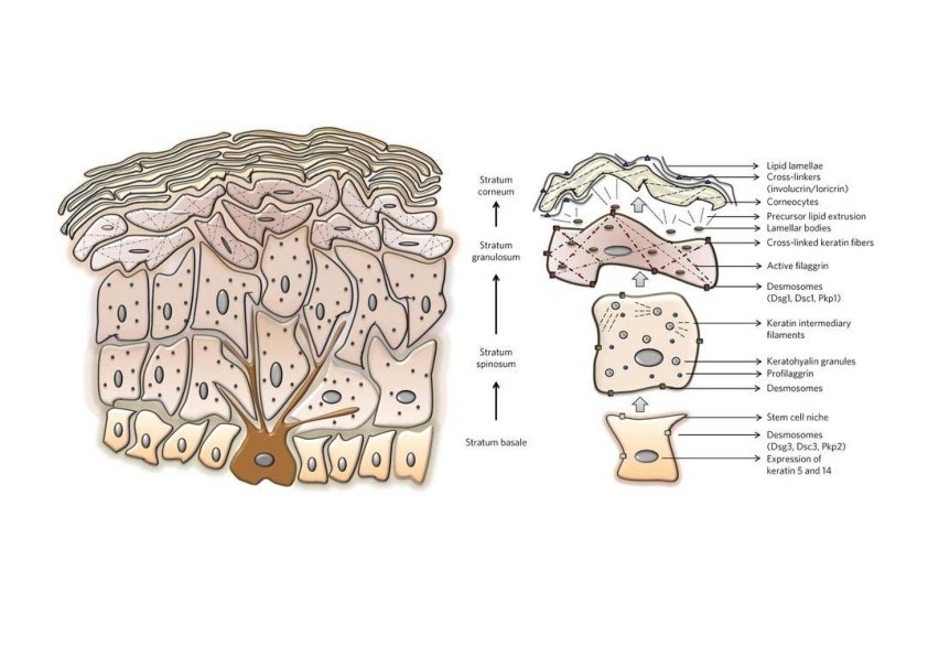
Layers (Strata) of the Epidermis • • • Stratum corneum Stratum lucidum Stratum granulosum Stratum spinosum Stratum basale
 • The mitotically active layer which generate the keratinocytes and their")
Stratum Basale (Germinativum) • The mitotically active layer which generate the keratinocytes and their derivatives (i. e. Merkel cells) • Cells anchored to basal lamina via anchoring fibrils and hemidesmosomes • Highest metabolic requirement of all layers • Active synthesis of tonofilaments, formation of extensive number of desmosomes • Cells assume cuboid profiles • Two major cell types: – Stem cells – Keratinocytes • This layer contain the most undifferentiated cells of epidermis

Stratum Spinosum • Polyhedral cells • Cells are firmly bound by the filament-filled cytoplasmic spines and desmosomes • Active synthesis of tonofilaments which provides cytoskeletal framework – Tonofilaments insert into the cytoplasmic part of the desmosomes • Appearance of membrane-coated granule (MCG) in upper portion of this layer – These are laminated structures – Key to formation of effective water barrier – MCG contain lamellar disks that are formed by lipid bilayers • Mitotic figure frequent (up to 3%) – All mitosis are confined to the malpighian layer (both stratum basale and stratum spinosum)

Stratum Granulosum • Presence of dense, refractile, basophilic granules of keratohyalin which contain profilaggrin • The basophilia is due to RNA within the granules (active sites of filaggrin synthesis) • Tonofilaments have condensed into tonofibrils which are anchored in the matrix of the granules • Many desmosomes present • MCG are released into extracellular space – thicken external surface of membrane and seal adjacent membranes thus forming a water barrier (note no zonula occludens)

Stratum Lucidum • Only in thickest skin • Appears as transparent layer, no visible nuclei or organelles • Sulfhnydryl groups (-SH) replaced by disulfide bonds (S-S) thus forming cystine groups that cross linked and stabilize the keratin molecules • Substance remaining in these cells called eleidin – Clear intracellular protein – Intermediary in formation of keratin from keratohyaline granules and tonofibrils

Stratum Corneum • Plasma membranes thickened by previous emptying of MCG into intercellular space in the subjacent layer • Desmosomes remain as membranous modifications • All organelles lost • Cells backed with keratin (tonofibrils embedded in matrix of keratohyalin) • Cell structure not evident

• Stages of keratinocytes development – Proliferation – Differentiation – Exfoliation Usually 28 -45 days for complete cycle • Thin skin lacks well developed: – Stratum corneum – Stratum lucidum – Stratum granulosum
 cells within st. basale. •")
Pigmentation system • Melanocytes – Appear as clear (neuron-like) cells within st. basale. • Do not form desmosomes with keratinocytes – Extend dendritic-like processes throughout st. spinosum – Same number of melanocytes in all races – Produce melanin – Transfer melanin to keratinocytes – Originate from neural crest • Tyrosinase catalyzes conversion of tyrosine to 3, 4 -dihydroxyphenyalanine (DOPA) and conversion of DOPA to dopaquinone which eventually forms melanin

Pigmentation system Stages of melanocyte melanogenesis (melanization of the melanosome; a membrane-limited body derived from Golgi apparatus) • Premelanosome – Circular vesicle – Tyrosinase activity – Fine peripheral granules • Immature melanosome – Ovoid vesicle – Tyrosinase activity – Parallel filaments 10 nm periodicity • Mature melanosome – Internal structure obscured by melanin – Tyrosinase activity still present • Melanin granule – Solid melanin – No tyrosinase activity

Pigmentation system • Transfer of melanin from melanocyte to keratinocytes is by a process termed “Cytocrine secretion” – Injection by melanocyte of melanin granules into a keratinocyte (exocytosis coupled with endocytosis) • Because of rapid transfer more melanin found in keratinocyte than melanocyte

Pigmentation system • Melanin accumulate on sunny side of nucleus – protection of nucleus from UV • Dark skin absorbs and disperse UV better than light skin • Racial differences due to: – Degree of melanin dispersion – Lysosomal action on melanosomes • Light races- melanin rapidly broken down • Dark races- melanin granules more resistant within st. spinosum • Tanning increase melanin concentration not number of melanosomes

Other causes of skin color • Carotene in dermis – Yellow-orange pigment (precursor of vitamin A) – Found in stratum corneum & dermis • Hemoglobin – Red, oxygen-carrying pigment in blood cells – If other pigments are not present, epidermis is translucent so pinkness will be evident

Langerhans cells • Appearance – Clear cells, generally has a dendritic appearance • Location – St. basale and spinosum, – Process extend toward surface • Act as antigen presenting cells with some phagocytic activity • Absence of tonofibrils and desmosomes • May contain melanin granules due to phagocytic activity of damaged keratinocytes • Responsible for the initial allergic reaction due to contact hypersensitivity • Bone marrow originated cell

Merkel’s cells • Merkel cell is modified keratinocyte – Has tonofibrils – Forms desmosome – Slow adapting • Mechanoreceptor – Associated with sensory nerve endings – Catecholamine containing granules on dark side of nucleus, mechanogated channels • Local release of neuropeptides can modulate vascular smooth muscle contraction. Hence Merkel cell has potential to play a paracrine* function in immediate response to stimulus * Change nearby cells by paracrine (signaling) factors

Dermis • Dermis – Corium – Papillary layer – loose CT – Reticular layer – dense Ct

Papillary Layer • Loose CT – Delicate collagenous fibers, – Fewer reticular and elastic fibers – Fibers attach to basement membrane hence perpendicular to surface • Passage for vessels – direct nutritive responsibility for epithelium • Reticular lamina produced by cells in this layer – has an inductive effect on overlying epidermis • Route for ducts of sweat glands

Papillary Layer • Dermal papillae – More papillae in thicker epidermis • Pegs (rete ridges) – Ridges alter topography of overlying epidermis to produce fingerprints • Site of Meissner’s corpuscles – Fine touch – Important use in reading braille
 of skin • Dense fibrous CT highly interwoven")
Reticular Layer • Major component (mass) of skin • Dense fibrous CT highly interwoven • Provide structural support for derivatives – Hair follicles – Associated glands
 • Loose CT • Serves for: – Protection – Fat storage")
Hypodermis (Subcutaneous Layer) • Loose CT • Serves for: – Protection – Fat storage – Houses • Secretory portion of eccrine sweat glands • Deep portions of the hair follicles – Frequent Pacinian corpuscles (pressure receptors) are found in this layer • Not truly component of skin – Corresponds to superficial fascia

Eccrine Sweat Gland • Not associated with hair follicles • Sweat is cell product – Regulation of body fluid • Sweat secretion may lead to total dehydration (1 -2 liter/hour) – Some elimination of body wastes – Regulate body temperature • Major source of evaporative heat loss • Distributed over entire body – Absent in: glands penis, clitoris, labia minora – High concentration in: palms and soles • Morphology – simple coiled tube (tubular) with ductal (smaller lumen) and secretory (larger lumen) portions

Eccrine Sweat Gland • Secretory component – only in deepest dermis or hypodermis. – Lined with a simple epithelium of (two types) pyramidal cells • Clear serous cell – – Base away from lumen Large nucleus, pale cytoplasm Involved in salt absorption and fluid transport Many mitochondria, glycogen, and intercellular canaliculi • Dark mucous cells – – – Inverted pyramid, base toward lumen Small nucleus Basophilic Involved in production of glycoproteins Golgi apparatus – Thus nuclei of secretory portion appear at two levels like pseudostratified epithelium

Eccrine Sweat Gland • Ductal component – Two concentric layers of cuboidal cells – Luminal surface appears thickened because of densely packed tonofilaments – Ductal cells separated from basement membrane (basal lamina) by incomplete layer of myoepithelial cells – Distal component of duct reabsorbs Na and Cl

Eccrine Sweat Gland Two types of eccrine glands in terms of stimulus sensitivity • Palmar – plantar – Increase secretion response to mental (emotional) state – Only minimal response to heat stress – Adrenergic • General eccrine – Response predominantly to heat stress – Cholinergic
 –")
Apocrine Sweat Glands • Component of Pilosebaceous Unit (hair follicle + associated structures) – Hormonal control • Develops from distal region of hair follicle in embryo • Atrophies on most follicles except in specific regions: axilla, external auditory meatus, eyelids, ano-genital areas

Apocrine Sweat Glands • Morphology – simple tubular glandular epithelium, secretory coil in deep dermis, duct opens into distal hair follicle – Lumen of tube varies with functional state, but much larger than lumen of an eccrine sweat gland – Simple epithelium • when contracted (empty) appears simple cuboidal to low columnar; • when distended appears almost like simple squamous – All cells attached to basal lamina; however attachment not clear because basal lamina partially covered by incomplete layer of myoepithelial cell
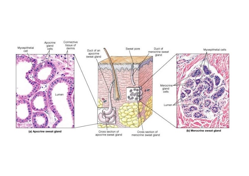

Apocrine Sweat Glands • Secretion appears apocrine “pinching off” of apical cytoplasm. EM reveals exocytosis – Secretion: viscous milky to clear, contains proteins, carbohydrates, ferric iron, fatty acids – Initially odorless – characteristic body odor formed after bacterial decomposition – Function unknown possibly bactericidal, or pheromone--amorous inclinations – Primarily adrenergic (emotional) stimulus for secretion, some cholinergic stimulus

Sebaceous Gland • No nerve supply, only hormonal regulation • Develops as portion of Pilosebaceous unit as a bud from the wall of a hair follicle below the apocrine gland within the dermis • Prominent distribution over face, neck, upper chest and back. • On upper back they are highly developed while the hair follicle remains rudimentary hence referred to as Sebaceous follicles
, almost entirely lipid – Secreted")
Sebaceous Gland • Sebum – whole cell product (holocrine), almost entirely lipid – Secreted continually, not diurnally – Acts as a permeability barrier, bacteriostatic, fungistatic, pheromone – Acne – androgen increase (during puberty) – increase sebum secretion • Germinal epithelium layer adjacent to basal lamina – As cells form sebum they take on foamy appearance, lose nuclei and most visible organelles; center of gland most highly differentiated

Hair Follicles • Pilosebaceous apparatus includes: hair follicle, sebaceous gland, apocrine sweat gland smooth muscle • Reduced importance of hair in human – – – Conserve body temperature Protect against physical trauma Provide sense of environment Personal adornment Functional reserve of epithelial cells after injury • Variation with sexes. Same number hair follicles males and females (estimated 2 million) 100, 000 on scalp. Difference in distribution between sexes
 – Lanugo – lost")
Hair types Three types of hairs (1 preterm, 2 mature) – Lanugo – lost shortly after birth – Vellus – covers most of the body before puberty • Very fine non pigmented equivalent to lanugo • Follicle extend to mid dermis • Converted to terminal follicles after puberty in axillary and anogenital regions in both sexes – In males also have conversion on face extremities and trunk – Terminal – • Coarse, pigmented, on scalp, eyelids, eyebrow • Follicle extends through dermis into hypodermis • Structure of terminal hair more complex (medulla and cortex) • Keratinocytes do not undergo desquamation, they form hard keratin

Structure of Hair • Shaft -- visible – Medulla, large, vacuolated and moderately keratinized cells – Cortex, heavily keratinized, compactly grouped fusiform cells – Cuticle, • Cuboidal midway up the bulb, then columnar. • Higher up, form flattened, heavily keratinized, shingle like cells • Root -- below the surface • Follicle surrounds root

Structure of Hair • Follicle surrounds root – Internal root sheath • Transient structure whose cells degenerate and disappear above the level of sebaceous gland – External root sheath – Continuous with the epidermal cells – Glassy membrane – Thickening of the basal lamina – Connective tissue sheath • Denser part of dermis surround the follicle

Structure of Hair • Base of follicle is bulb – Dermal papilla • Blood vessels – Germinal cell layer

Hair Related Structures • Arrector pili – Smooth muscle in dermis contracts with cold or fear. – Forms goosebumps as hair is pulled vertically • Hair root plexus – Detect hair movement

Hair Growth • The hair growth cycle consists of a growing stage and a resting stage. – Growth cycle = growth stage & resting stage • Growth stage (anagen) – lasts for 2 to 6 years – matrix cells at base of hair root producing length • Resting stage (catagen and telogen) – lasts for 3 months – matrix cells inactive & follicle atrophies – Old hair falls out as growth stage begins again • normal hair loss is 70 to 100 hairs per day
 influence terminal hair follicles. In contrast, estrogen")
Hair Growth • Male sex hormone (androgen) influence terminal hair follicles. In contrast, estrogen influences melanocytes • Both rate of growth and the replacement cycle can be altered by illness, diet, high fever, surgery, blood loss, severe emotional stress, and gender. • Chemotherapeutic agents affect the rapidly dividing matrix hair cells resulting in hair loss.

Hair Color • Hair color is due primarily to the amount and type of melanin. • Graying of hair occurs because of a progressive decline in tyrosinase. – Dark hair contains true melanin – Blond and red hair contain melanin with iron and sulfur added – Graying hair is result of decline in melanin production – White hair has air bubbles in the medullary shaft

Structure of Nails • Tightly packed keratinized cells • Nail body – visible portion pink due to underlying capillaries – free edge appears white • Nail root – buried under skin layers – lunula is white due to thickened stratum basale • Eponychium (cuticle) – stratum corneum layer

Nail Growth • Nail matrix is below nail root -- produces growth • Cells transformed into tightly packed keratinized cells • 1 mm per week • Certain nail conditions may indicate disease
- Slides: 53