Epidemiologists and Laboratory Science Najib Aziz M D

Epidemiologists and Laboratory Science Najib Aziz, M. D. Adjunct Associate Professor Department of Epidemiology UCLA, Fielding School of Public Health January 2014

Introduction: • Advance in the laboratory science technologies enable us to detect trace amount of material of interest and determine from what organism it came from. • Epidemiology describes the distribution of health and disease in a population and determinant of that distribution. • The laboratory science tools enable the epidemiologist to moves the epidemiologic studies beyond the detection of risk factors and probable transmission mode, to identify mechanisms of disease pathogenesis and describe transmission systems. • Every laboratory measurement is subject to error from variety source and the goal should be to reduce the error source so the true biological variation can be observed. • Laboratory test is only useful if it is reliable, valid and interpreted appropriately. The validity and reliability of the test assured by quality control and quality assurance programs.
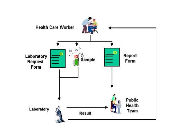

Ø Planning -Define the possible causes of the outbreak -Decide which clinical specimens are required to confirm the cause of the outbreak. Ø Selection of the laboratory for testing -Decide who will collect, process and transport the specimens -Define the procedures necessary for specimen management
 Patient or subject Preparation b)")
Quality Assurance Cycle of Laboratory Work 1. Pre-Analytic: a) Patient or subject Preparation b) Sample Collection c) Personnel Competency Test Evaluations d) Sample Receipt and Accessioning e) Sample Transport 1. Analytic: a) Quality Control b) Testing 2. Post-Analytic: a) Reporting b) Record Keeping

The Quality Assurance Cycle Patient/Client Prep Sample Collection Personnel Reportin Competency • Data and Lab g Management Test Evaluations • Safety • Customer Service Sample Receipt and Accessioning Record Sample Keepin Transport g Quality Control Testing CDC

Optimal laboratory work flow for an investigational or outbreak project Minimizing of the potential error Specimens collection Transportation Processing Storage Data analyze

1. Specimen collection • Type of specimen • Time of collection( effect of circadian rhythms) • Who collect the specimen ( Subject, Research associate, investigator) trained and retrained periodically to ensure protocols are followed • How collect the specimen ( e. g. clean-catch midstream of urine) • Quality of specimen collected • Specimen labeling

Specimen Type: - Blood for smears - Blood for culture - Blood for Serum - Blood for plasma - Cerebrospinal Fluid (CSF) - Sputum - Stool samples - Throat swabs - Nasopharyngeal swabs - Rectal swabs - Urine - Saliva - Water sample - Food sample

Vacutainer Venous Blood collection tubes Gold Gray Light Tige green r gree n Red gray Ti n Lavend er Red top Whit e Orange Yello w Royal Blue Pin k Green Light Blue

Urine collection Device Saliva collection Device swabs collection Device

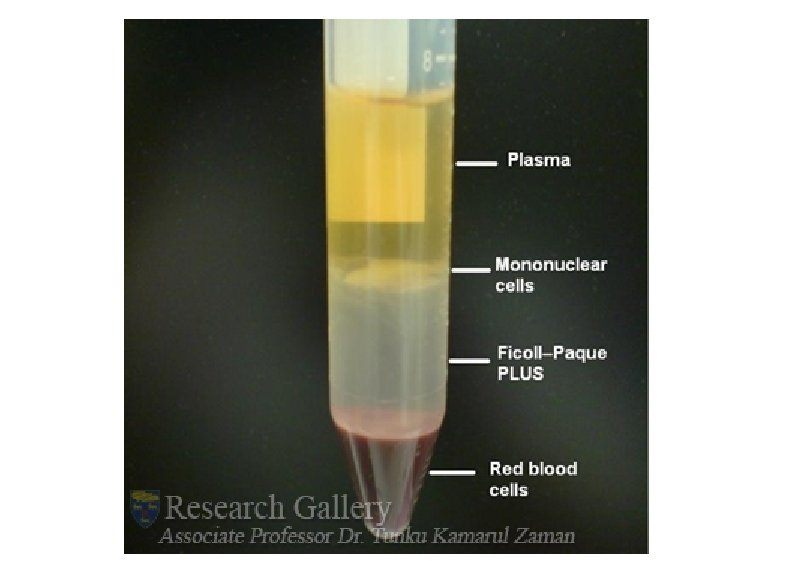
Whole blood : v Cellular elements (red blood cells or RBC, white blood cells or WBC, and platelets or PLT) v Liquid component, which is either serum or plasma. v In general, adult blood has about 40% cellular elements and 60% serum or plasma Serum is the liquid expressed from clotted blood does not contain fibrinogen or other coagulation factors, preferred specimen for most chemistry, blood bank and serology tests Plasma is the liquid portion of the blood present in anticoagulated specimens. Plasma contains all the coagulation factors, including fibrinogen

Common errors affecting all types of specimens v Failure to label a specimen correctly and to provide all pertinent information v Failure to use the correct container/tube for appropriate specimen preservation v Insufficient quantity of specimen to run test or QNS (quantity not sufficient). v Inaccurate and incomplete subject instructions prior to collection. v Failure to tighten specimen container lids, resulting in leakage and/or contamination of specimens

SERUM PREPARATION ERRORS v Failure to separate serum from red cells within 60 minutes of venipuncture v Failure to allow the specimens to clot before centrifugation. v Hemolysis: red blood cells break down and components spill into serum v Lipemia: cloudy or milky serum sometimes due to the patient's diet

PLASMA PREPARATION ERRORS Ø Failure to collect specimen in correct additive. Ø Failure to mix specimen with additive immediately after collection. Ø Hemolysis or damage to red blood cells breakdown. Ø Incomplete filling of the tube, thereby creating a dilution factor excessive for total specimen volume (QNS). Ø Failure to separate plasma from cells within 30 -45 minutes of draw. Ø Failure to label transport tubes as "plasma". Ø Failure to indicate type of anticoagulant (eg, "EDTA", "citrate", etc. )

URINE COLLECTION ERRORS Ø Failure to obtain a clean-catch, midstream specimen. Ø Failure to refrigerate specimen or store in a cool place. Ø Failure to provide a complete 24 -hour collection/aliquot Ø Failure to add the proper preservative to the urine collection container prior to collection of the specimen. Ø Failure to provide a sterile collection container and refrigerate specimen when bacteriological examination of the specimen is required. Ø Failure to tighten specimen container lids, resulting in leakage of specimen. Ø Failure to provide patients with adequate instructions for 24 -hr urine collection.

2. Transport of specimen to laboratory Ø Inaccuracy in labeling of specimen Ø Labeling lost during transport Ø Transport media improperly prepared Ø Specimen heated or cooled during transport Ø Delays or inconsistent transport time Ø Specimen lost during transport to laboratory

3. Specimen Processing v Specimen Processing is one of the first critical preanalytical steps in laboratory Science. v Each specimen checked for proper identification, specimen integrity, and then accessioned into laboratory computer system. v Specimens are processed per the study SOP, stored or distributed to the appropriate laboratory for testing

Blood after centrifugation

v. Regardless of who collects the specimens, having an easy-to-follow instructions, color coding or numbering materials to match steps, and providing packets containing all materials and instructions together will reduce errors in specimen collection. v. Determining the required storage conditions and tolerance for storage is an important component of protocol development. v. A small amount of specimen stored in a large vial may result in specimen loss due to evaporation. v. The label should be able to tolerate the storage conditions, because some labels fall off when a vial is frozen and thawed.

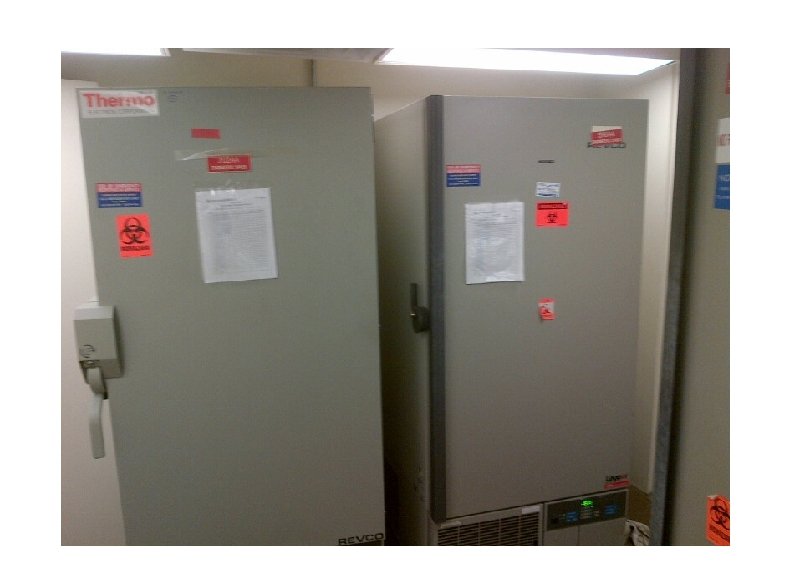
4. Storage, Repository or Biobank The process of collecting, maintaining, and sharing biological samples is viewed as an essential tool in the modern landscape of the genetic, medical, and behavioral sciences and we should; Ø Keep precious biological samples safe and in a secure place Ø Check and log freezers temperature daily. Ø Locate samples quickly and easily through computer base program to access sample reporting and retrieval easily. Ø Retrieval and shipping of biological samples while maintaining the cold environment. Ø Trained and certified personnel with compliance to IATA, DOT and ICAO regulations should ship the specimen.

Diagnostics test for infectious disease

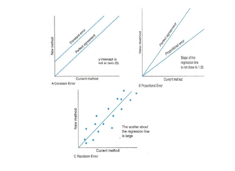
5. Laboratory Test • The laboratory test should reflect the true value and be valid and It depends on two major class of errors. 1. Random error : It occurs without prediction or regularity. factors affecting the precision of an assay include: -Temperature fluctuations -Unstable instrumentation -Change in reagents -Manual techniques variation ( pipetting, mixing, timing) -Operator variation
: (occur one direction , over or under estimation) It is")
2. Systemic error (bias): (occur one direction , over or under estimation) It is defined as error that the results consistently low or high. A) Constant error: If the error is consistently low or high by the same amount over the entire concentration range. B) Proportional Error: If the error is consistently low or high by an amount Proportional to the concentration of the analyte. (e. g. incorrect assignment of calibrator) Avoid of systemic error: • Set of inclusionary and exclusionary criteria • As similar as possible, collect, store, and process of specimen for all participants. • Arranging and selecting a laboratory procedure so that any effects of storage and testing equally impact all specimens
 Repository Quality Assurance")
UCLA Multicenter AIDS Cohort Study (MACS) Repository Quality Assurance
 6 x 10 m. L Green top tube (sodium")
Blood Collection for MACS A) 6 x 10 m. L Green top tube (sodium heparin) for local and national cells and plasma storage B) 7 x 10 m. L (SST) Red top or gold blood tube for local, national serum storage, HIV-1 and Chemistry testing. C) 3 x 5 m. L Lavender top blood tube for cell phenotyping by Flow cytomtery, Complete blood count and Hb. A 1 c testing. D) 2 x 10 m. L lavender top blood tube for plasma storage and HIV viral load testing.

Specimen allocation Plasma UCLA Repository NIH Repository Heparin 3 vials of 3 m. L EDTA 3 vials of 1. 0 m. L 1 vial of 3 m. L 6 vials of 0. 5 m. L 4 vials of 0. 5 and 2 vials of 1. 0 m. L Serum UCLA Repository NIH Repository 3 vials of 3 m. L 2 vial of 3 m. L 1 vial of 1 m. L for HIV 6 vials of 0. 5 m. L PBMC UCLA Repository NIH Repository Viable cells 3 vials at 10^7 cells/m. L 2 vials at 2. 5 x 10^6 Cell pellets
 Internal QC 1. QC of cells counter: Every other week")
PBMC Quality Control A) Internal QC 1. QC of cells counter: Every other week two samples of PBMC are counted using Lab Z 1 Coulter counter and CTRL Sysmex Hematology analyzer and result from both methods are evaluated and the %CV of both methods should be within 15. 2. QC of laboratory tech: Every other week 10 m. L of blood from a normal donor process and freeze and then thaw by the tech and evaluate for cell viability and viable cell recovery.

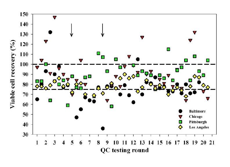
B. External Quality Assurance 1. To ensure the adequate recovery of functionally viable cells from each vial, the MACS established a prospective quality assessment (QA) program in July 2004 to evaluate the PBMC cryopreserved in the four MACS laboratories. 2. In this program, randomly selected vials of recently cryopreserved PBMC from all MACS laboratories are evaluated three times a year, not only for viable cell recovery but also for phenotype and function of the recovered cells.

Viable Cell
")
Cells Function after thaw: Proliferative responses of cryopreserved PBMC (n = 28 per group) to phytohemagglutinin (PHA). The stimulation index was defined as the counts per minute in the presence of PHA divided by the counts per minute in the absence of PHA.

T lymphocyte phenotypes in fresh wholeblood and cryopreserved PBMC Aziz et al.
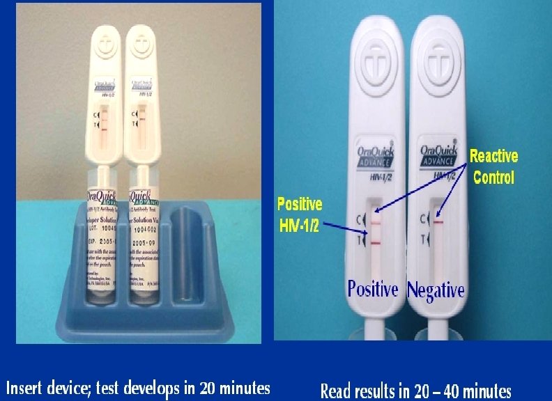
. As screen test ØFollowing viral exposure, the first antibody")
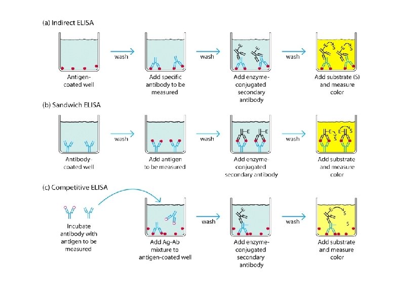
Enzyme linked immunosorbent assay (ELISA). As screen test ØFollowing viral exposure, the first antibody to appear is Ig. M, which is followed by a much higher titre of Ig. G. Ø In cases of reinfection, the level of specific Ig. M either remain the same or rises slightly. But Ig. G shoots up rapidly and far more earlier than in a primary infection. ØMany different types of serological tests are available such as ELISA, RIA , etc. ØNewer techniques such as ELISA offer better sensitivity, specificity and reproducibility than classical techniques
 • Principle: • Use of enzyme-labelled immunoglobulin to detect antigens or antibodies")
(ELISA continu) • Principle: • Use of enzyme-labelled immunoglobulin to detect antigens or antibodies • signals are developed by the action of hydrolyzing enzyme on chromogenic substrate • optical density measured by micro-plate reader

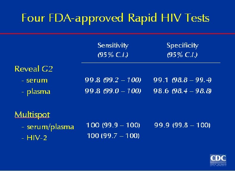
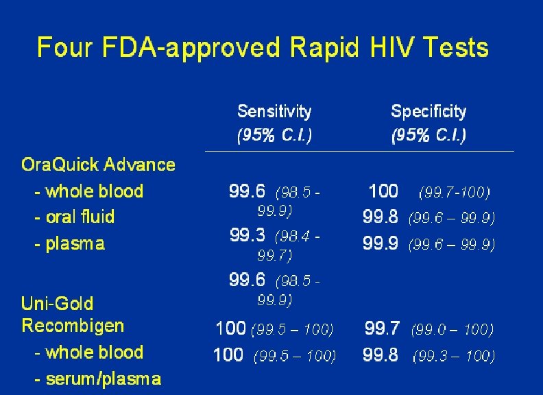
HIV Serological Profile
 HIV-1, 2")
HIV-1/2 Plus 0 ELISA Screen test: • Sandwich enzyme-linked immunosorbent assay (ELISA) HIV-1, 2 +O based on the principle of the direct antibody sandwich. • Microwell strip plates (Solid phase) are coated with purified antigens: gp 160 and p 24 recombinant proteins derived from HIV 1 , peptide from HIV-2 (gp 36) and synthetic ploypeptide of HIV-1 group O. • Sample and controls are added to the wells along with specimen diluents and incubated.
 GS HIV Combo Ag/Ab EIA The GS HIV Combo Ag/Ab EIA is an")
1) GS HIV Combo Ag/Ab EIA The GS HIV Combo Ag/Ab EIA is an enzyme immunoassay kit for the simultaneous qualitative detection of HIV p 24 antigen and antibodies to HIV Type 1 (HIV-1 groups M and O) and HIV Type 2 (HIV-2) in human serum and plasma 2) GS HIV-1/HIV-2 PLUS O EIA This recombinant and synthetic peptide EIA test is used to detect antibodies to human immunodeficiency virus type 1 (HIV-1 groups M and O) and/or type 2 (HIV-2) in human serum, plasma, or cadaveric serum specimens 3) HIV-1 Western Blot Confirmatory Test

Clear determination of positive and negative Results

GS HIV-1 Western Blot is a qualitative assay for the detection and identification of antibodies to human immunodeficiency virus type 1 (HIV-1) that allows same-day results

. HIV-1 Western blot results at three MACS clinic visits. K 0, K 1, and K 2 are negative, weak positive, and strong positive controls, respectively. P 1, P 2, and P 3 are before, during (6 months later), and 7 months after seroconversion, respectively. NC is an HIV-1 negative-control plasma sample.

Interpretation: NEGATIVE: No bands are present POSITIVE : At least TWO of the major bands: gp 160/120, gp 41 or p 24 must be present. Bands must be at least as intense as the Low Positive control gp 120 band (a reactivity score of + or greater) to be considered POSITIVE. The band at gp 41 must be broad and diffuse. INDETERMINATE: Any pattern of one or more bands are present but the blot does not meet the criteria for positive results. Indeterminate results should not be considered either POSITIVE or NEGATIVE. Repeat the indeterminate immunoblot using the original specimen. If the sample is still indeterminate, retest using fresh sample drawn 2 -4 weeks after and again three and six months after the original draw
 Internal QC 1) Three Kit controls (Negative, HIV-1 Low Positive Control")
Quality Control A) Internal QC 1) Three Kit controls (Negative, HIV-1 Low Positive Control and HIV-1 High Positive Control) 2) Two Lab control ( negative and HIV-1 high positive control) B) External QC CAP sends 5 serum/plasma samples quarterly

HIV Viral load test: ØTest is a nucleic acid amplification test for the quantitation of Human Immunodeficiency Virus Type 1 (HIV-1) RNA in human plasma ØThis test can quantitate HIV-1 RNA over the range of 48 - 10, 000 copies/m. L Øspecimen volume required for this method is 1000 μL. ØUpon loading the sample in appropriate racks, nucleic acid extraction, amplification and detection are performed using the COBAS Taq. Man HIV-1 Test, v 2. 0 software

COBAS® Taq. Man® Analyzer • COBAS® Taq. Man® Analyzer or the COBAS® Taq. Man® 48 Analyzer

Multispot HIV-1 and HIV-2 Rapid Test

UCLA MACS Laboratory Shaun Hsueh, Chantel Delshad , Yegermal Asnake

Wilshire LAMS Clinic Photo N/A MACS Harbor UCLA Clinic Los Angeles GLC Carlos Aquino, Lisa Siqueiros, Pedro Chavez Ray Mercado , Eduardo Mercado. GLC staffs
- Slides: 59