Enzymology Dr Dhananjay Bhale MBBS MD PROFESSOR AND

Enzymology Dr Dhananjay Bhale MBBS, MD PROFESSOR AND HEAD BIOCHEMISTRY MGMMC Aurangabad NINTH E D I T I O N

Specific Learning Objectives The learner will be able to: • Classify enzymes • Explain the basic concepts and mode of action of enzymes • Describe the active center of enzymes and specificity of enzymes • Outline Michaelis-Menten theory, Fischer’s template theory and Koshland’s induced fit theory • Define Michaelis constant, Km value, Vmax • Enumerate factors influencing enzyme activity • Classify the coenzymes and cofactors • Describe different types of enzyme inhibition with examples • Explain the mechanisms of regulation, covalent modification, repression, and induction

Classification of Enzymes Class 1. Oxidoreductases: Transfer of hydrogen or addition of oxygen; e. g. Lactate dehydrogenase (coenzyme NAD); Glucose-6 -phosphate dehydrogenase (coenzyme NADP); Succinate dehydrogenase (coenzyme FAD); dioxygenases. Class 2. Transferases: Transfer of groups other than hydrogen. Example, Aminotransferase. (Subclass: Kinase, transfer of phosphoryl group from ATP; e. g. Hexokinase) Class 3. Hydrolases: Hydrolysis of the bond (Cleave bond add water); e. g. Acetylcholine esterase; Trypsin

Class 4. Lyases: Cleave without adding water, e. g. Aldolase; HMG Co. A lyase; ATP Citrate lyase. Class 5. Isomerases: Intramolecular transfers. They include racemases and epimerases. Example, Triose phosphate isomerase. Class 6. Ligases: ATP dependent condensation of two molecules, e. g. Acetyl-Co. A carboxylase; Glutamine synthetase; PRPP synthetase

Salient features of Coenzymes 1. The protein part of the enzyme gives the necessary three dimensional infrastructure for chemical reaction; but the group is transferred from or accepted by the coenzyme. 2. The coenzyme is essential for the biological activity of the enzyme. 3. Coenzyme is a low molecular weight organic substance. It is heat stable. 4. Generally, the coenzymes combine loosely with the enzyme molecules. The enzyme and coenzyme can be separated easily by dialysis. 5. Inside the body, when the reaction is completed, the coenzyme is released from the apoenzyme, and can bind to another enzyme molecule.

6. One molecule of the coenzyme is able to convert a large number of substrate molecules with the help of enzyme. 7. Most of the coenzymes are derivatives of vitamin B complex group of substances.

One coenzyme molecule can work with different enzymes.

Synthetase and Synthase are Different Synthetases are ATP dependent enzymes catalyzing biosynthetic reactions; they belong to Ligases (class 6). Examples are Carbamoyl phosphate synthetase; Argininosuccinate synthetase; PRPP synthetase and glutamine synthetase. Synthases are enzymes catalyzing biosynthetic reactions; but they do not require ATP directly; they belong to classes other than ligases. Examples are glycogen synthase and ALA synthase.

Reaction of lactate dehydrogenase needs the help of NAD+

NAD+ accepts hydrogen
 Hydroxy ethyl Pyridoxal phosphate (PLP)")
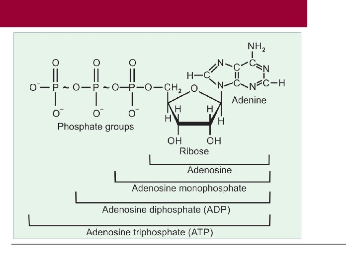
Examples of Coenzymes Coenzyme Group transferred Thiamine pyrophosphate (TPP) Hydroxy ethyl Pyridoxal phosphate (PLP) Amino group Biotin Carbon dioxide Coenzyme A (Co A) Acyl groups Tetrahydrofolate (FH 4) One carbon group Adenosine triphosphate (ATP) Phosphate

Metalloenzymes Metal Enzyme containing the metal Zinc Carbonic anhydrase, carboxy peptidase, alcohol dehydrogenase Magnesium Hexokinase, phosphofructokinase, enolase, pyruvate kinase Manganese Phophoglucomutase, glycosyl transferases Copper Tyrosinase, cytochrome oxidase, lysyl oxidase, superoxide dismutase Iron Cytochrome oxidase, catalase, peroxidase, xanthine oxidase Calcium Phospholipase, lipase Molybdenum Xanthine oxidase

Theories Explaining the Mode of Action of Enzymes 1. 2. 3. 4. 5. 6. 7. 8. 9. Lowering of activation energy Acid base catalysis Substrate strain Covalent catalysis Entropy effect Product substrate orientation theory Michaelis-Menten theory Fischer’s template theory Koshland’s induced fit theory

Lowering of activation energy by enzymes. Red circle = substrate; D = energy level of product. C to A = activation energy in the absence of enzyme; C to B is activation energy in presence of enzyme; B to A = lowering of activation energy by enzyme.

Acid base catalysis with the help of histidine

Correct alignment of amino acids in the active center of the enzyme.

Enzyme substrate complex

Fischer’s Template Theory This theory states that the three dimensional structure of the active site of the enzyme is complementary to the substrate. Thus enzyme and substrate fit each other. Substrate fits on the enzyme, similar to lock and key. The lock can be opened only by its specific key. However, Fischer envisaged a rigid structure for enzymes, which could not explain the flexibility shown by enzymes.
 Enzyme has shallow grooves; substrate alignment is not correct.")
Koshland’s induced fit theory. (A) Enzyme has shallow grooves; substrate alignment is not correct. (B) Fixing of substrate induces structural changes in enzyme. (C) Now substrate correctly fits into the active site of enzyme. (D) Substrate is cleaved into two products

Active center of enzyme 1. The region of the enzyme where substrate binding and catalysis occurs is referred to as active site or active center 2. Although all parts are required for keeping the exact three dimensional structure of the enzyme, the reaction is taking place at the active site. The active site occupies only a small portion of the whole enzyme. 3. Generally active site is situated in a crevice or cleft of the enzyme molecule. To the active site, the specific substrate is bound. The binding of substrate to active site depends on the alignment of specific groups or atoms at active site. 4. During the binding, these groups may realign themselves to provide the unique conformational orientation so as to promote exact fitting of substrate to the active site.

5. The substrate binds to the enzyme at the active site by noncovalent bonds. These forces are hydrophobic in nature. 6. The amino acids or groups that directly participate in making or breaking the bonds (present at the active site) are called catalytic residues or catalytic groups. 7. he active site contains substrate binding site and catalytic site; sometimes these two may be separate.
.")
Active center of Lysozyme. A, B, C, D, E, F are carbohydrate units (substrate). D = N-acetyl muramic acid; E= N-acetyl glucosamine. Bond is broken between D and E, with the help of Glu and Asp residues in the enzyme, which are opposite to each other

Proteases Active Centre Type Example Serine proteases Trypsin, chymotrypsin, clotting factors Aspartic proteases Pepsin Cysteinyl aspartic proteases Caspases in apoptosis Metalloproteases Carboxy peptidases Protease inhibitors used as drugs ACE inhibitor (Captopril), HIV protease inhibitor (Retonavir)

1. Exergonic or Exothermic Reaction Energy is released from the reaction, and therefore reaction goes to completion, e. g. urease enzyme: Urea → ammonia + CO 2 + energy 2. Isothermic Reaction When energy exchange is negligible, and the reaction is easily reversible, e. g. Pyruvate + 2 H Lactate 3. Endergonic or Endothermic Reaction Energy is consumed and external energy is to be supplied for these reactions. In the body this is usually accomplished by coupling the endergonic reaction with an exergonic reaction, e. g. Hexokinase catalyzes the following reaction: Glucose + ATP → Glucose-6 -phosphate + ADP

Velocity or rate of enzyme reaction is assessed by the rate of change of substrate to product per unit time. In practice, initial velocity is determined.

1. The equilibrium constant of the reaction is the ratio of reaction rate constants of forward and backward reactions. 2. At equilibrium, forward and backward reactions are equal. Equilibrium is a dynamic state. Even though no net change in concentrations of substrate and product occurs, molecules are always interconverted. 3. Numerical value of the constant can be calculated by finding the concentrations of substrates and products. 4. If Keq is more than 1, the forward reaction is favored. In such instances, the reaction is spontaneous and exothermic. 5. Concentration of enzyme does not affect the Keq. Concentration of enzyme certainly increases the rate of reaction; but not the Keq or the ultimate state. In other words, enzyme makes it quicker to reach the equilibrium. Catalysts increase the rate of reaction, but do not alter the equilibrium.

Factors Affecting Enzyme Activity 1. Enzyme concentration 2. Substrate concentration 3. Product concentration 4. Temperature 5. Hydrogen ion concentration (p. H) 6. Presence of activators 7. Presence of inhibitors 8. Presence of repressor or derepressor 9. Covalent modification
 is directly proportional to the enzyme concentration,")
Rate of a reaction or velocity (V) is directly proportional to the enzyme concentration, when sufficient substrate is present. Velocity of reaction increases proportionately with the concentration of enzyme, provided substrate concentration is unlimited.
. As substrate concentration is increased, the velocity")
Effect of substrate concentration (substrate saturation curve). As substrate concentration is increased, the velocity is also correspondingly increased in the initial phases; but the curve flattens afterwards.

Effect of substrate concentration on enzyme activity. Enzyme molecular are shown as half-circles. Substrate molecules are red dots. (A) Substrate moleculars are low; so only a few enzyme molecules are working and velocity is less. (B) At half-maximal velocity (Km). 50% enzyme molecules are bound with substrate. (C) As a lot of substrate molecules are available, all enzyme molecules are bound. (D) Further increases in the sustrate will not increase the velocity further.

50% velocity in Y axis is extrapolated to the corresponding point on X-axis, which gives the numerical value of Km. Km is independent of enzyme concentration. If enzyme concentration is doubled, the Vmax will be double. But the Km will remain exactly same. Thus, irrespective of enzyme concentration, 50% molecules are bound to substrate at that particular substrate concentration. The lesser the numerical value of Km, the affinity of the enzyme for the substrate is more. To cite an example, Km of glucokinase is 10 mmol/L and that of hexokinase is 0. 05 mmol/L. Therefore, 50% molecules of hexokinase are saturated even at a lower concentration of glucose. In other words, hexokinase has more affinity for glucose than glucokinase.
")
Salient Features of Km 1. Km value is substrate concentration (expressed in moles/ L) at half-maximal velocity. 2. It denotes that 50% of enzyme molecules are bound with substrate molecules at that particular substrate concentration. 3. Km is independent of enzyme concentration. If enzyme concentration is doubled, the Vmax will be double. But the Km will remain exactly same. In other words, irrespective of enzyme concentration, 50% molecules are bound to substrate at that particular substrate concentration. 4. Km is the signature of the enzyme. Km value is thus a constant for an enzyme. It is the characteristic feature of a particular enzyme for a specific substrate. 5. Km denotes the affinity of enzyme for substrate. The lesser the numerical value of Km, the affinity of the enzyme for the substrate is more

Effect of enzyme concentration on Km. Km is independent of enzyme concentration. If enzyme concentration is doubled, the Vmax will be double. But the Km will remain exactly same.

Lineweaver-Burk Plot or Double Reciprocal Plot

Sigmoid substrate-saturation kinetics of cooperative binding. When the enzyme has many subunits, the binding of substrate to one unit enhances the affinity for binding to other subunits. This is called cooperative binding. In such cases, the saturation curve will be sigmoid shaped. Oxygen binding to hemoglobin is a good example.

Effect of Concentration of Products In a reversible reaction, S < P, when equilibrium is reached, as per the law of mass action, the reaction rate is slowed down. So, when product concentration is increased, the reaction is slowed, stopped or even reversed. In inborn errors of metabolism, one enzyme of a metabolic pathway is blocked. For example, E 1 E 2 E 3 A B C || D If E 3 enzyme is absent, C will accumulate, which in turn, will inhibit E 2. Consequently, in course of time, the whole pathway is blocked.

Effect of temperature on velocity.

Effect of p. H on enzyme velocity.

Enzyme activity can be regulated by the following methods: 1. Competitive and noncompetitive inhibitions 2. Allosteric control 3. Multiple forms of enzymes (isozymes) 4. Reversible covalent modification 5. Proteolytic activation, and 6. Controlling the amount of enzyme present.

Competitive Inhibition Inhibitor molecules are competing with the normal substrate molecules for binding to the active site of the enzyme, because the inhibitor is a structural analog of the substrate. E + S <-> E–S → E + P E + I <-> E–I Since E-I (enzyme–inhibitor complex) can react only to reform the enzyme and inhibitor, the number of enzyme molecules available for E-S formation is reduced. Suppose 100 molecules of substrate and 100 molecules of inhibitor are competing for 100 molecules of the enzyme. So, half of enzyme molecules are trapped by the inhibitor and only half the molecules are available for catalysis to form the product. Since effective concentration of enzyme is reduced, the reaction velocity is decreased.

Malonate inhibits succinate dehydrogenase enzyme

In competitive inhibition, the inhibitor will be a structural analog of the substrate. Competitive inhibition is usually reversible. Or, excess substrate abolishes the inhibition. In the case of competitive inhibition, the Km is increased in presence of competitive inhibitor. Thus competitive inhibitor apparently increases the Km. In other words, the affinity of the enzyme towards substrate is apparently decreased in presence of the inhibitor. But Vmax is not changed.
 Lineweaver-Burk Plot; (B)")
Substrate saturation curve in presence and absence of competitive inhibitor. (A) Lineweaver-Burk Plot; (B) Michaelis Menten Saturation Curve.

Clinical importance of competitive inhibition 1. Sulfonamides: They are commonly employed antibacterial agents. Bacteria synthesize folic acid by combining PABA with pteroyl glutamic acid. Sulfa drugs, being structural analog of PABA, will inhibit the folic acid synthesis in bacteria, and they die. 2. Methotrexate is a structural analogue of folic acid, and so can competitively inhibit folate reductase enzyme. Therefore, methotrexate is used as an anticancer drug. 3. Dicoumarol: It is structurally similar to vitamin K and can act as an anticoagulant by competitively inhibiting the vitamin K activity. 4. Isonicotinic acid hydrazide (INH) is an antituberculous drug. It is structurally similar to pyridoxal, and prolonged use of INH may cause pyridoxal deficiency and peripheral neuropathy.

Clinically Useful Competitive Inhibitors Drug Enzyme inhibited Clinical use Allopurinol Xanthine oxidase Gout Dicoumarol Vitamin K epoxide reductase Anticoagulant Penicillin Transpeptidase Antibacterial Sulfonamide Pteroid synthetase Antibacterial Trimethoprim FH 2 -reductase Antibacterial Pyrimethamine FH 2 -reductase Antimalarial Methotrexate FH 2 -reductase Anticancer 6 mercaptopurine Adenylosuccinate synthetase Anticancer 5 -fluorouracil Thymidylate synthase Anticancer

Clinically Useful Competitive Inhibitors Drug Enzyme inhibited Clinical use Azaserine Phosphoribosylamidotransferase Anticancer Cytosine arabinoside DNA polymerase Anticancer Acyclovir DNAP of virus Antiviral Neostigmine ACh-esterase Myasthenia Alpha-methyldopa Dopa decarboxylase Hypertension Lovastatin HMGCo. A reductase Cholesterol lowering Oseltamiver (Tamiflu) Neuraminidase Influenza
 There is no competition between substrate and inhibitor. The inhibitor usually")
Noncompetitive Inhibition (Irreversible) There is no competition between substrate and inhibitor. The inhibitor usually binds to a different domain on the enzyme, other than the substrate binding site. Since these inhibitors have no structural resemblance to the substrate, an increase in the substrate concentration generally does not relieve this inhibition. Examples are: (a) Cyanide inhibits cytochrome oxidase. (b) Fluoride will remove magnesium and manganese ions and so will inhibit the enzyme, enolase, and consequently the glycolysis. (c) Iodoacetate would inhibit enzymes having -SH group in their active centers. (d) BAL (British Anti Lewisite; dimercaprol) is used as an antidote for heavy metal poisoning.

Non-competitive inhibition
 is remaining substrate. curve of non-competitive inhibition. (A) Lineweaver(B) Michaelis-Menten")
Saturation Burk Plot; (Vmax) is remaining substrate. curve of non-competitive inhibition. (A) Lineweaver(B) Michaelis-Menten Saturation Curve. The velocity reduced. But Km value is not changed, because the enzyme molecules have the same affinity for the

Comparison of Two Types of Inhibition Competitive inhibition Noncompetitive inhibition Acting on Active site May or may not Structure of inhibitor Substrate analog Unrelated molecule Inhibition is Reversible Generally irreversible Excess substrate Inhibition relieved No effect Km Increased No change Vmax No change Decreased Significance Drug action Toxicological

Uncompetitive Inhibition Inhibitor does not have any affinity for free enzyme. Inhibitor binds to enzyme–substrate complex; but not to the free enzyme. In such cases both Vmax and Km are decreased. Inhibition of placental alkaline phosphatase (Regan isoenzyme) by phenylalanine is an example of uncompetitive inhibition.

Suicide Inhibition It is also known as mechanism-based inactivation. The inhibitor makes use of the enzyme’s own reaction mechanism to inactivate it. In suicide inhibition, the structural analog is converted to a more effective inhibitor with the help of the enzyme to be inhibited. For example, ornithine decarboxylase (ODC) catalyzes the conversion of ornithine to putrescine which is necessary for polyamine synthesis. When the ODC in trypanosoma is inhibited multiplication of the parasite is arrested. Therefore, inhibitors of ODC enzyme such as difluoro methyl ornithine (DFMO) have been found to be effective against trypanosomiasis (sleeping sickness). A similar mechanism is observed in the case of Allopurinol which is oxidized by xanthine oxidase to alloxanthine that is a strong inhibitor of xanthine oxidase. The anti-inflammatory action of Aspirin is also based on the suicide inhibition. Arachidonic acid is converted to prostaglandin by the enzyme Cyclo-oxygenase. Aspirin acetylates a serine residue in the active center of cyclo-oxygenase, thus prostaglandin synthesis is inhibited, and so inflammation subsides.

Allosteric Regulation Allosteric enzyme has one catalytic site where the substrate binds and another separate allosteric site where the modifier binds (allo = other). Allosteric and substrate binding sites may or may not be physically adjacent. The binding of the regulatory molecule can either enhance the activity of the enzyme (allosteric activation) or inhibit the activity of the enzyme (allosteric inhibition). The binding of substrate to one of the subunits of the enzyme may enhance substrate binding by other subunits.

Action of allosteric enzymes. The enzyme has separate active site and allosteric site. Figure 1 depicts that the activator is fixed at the allosteric site. When active site has correct conformation, and the substrate is correctly fixed. Figure 2 shows that the inhibitor is fixed at the allosteric site when active site is deformed and the substrate could not fix. (A: active site; Allo: allosteric; S: substrate; AA: allosteric activator; In: inhibitor site; Aln: allosteric inhibitor).

Saturation curve of allosteric inhibition

Salient features of allosteric inhibition 1. The inhibitor is not a substrate analog. 2. It is partially reversible, when excess substrate is added. 3. Km is usually increased. 4. Vmax is reduced. 5. The effect of allosteric modifier is maximum at or near substrate concentration equivalent to Km. 6. Most allosteric enzymes possess quaternary structure. They are made up of subunits, e. g. Aspartate transcarbamoylase has 6 subunits and pyruvate kinase has 4 subunits.

Examples of allosteric enzymes Enzyme Allosteric inhibitor Allosteric activator ALA synthase Heme -- Aspartate transcarbamoylase CTP ATP HMGCo. A reductase Cholesterol -- Phosphofructokinase ATP, citrate AMP, F-2, 6 -P Pyruvate carboxylase ADP Acetyl Co. A carboxylase Acyl Co. A Citrate synthase ATP Carbamoyl phosphate synthetase I NAG Carbamoyl phosphate synthetase II UTP

Feedback Inhibition The term feedback inhibition means that the activity of the enzyme is inhibited by the product of the reaction. Inhibition of hexokinase by glucose-6 -phosphate is an example. Glucose + ATP → Glucose-6 -phosphate + ADP End product inhibition Aspartate Carbamoyl ----------- Carbamoyl aspartate + Pi transcarbamoylase phosphate + Aspartate (CTP, the end product will allosterically inhibit aspartate transcarbamoylase) ALA synthase Succinyl Co. A + Glycine ----------- delta aminolevulinic acid (ALA) (The end product, heme will allosterically inhibit the ALA synthase).

Induction is effected through the process of derepression. The inducer will relieve the repression on the operator site and will remove the block on the biosynthesis of the enzyme molecules. Classical example is the induction of lactose-utilizing enzymes in the bacteria when the media contains lactose in the absence of glucose.

Repression Repressor acts at the gene level; the effect is noticeable only after a lag period of hours or days; and the number of enzyme molecules is reduced in the presence of repressor molecule.

Covalent Modification The activity of enzymes may be increased or decreased by covalent modification. It means, either addition of a group to the enzyme protein by a covalent bond; or removal of a group by cleaving a covalent bond. Zymogen activation by partial proteolysis is an example of covalent activation. Commonest type of covalent modification is the reversible protein phosphorylation. The phosphate group may be attached to serine, threonine or tyrosine residues. When hormone binds to the membrane bound receptor, hormonereceptor complex (HR) is formed. The enzyme adenylate cyclase is activated. This activation is mediated through a G protein.

Examples of Covalent Modification Enzyme Phosphorylated enzyme Acetyl-Co. A carboxylase Inactive Glycogen synthase Inactive Pyruvate dehydrogenase Inactive HMG-Co. A reductase Inactive Pyruvate kinase Inactive PFK 2 Inactive Glycogen phosphorylase Active Citrate lyase Active Phosphorylase b kinase Active HMG-Co. A reductase kinase Active Fructose-2, 6 -bisphosphatase Active

Absolute Specificity For example, hydrolysis of urea to ammonia and carbon dioxide is catalyzed by urease. Urea is the only substrate for urease. Similarly glucose oxidase will oxidize only beta-D-glucose and no other isomeric form. Bond Specificity Most of the proteolytic enzymes are showing bond specificity. For example, trypsin can hydrolyze peptide bonds formed by carboxyl groups of arginine or lysine residues in any proteins.

Group Specificity One enzyme can catalyze the same reaction on a group of structurally similar compounds, e. g. hexokinase can catalyze phosphorylation of glucose, galactose and mannose. Stereospecificity Human enzymes are specific for L amino acids and D carbohydrates. Fumarase will hydrate fumaric acid (transform) to malic acid; but the corresponding cis form, maleic acid will not be acted upon Lactate dehydrogenase, acting on pyruvate will form only L lactate, but not the D variety. Textbook of Biochemistry for Medical Students, 9/e by DM Vasudevan, et al. © Jaypee Brothers Medical Publishers

Example of stereospecificity.
- Slides: 66