
Enzymes Significance of enzyme study 1 Normal enzyme







- Slides: 102
Enzymes Significance of enzyme study: 1. Normal enzyme function is required for life maintenance 2. Medical treatment and diagnostic 3. Drug development
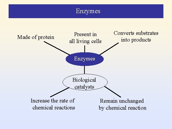
Enzymes Made of protein Present in all living cells Converts substrates into products Enzymes Biological catalysts Increase the rate of chemical reactions Remain unchanged by chemical reaction
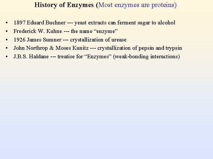
History of Enzymes (Most enzymes are proteins) • • • 1897 Eduard Buchner --- yeast extracts can ferment sugar to alcohol Frederick W. Kuhne --- the name “enzyme” 1926 James Sumner --- crystallization of urease John Northrop & Moses Kunitz --- crystallization of pepsin and trypsin J. B. S. Haldane --- treatise for “Enzymes” (weak-bonding interactions)
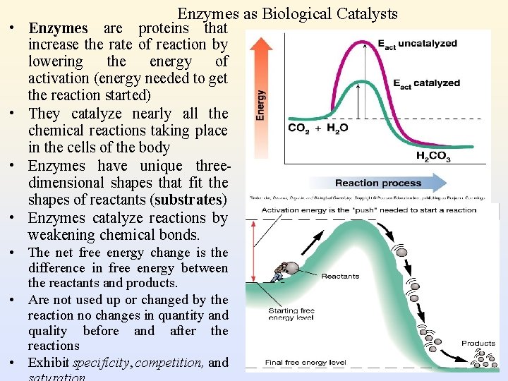
Enzymes as Biological Catalysts • Enzymes are proteins that increase the rate of reaction by lowering the energy of activation (energy needed to get the reaction started) • They catalyze nearly all the chemical reactions taking place in the cells of the body • Enzymes have unique threedimensional shapes that fit the shapes of reactants (substrates) • Enzymes catalyze reactions by weakening chemical bonds. • The net free energy change is the difference in free energy between the reactants and products. • Are not used up or changed by the reaction no changes in quantity and quality before and after the reactions • Exhibit specificity, competition, and
• Enzymes bind to their substrates and bring them together in the proper orientation for the reaction to begin. • The binding and aligning of the substrates lowers the activation energy of the reaction. • Enzyme-catalyzed reactions take place usually under relatively mild conditions. • Do not change the equilibrium points: only enhance the reaction rates. • Enzyme-catalyzed reactions have very high catalytic efficiency. • Enzymatic activities are highly regulated in response to the external changes satisfying the needs of biological processes. • Regulations can be accomplished through varying the enzyme quantity, adjusting the enzymatic activity or changing the substrate concentration • Unlike conventional catalysts, enzymes demonstrate the ability to distinguish different substrates. There are three types of substrate specificities, Absolute specificity Relative specificity Stereospecificity
Absolute specificity Enzymes can recognize only one type of substrate and implement their catalytic functions.
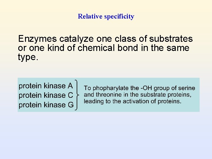
Relative specificity Enzymes catalyze one class of substrates or one kind of chemical bond in the same type.
Stereospecificity The enzyme can act on only one form of isomers of the substrates. Lactate dehydrogenase can recognize only the L -form but the D-form lactate.
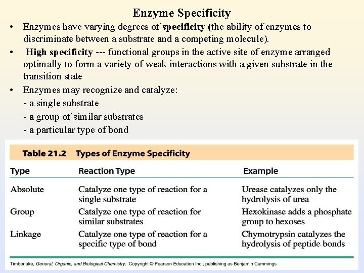
Enzyme Specificity • Enzymes have varying degrees of specificity (the ability of enzymes to discriminate between a substrate and a competing molecule). • High specificity --- functional groups in the active site of enzyme arranged optimally to form a variety of weak interactions with a given substrate in the transition state • Enzymes may recognize and catalyze: - a single substrate - a group of similar substrates - a particular type of bond
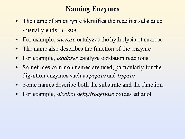
Naming Enzymes • The name of an enzyme identifies the reacting substance - usually ends in –ase • For example, sucrase catalyzes the hydrolysis of sucrose • The name also describes the function of the enzyme • For example, oxidases catalyze oxidation reactions • Sometimes common names are used, particularly for the digestion enzymes such as pepsin and trypsin • Some names describe both the substrate and the function • For example, alcohol dehydrogenase oxides ethanol
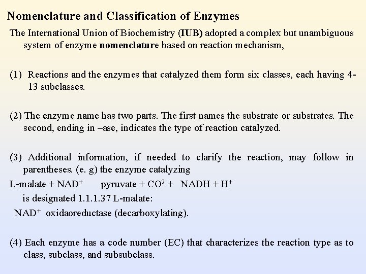
Nomenclature and Classification of Enzymes The International Union of Biochemistry (IUB) adopted a complex but unambiguous system of enzyme nomenclature based on reaction mechanism, (1) Reactions and the enzymes that catalyzed them form six classes, each having 413 subclasses. (2) The enzyme name has two parts. The first names the substrate or substrates. The second, ending in –ase, indicates the type of reaction catalyzed. (3) Additional information, if needed to clarify the reaction, may follow in parentheses. (e. g) the enzyme catalyzing L-malate + NAD+ pyruvate + CO 2 + NADH + H+ is designated 1. 1. 1. 37 L-malate: NAD+ oxidaoreductase (decarboxylating). (4) Each enzyme has a code number (EC) that characterizes the reaction type as to class, subclass, and subsubclass.
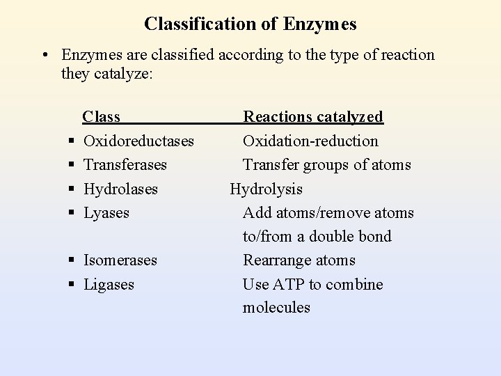
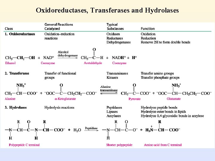
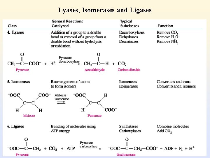
Classification of Enzymes • Enzymes are classified according to the type of reaction they catalyze: § § Class Oxidoreductases Transferases Hydrolases Lyases § Isomerases § Ligases Reactions catalyzed Oxidation-reduction Transfer groups of atoms Hydrolysis Add atoms/remove atoms to/from a double bond Rearrange atoms Use ATP to combine molecules
Oxidoreductases, Transferases and Hydrolases
Lyases, Isomerases and Ligases
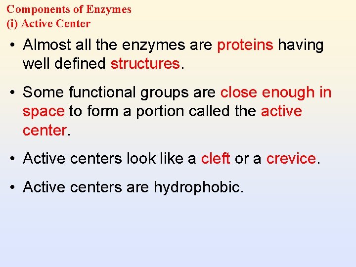
Components of Enzymes (i) Active Center • Almost all the enzymes are proteins having well defined structures. • Some functional groups are close enough in space to form a portion called the active center. • Active centers look like a cleft or a crevice. • Active centers are hydrophobic.
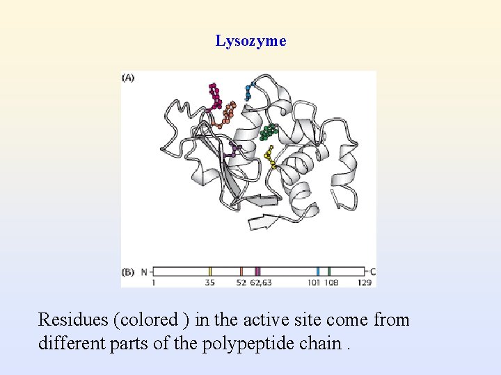
Lysozyme Residues (colored ) in the active site come from different parts of the polypeptide chain.
Two essential groups The active center has two essential groups in general. • Binding group: to associate with the reactants to form an enzyme-substrate complex • Catalytic group: to catalyze the reactions and convert substrates into products
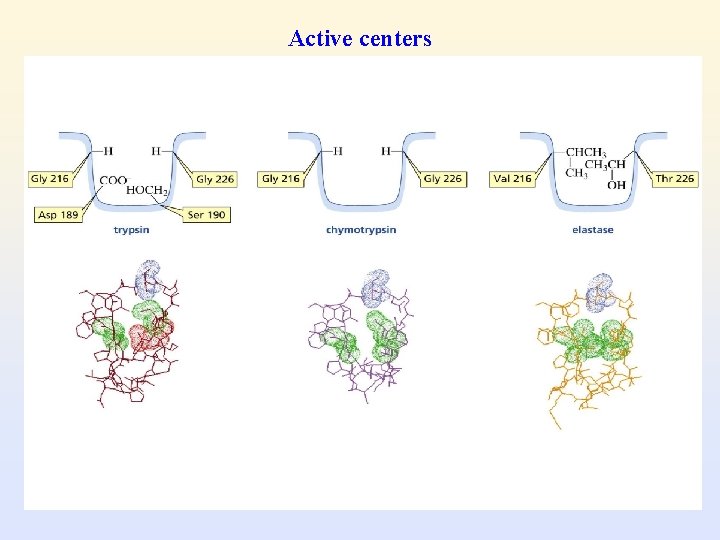
Active centers
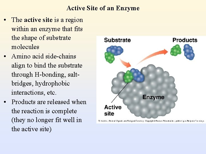
Active Site of an Enzyme • The active site is a region within an enzyme that fits the shape of substrate molecules • Amino acid side-chains align to bind the substrate through H-bonding, saltbridges, hydrophobic interactions, etc. • Products are released when the reaction is complete (they no longer fit well in the active site)
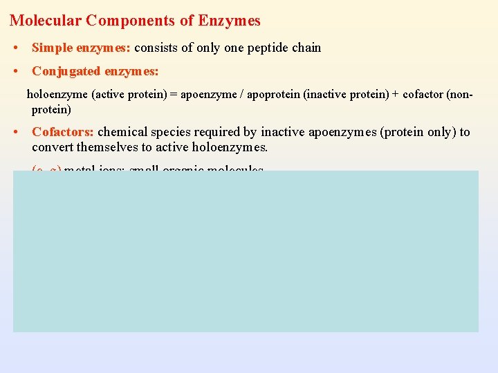
Molecular Components of Enzymes • Simple enzymes: consists of only one peptide chain • Conjugated enzymes: holoenzyme (active protein) = apoenzyme / apoprotein (inactive protein) + cofactor (nonprotein) • Cofactors: chemical species required by inactive apoenzymes (protein only) to convert themselves to active holoenzymes. (e. g) metal ions; small organic molecules
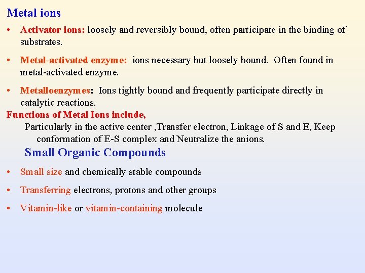
Metal ions • Activator ions: loosely and reversibly bound, often participate in the binding of substrates. • Metal-activated enzyme: ions necessary but loosely bound. Often found in metal-activated enzyme. • Metalloenzymes: Ions tightly bound and frequently participate directly in catalytic reactions. Functions of Metal Ions include, Particularly in the active center , Transfer electron, Linkage of S and E, Keep conformation of E-S complex and Neutralize the anions. Small Organic Compounds • Small size and chemically stable compounds • Transferring electrons, protons and other groups • Vitamin-like or vitamin-containing molecule
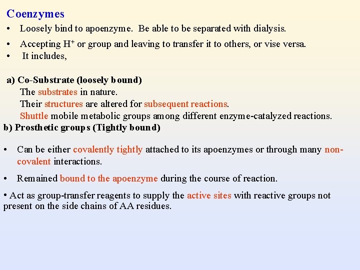
Coenzymes • Loosely bind to apoenzyme. Be able to be separated with dialysis. • Accepting H+ or group and leaving to transfer it to others, or vise versa. • It includes, a) Co-Substrate (loosely bound) The substrates in nature. Their structures are altered for subsequent reactions. Shuttle mobile metabolic groups among different enzyme-catalyzed reactions. b) Prosthetic groups (Tightly bound) • Can be either covalently tightly attached to its apoenzymes or through many noncovalent interactions. • Remained bound to the apoenzyme during the course of reaction. • Act as group-transfer reagents to supply the active sites with reactive groups not present on the side chains of AA residues.
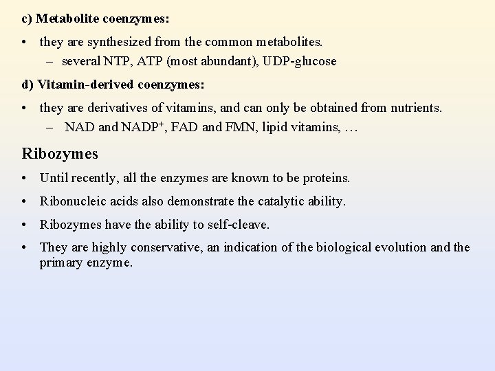
c) Metabolite coenzymes: • they are synthesized from the common metabolites. – several NTP, ATP (most abundant), UDP-glucose d) Vitamin-derived coenzymes: • they are derivatives of vitamins, and can only be obtained from nutrients. – NAD and NADP+, FAD and FMN, lipid vitamins, … Ribozymes • Until recently, all the enzymes are known to be proteins. • Ribonucleic acids also demonstrate the catalytic ability. • Ribozymes have the ability to self-cleave. • They are highly conservative, an indication of the biological evolution and the primary enzyme.
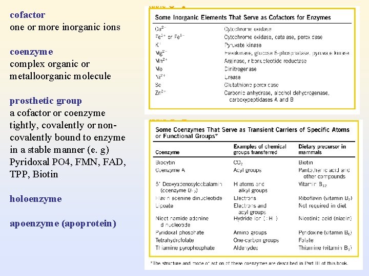
cofactor one or more inorganic ions coenzyme complex organic or metalloorganic molecule prosthetic group a cofactor or coenzyme tightly, covalently or noncovalently bound to enzyme in a stable manner (e. g) Pyridoxal PO 4, FMN, FAD, TPP, Biotin holoenzyme apoenzyme (apoprotein)
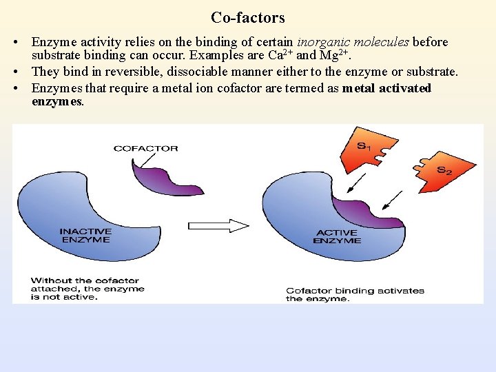
Co-factors • Enzyme activity relies on the binding of certain inorganic molecules before substrate binding can occur. Examples are Ca 2+ and Mg 2+. • They bind in reversible, dissociable manner either to the enzyme or substrate. • Enzymes that require a metal ion cofactor are termed as metal activated enzymes.
Co-enzymes (Recyclable substrate shuttles / group transfer agents) • Coenzymes are organic molecules. • Do not alter the enzyme’s binding site. • Accept electrons from an enzyme in one metabolic pathway and transfer them to an enzyme in another. • They are not needed in large amounts. • It also stabilizes substrates such as hydrogen / hydride ions that are unstable in the aqueous environment of the cell. • The water soluble vitamins (thiamine (B 1) niacin, riboflavin (B 2)) are all good examples of cofactors. – NAD (nicotinamide adenine dinucleotide) – FAD (flavine adenine dinucleotide)
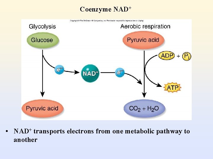
Coenzyme NAD+ • NAD+ transports electrons from one metabolic pathway to another
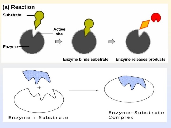
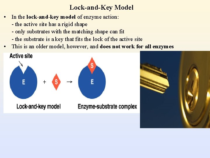
Lock-and-Key Model • In the lock-and-key model of enzyme action: - the active site has a rigid shape - only substrates with the matching shape can fit - the substrate is a key that fits the lock of the active site • This is an older model, however, and does not work for all enzymes
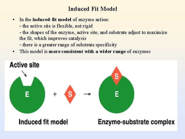
Induced Fit Model • In the induced-fit model of enzyme action: - the active site is flexible, not rigid - the shapes of the enzyme, active site, and substrate adjust to maximize the fit, which improves catalysis - there is a greater range of substrate specificity • This model is more consistent with a wider range of enzymes
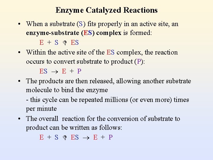
Enzyme Catalyzed Reactions • When a substrate (S) fits properly in an active site, an enzyme-substrate (ES) complex is formed: E + S ES • Within the active site of the ES complex, the reaction occurs to convert substrate to product (P): ES E + P • The products are then released, allowing another substrate molecule to bind the enzyme - this cycle can be repeated millions (or even more) times per minute • The overall reaction for the conversion of substrate to product can be written as follows: E + S E + P
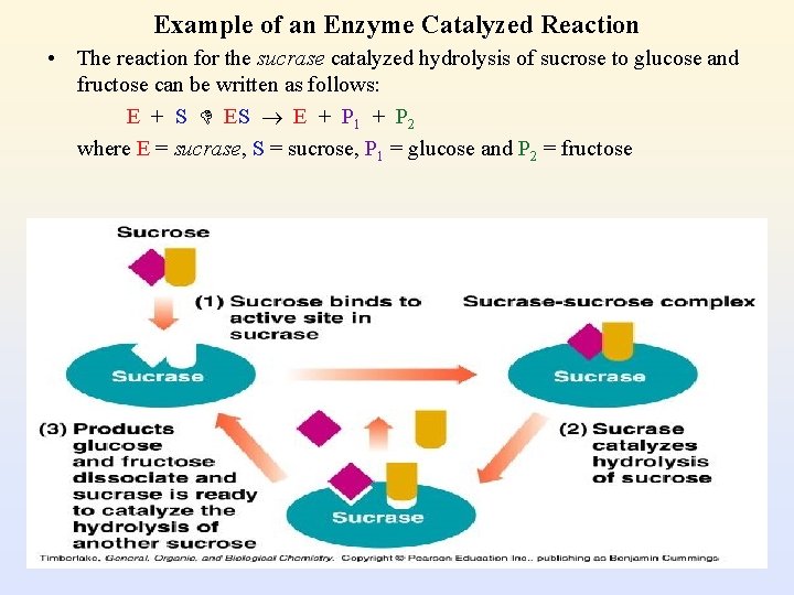
Example of an Enzyme Catalyzed Reaction • The reaction for the sucrase catalyzed hydrolysis of sucrose to glucose and fructose can be written as follows: E + S E + P 1 + P 2 where E = sucrase, S = sucrose, P 1 = glucose and P 2 = fructose
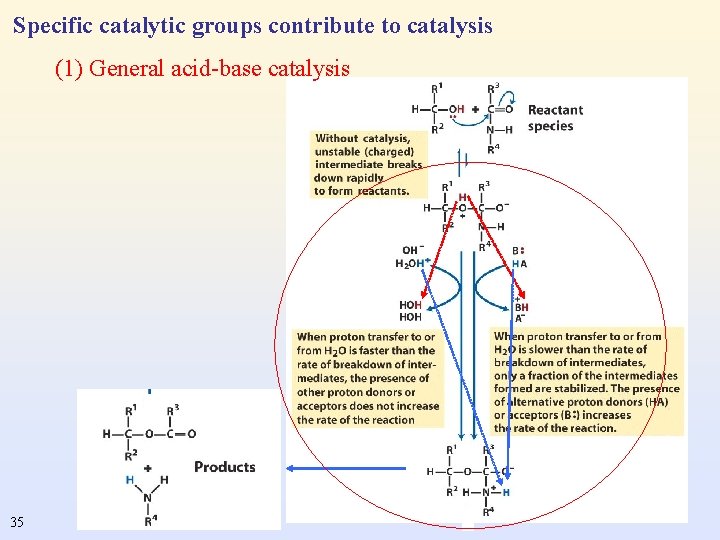
Specific catalytic groups contribute to catalysis (1) General acid-base catalysis 35
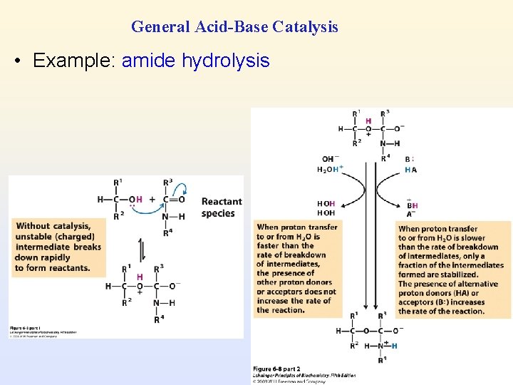
General Acid-Base Catalysis • Example: amide hydrolysis
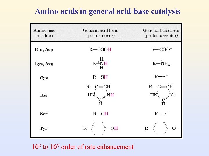
Amino acids in general acid-base catalysis 102 to 105 order of rate enhancement
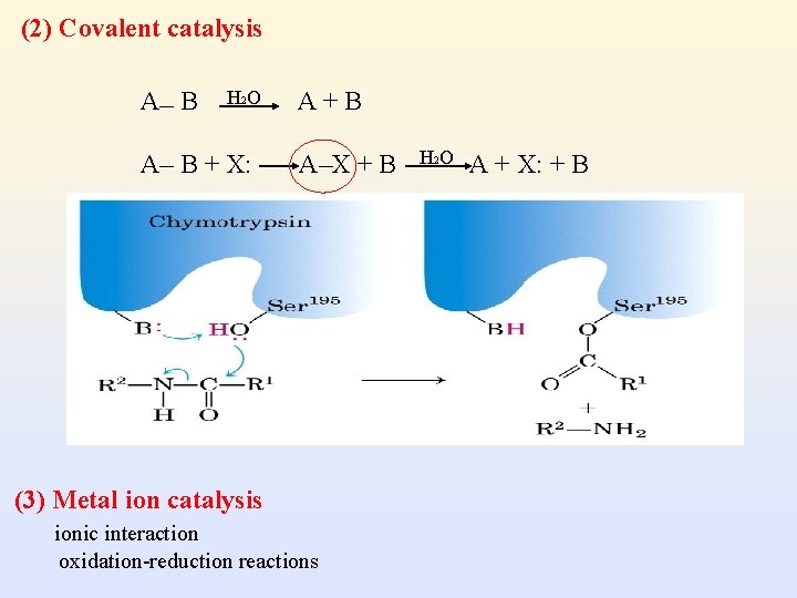
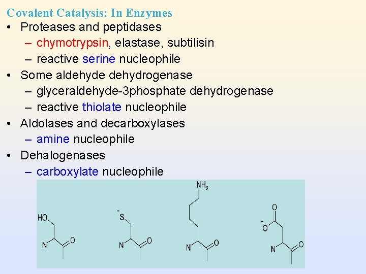
(2) Covalent catalysis A B H 2 O A B + X: A+B A X+B (3) Metal ion catalysis ionic interaction oxidation-reduction reactions H 2 O A + X: + B
Covalent Catalysis: In Enzymes • Proteases and peptidases – chymotrypsin, elastase, subtilisin – reactive serine nucleophile • Some aldehyde dehydrogenase – glyceraldehyde-3 phosphate dehydrogenase – reactive thiolate nucleophile • Aldolases and decarboxylases – amine nucleophile • Dehalogenases – carboxylate nucleophile
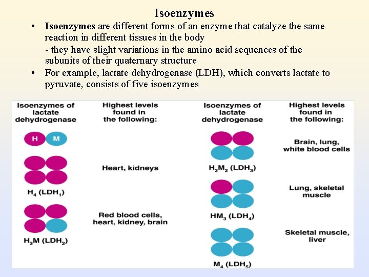
Isoenzymes • Isoenzymes are different forms of an enzyme that catalyze the same reaction in different tissues in the body - they have slight variations in the amino acid sequences of the subunits of their quaternary structure • For example, lactate dehydrogenase (LDH), which converts lactate to pyruvate, consists of five isoenzymes
• Due to gene differentiation: the different gene products or different peptides of the same gene • Present in different tissues of the same system, or subcellular components of the same cell. • Synthesized from different genes (malate dehydrogenase in cytosol versus in mitochondria) • Oligomeric forms of more than one type of subunits (lactate dehydrogenase) • Different carbohydrate content (alkaline phosphatase) (e. g) Creatine Phosphokinase 3 isoenzymes, BB, BM, MM • Dimeric form: M (muscle) or B (brain) • CPK 2 is undetectable (<2%) in serum for healthy individuals, and elevated to 20% in the first 6 -18 hrs after myocardial infarction. • Used as a earliest reliable indicator of myocardial infarction.
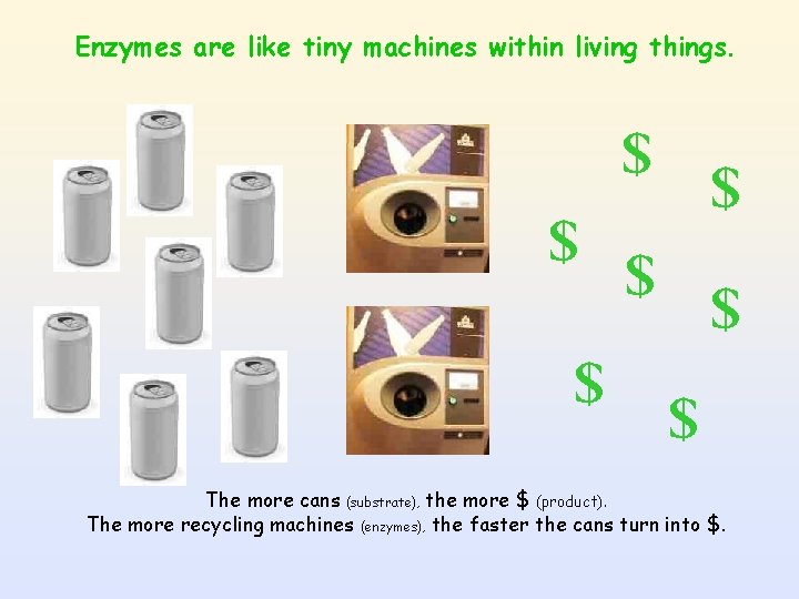
Enzymes are like tiny machines within living things. $ $ $ $ The more cans (substrate), the more $ (product). The more recycling machines (enzymes), the faster the cans turn into $.
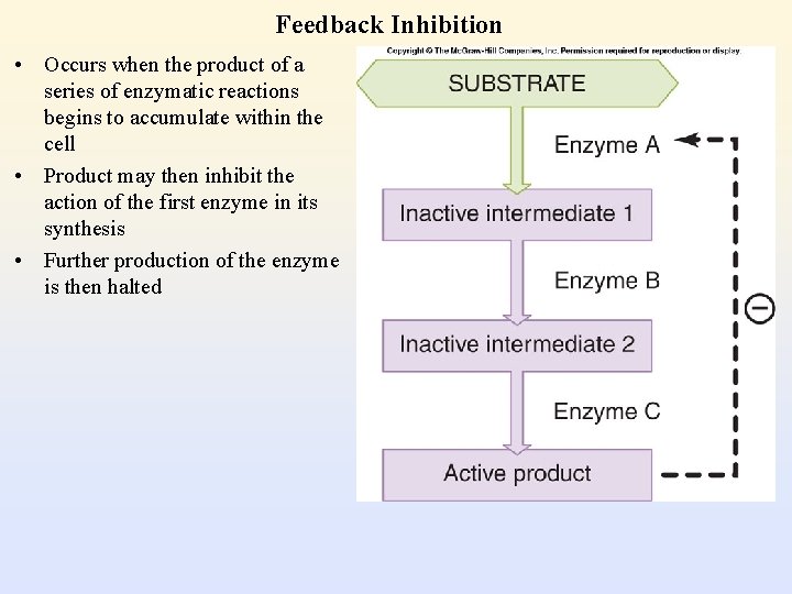
Feedback Inhibition • Occurs when the product of a series of enzymatic reactions begins to accumulate within the cell • Product may then inhibit the action of the first enzyme in its synthesis • Further production of the enzyme is then halted
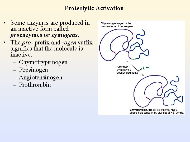
Proteolytic Activation • Some enzymes are produced in an inactive form called proenzymes or zymogens. • The pro- prefix and -ogen suffix signifies that the molecule is inactive. – Chymotrypsinogen – Pepsinogen – Angiotensinogen – Prothrombin
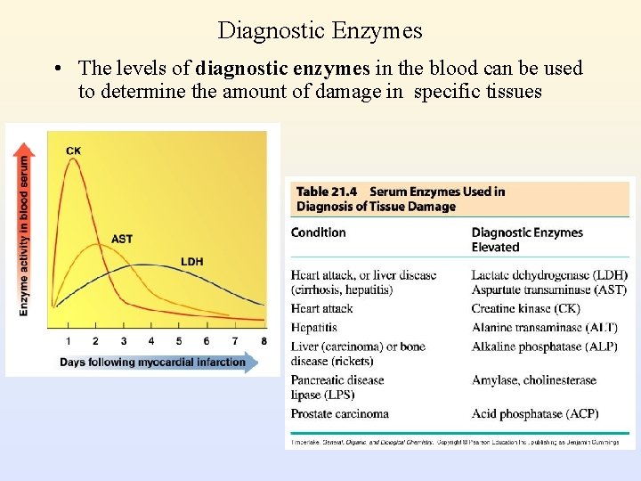
Diagnostic Enzymes • The levels of diagnostic enzymes in the blood can be used to determine the amount of damage in specific tissues
Factors affecting enzyme-catalyzed reaction • Substrate concentration • Enzyme concentration • Temperature • p. H • Inhibitors • Activators
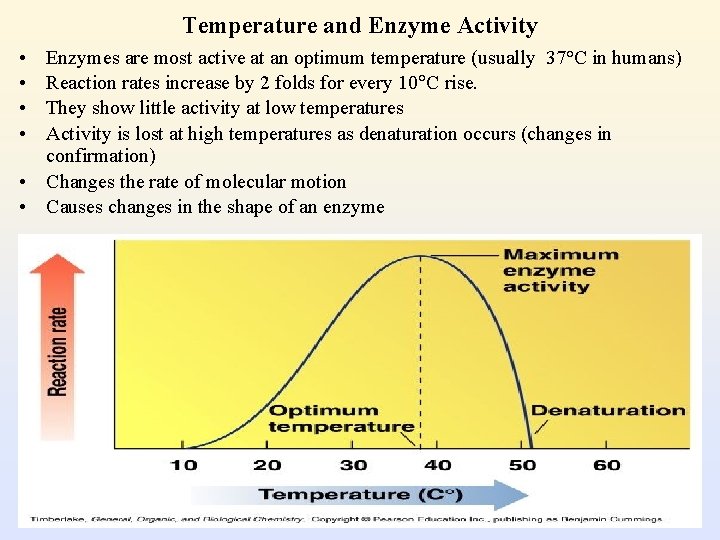
Temperature and Enzyme Activity • • Enzymes are most active at an optimum temperature (usually 37°C in humans) Reaction rates increase by 2 folds for every 10 C rise. They show little activity at low temperatures Activity is lost at high temperatures as denaturation occurs (changes in confirmation) • Changes the rate of molecular motion • Causes changes in the shape of an enzyme
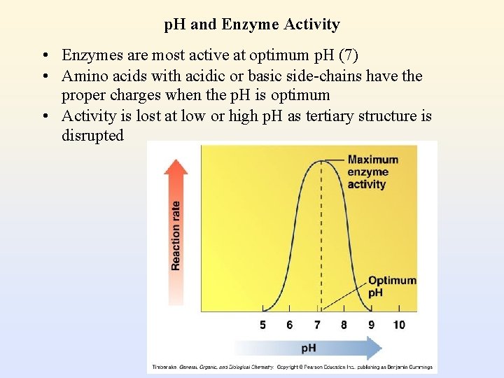
p. H and Enzyme Activity • Enzymes are most active at optimum p. H (7) • Amino acids with acidic or basic side-chains have the proper charges when the p. H is optimum • Activity is lost at low or high p. H as tertiary structure is disrupted
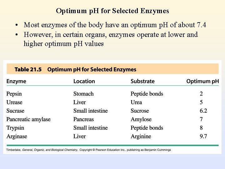
Optimum p. H for Selected Enzymes • Most enzymes of the body have an optimum p. H of about 7. 4 • However, in certain organs, enzymes operate at lower and higher optimum p. H values
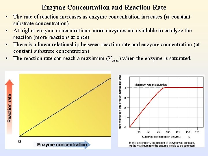
Enzyme Concentration and Reaction Rate • The rate of reaction increases as enzyme concentration increases (at constant substrate concentration) • At higher enzyme concentrations, more enzymes are available to catalyze the reaction (more reactions at once) • There is a linear relationship between reaction rate and enzyme concentration (at constant substrate concentration) • The reaction rate can reach a maximum (Vmax) when the enzyme is saturated.
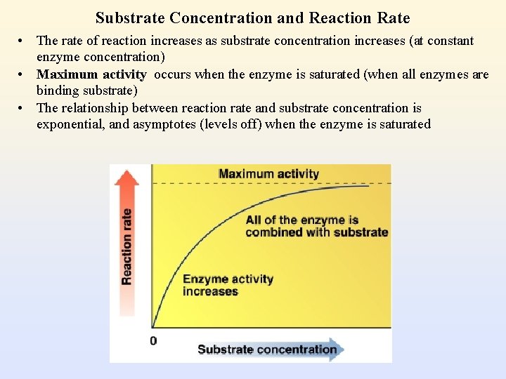
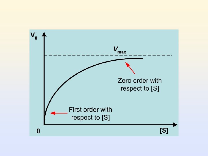
Substrate Concentration and Reaction Rate • The rate of reaction increases as substrate concentration increases (at constant enzyme concentration) • Maximum activity occurs when the enzyme is saturated (when all enzymes are binding substrate) • The relationship between reaction rate and substrate concentration is exponential, and asymptotes (levels off) when the enzyme is saturated
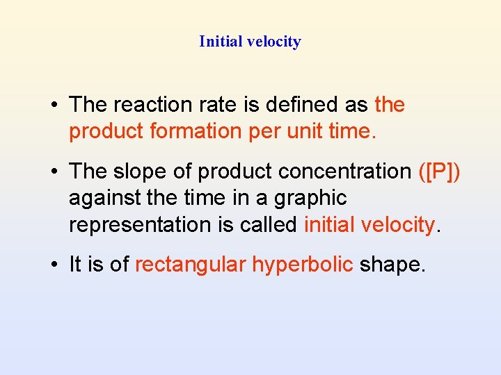
Initial velocity • The reaction rate is defined as the product formation per unit time. • The slope of product concentration ([P]) against the time in a graphic representation is called initial velocity. • It is of rectangular hyperbolic shape.
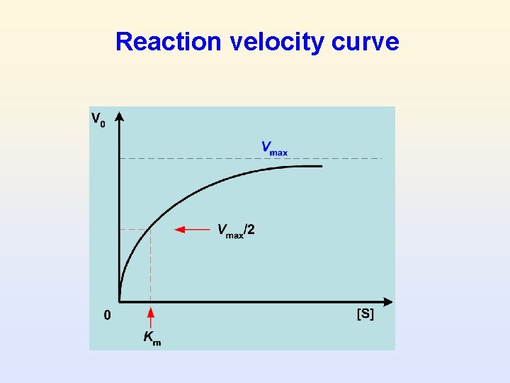
Reaction velocity curve
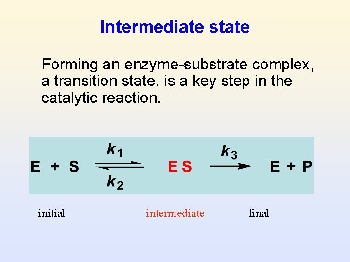
Intermediate state Forming an enzyme-substrate complex, a transition state, is a key step in the catalytic reaction. initial intermediate final
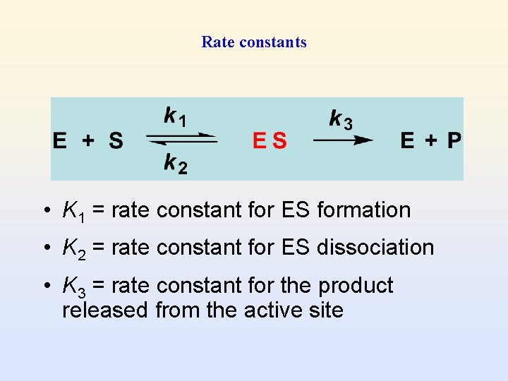
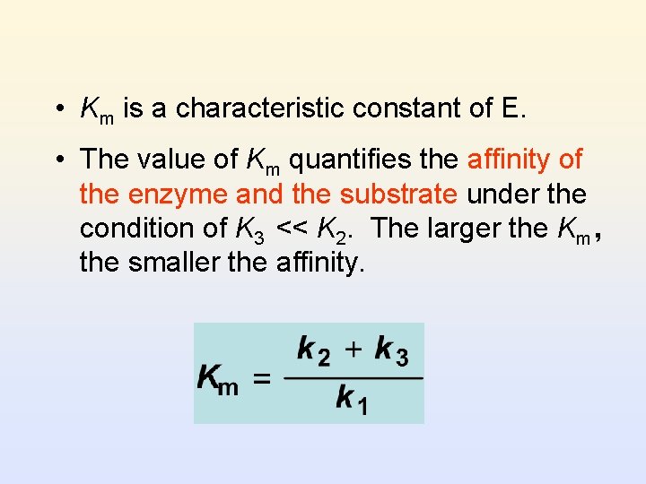
Rate constants • K 1 = rate constant for ES formation • K 2 = rate constant for ES dissociation • K 3 = rate constant for the product released from the active site
Michaelis-Menten Equation • The mathematical expression of the product formation with respect to the experimental parameters • Michaelis-Menten equation describes the relationship between the reaction rate and substrate concentration [S].
Assumptions • [S] >> [E], changes of [S] is negligible. • K 2 is negligible compared with K 1. • Steady-state: the rate of E-S complex formation is equal to the rate of its disassociation (backward E + S and forward to E + P)
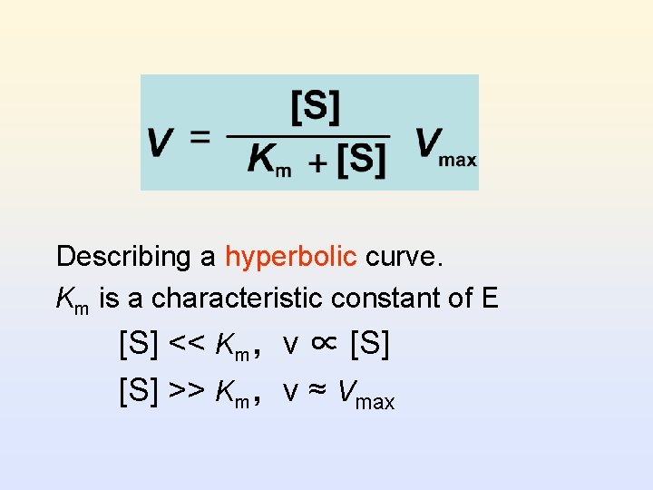
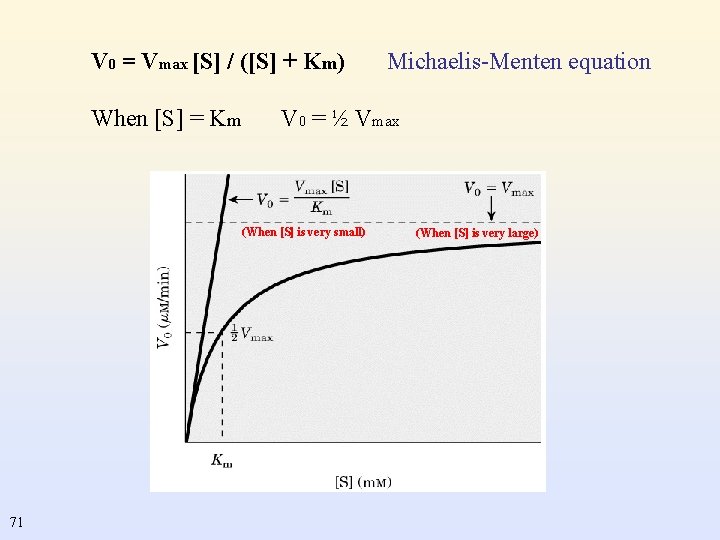
Describing a hyperbolic curve. Km is a characteristic constant of E [S] << Km,v ∝ [S] >> Km,v ≈ Vmax
Significance of Km • the substrate concentration at which enzyme-catalyzed reaction proceeds at one-half of its maximum velocity • Km is independent of [E]. It is determined by the structure of E, the substrate and environmental conditions (p. H, T, ionic strength, …)
• Km is a characteristic constant of E. • The value of Km quantifies the affinity of the enzyme and the substrate under the condition of K 3 << K 2. The larger the Km, the smaller the affinity.
Significance of Vmax • The reaction velocity of an enzymatic reaction when the binding sites of E are saturated with substrates. • It is proportional to [E].
Turnover number k 3 = Vmax / [E] • Vmax is the reaction rate when the enzymes are saturated, and is independent of the enzyme concentration. • The number of the products converted in a unit time by one enzyme molecule which is saturated.
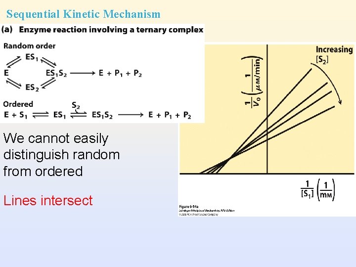
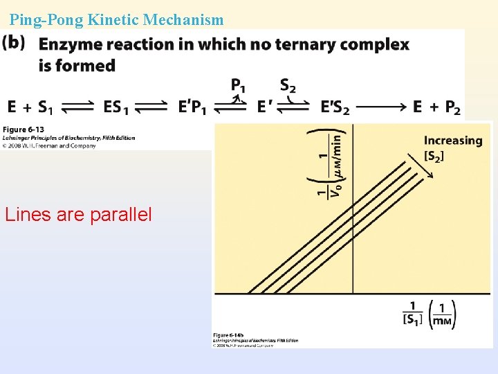
Two-substrate Reactions • Kinetic mechanism: the order of binding of substrates and release of products • When two or more reactants are involved, enzyme kinetics allows to distinguish between different kinetic mechanisms – – Sequential mechanism Ping-Pong mechanism
Sequential Kinetic Mechanism We cannot easily distinguish random from ordered Lines intersect
Ping-Pong Kinetic Mechanism Lines are parallel
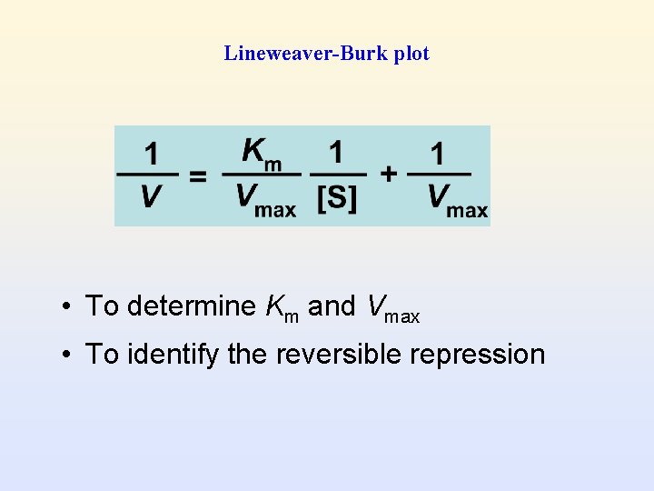
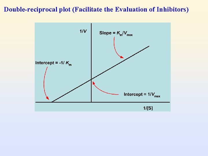
Lineweaver-Burk plot • To determine Km and Vmax • To identify the reversible repression
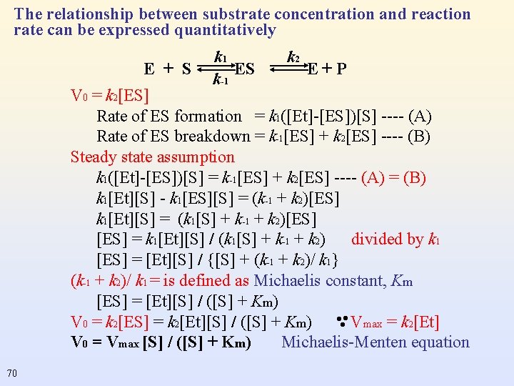
The relationship between substrate concentration and reaction rate can be expressed quantitatively E + S k 1 ES k-1 k 2 E+P V 0 = k 2[ES] Rate of ES formation = k 1([Et]-[ES])[S] ---- (A) Rate of ES breakdown = k-1[ES] + k 2[ES] ---- (B) Steady state assumption k 1([Et]-[ES])[S] = k-1[ES] + k 2[ES] ---- (A) = (B) k 1[Et][S] - k 1[ES][S] = (k-1 + k 2)[ES] k 1[Et][S] = (k 1[S] + k-1 + k 2)[ES] = k 1[Et][S] / (k 1[S] + k-1 + k 2) divided by k 1 [ES] = [Et][S] / {[S] + (k-1 + k 2)/ k 1} (k-1 + k 2)/ k 1 = is defined as Michaelis constant, Km [ES] = [Et][S] / ([S] + Km) V 0 = k 2[ES] = k 2[Et][S] / ([S] + Km) Vmax = k 2[Et] V 0 = Vmax [S] / ([S] + Km) Michaelis-Menten equation . . . 70
V 0 = Vmax [S] / ([S] + Km) When [S] = Km V 0 = ½ Vmax (When [S] is very small) 71 Michaelis-Menten equation (When [S] is very large)
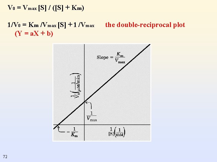
V 0 = Vmax [S] / ([S] + Km) 1/V 0 = Km /Vmax [S] + 1 /Vmax (Y = a. X + b) 72 the double-reciprocal plot
Double-reciprocal plot (Facilitate the Evaluation of Inhibitors)
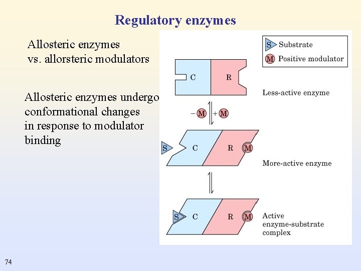
Regulatory enzymes Allosteric enzymes vs. allorsteric modulators Allosteric enzymes undergo conformational changes in response to modulator binding 74
Enzyme Inhibitors • Inhibitors (I) are molecules that cause a loss of enzyme activity. • Inhibitors can be normal body metabolites and foreign substances (drugs and toxins). • They prevent substrates from fitting into the active site of the enzyme: E + S E + P E + I EI no P formed
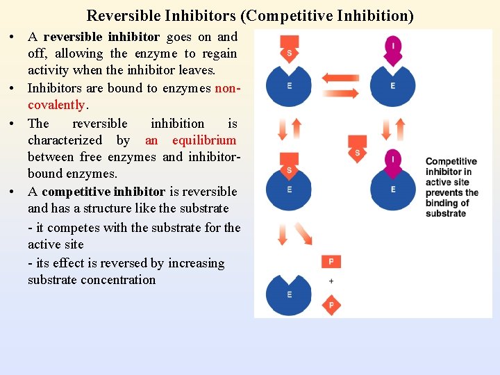
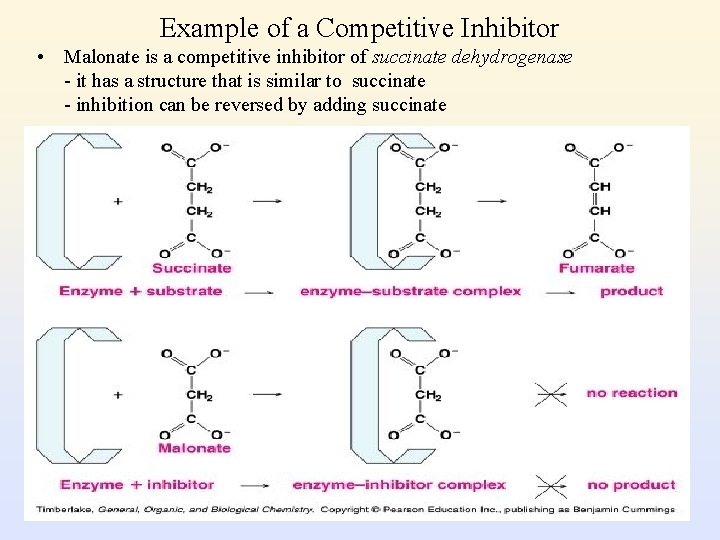
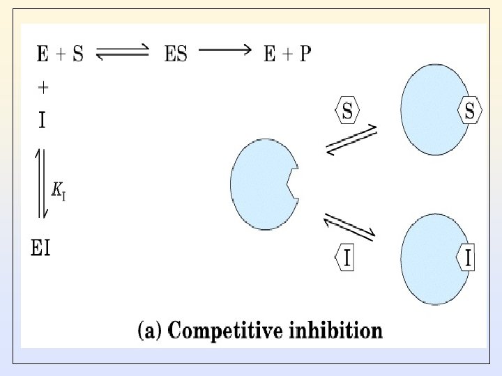
Reversible Inhibitors (Competitive Inhibition) • A reversible inhibitor goes on and off, allowing the enzyme to regain activity when the inhibitor leaves. • Inhibitors are bound to enzymes noncovalently. • The reversible inhibition is characterized by an equilibrium between free enzymes and inhibitorbound enzymes. • A competitive inhibitor is reversible and has a structure like the substrate - it competes with the substrate for the active site - its effect is reversed by increasing substrate concentration
Example of a Competitive Inhibitor • Malonate is a competitive inhibitor of succinate dehydrogenase - it has a structure that is similar to succinate - inhibition can be reversed by adding succinate
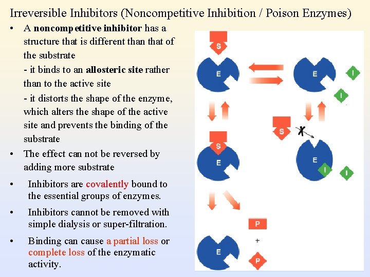
Irreversible Inhibitors (Noncompetitive Inhibition / Poison Enzymes) • A noncompetitive inhibitor has a structure that is different than that of the substrate - it binds to an allosteric site rather than to the active site - it distorts the shape of the enzyme, which alters the shape of the active site and prevents the binding of the substrate • The effect can not be reversed by adding more substrate • Inhibitors are covalently bound to the essential groups of enzymes. • Inhibitors cannot be removed with simple dialysis or super-filtration. • Binding can cause a partial loss or complete loss of the enzymatic activity.
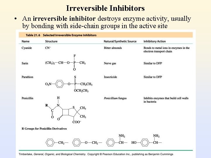
Irreversible Inhibitors • An irreversible inhibitor destroys enzyme activity, usually by bonding with side-chain groups in the active site
Competitive Inhibition
Non-Competitive Inhibition
Non-Competitive Inhibition
Un-Competitive Inhibition
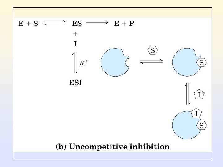
• Uncompetitive inhibitors bind only to the enzyme-substrate complexes. • The E-I-S complexes do not proceed to form products. • The E-I-S complexes do not backward to the substrates and enzymes. • This inhibition has the effects on reducing both Vmax and Km. • Commonly in the multiple substrate reactions.
Suicide Inactivator (Mechanism based inactivator) • These compounds are relatively unreactive until they bind to the active site of a specific enzyme. • Undergoes the first few chemical steps of the normal enzymatic reaction, but instead of being transformed into normal product, the inactivator is converted to a very reactive compound that combines irreversibly with the enzyme.
Activators • Activators are the compounds which bind to an enzyme or an enzyme-substrate complex to enhance the enzymatic activity without being modified by the enzymes. (e. g) Metal ions essential activators: no enzymatic activity without it Mg 2+ of hexokinase non-essential activators: enhancing the catalytic power. • Enzymatic activity is a measure of the capability of an enzyme of catalyzing a chemical reaction. • It directly affects the reaction rate. • International unit (IU): the amount of enzyme required to convert 1 µmol of substrate to product per minute under a designated condition. • Determination of the enzymatic activity requires proper treatment of enzymes, excess amount of substrate, optimal T and p. H, … • One katal is the amount of enzyme that converts 1 mol of substrate per second. • IU = 16. 67× 10 -9 kat
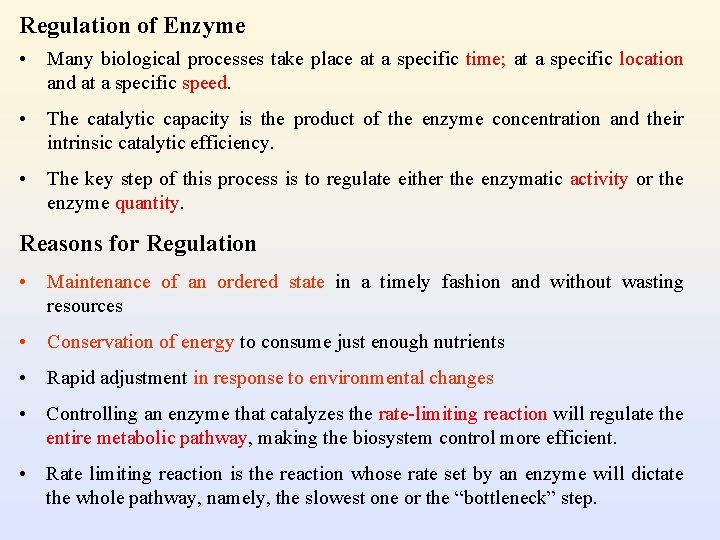
Regulation of Enzyme • Many biological processes take place at a specific time; at a specific location and at a specific speed. • The catalytic capacity is the product of the enzyme concentration and their intrinsic catalytic efficiency. • The key step of this process is to regulate either the enzymatic activity or the enzyme quantity. Reasons for Regulation • Maintenance of an ordered state in a timely fashion and without wasting resources • Conservation of energy to consume just enough nutrients • Rapid adjustment in response to environmental changes • Controlling an enzyme that catalyzes the rate-limiting reaction will regulate the entire metabolic pathway, making the biosystem control more efficient. • Rate limiting reaction is the reaction whose rate set by an enzyme will dictate the whole pathway, namely, the slowest one or the “bottleneck” step.
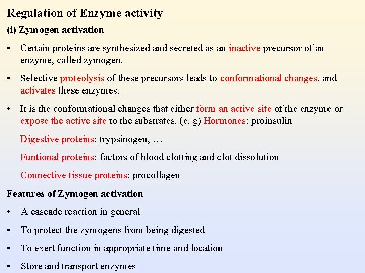
Regulation of Enzyme activity (i) Zymogen activation • Certain proteins are synthesized and secreted as an inactive precursor of an enzyme, called zymogen. • Selective proteolysis of these precursors leads to conformational changes, and activates these enzymes. • It is the conformational changes that either form an active site of the enzyme or expose the active site to the substrates. (e. g) Hormones: proinsulin Digestive proteins: trypsinogen, … Funtional proteins: factors of blood clotting and clot dissolution Connective tissue proteins: procollagen Features of Zymogen activation • A cascade reaction in general • To protect the zymogens from being digested • To exert function in appropriate time and location • Store and transport enzymes
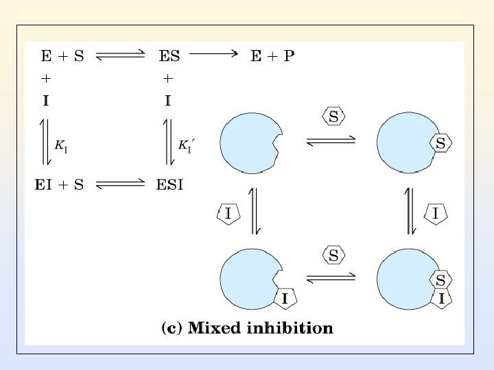
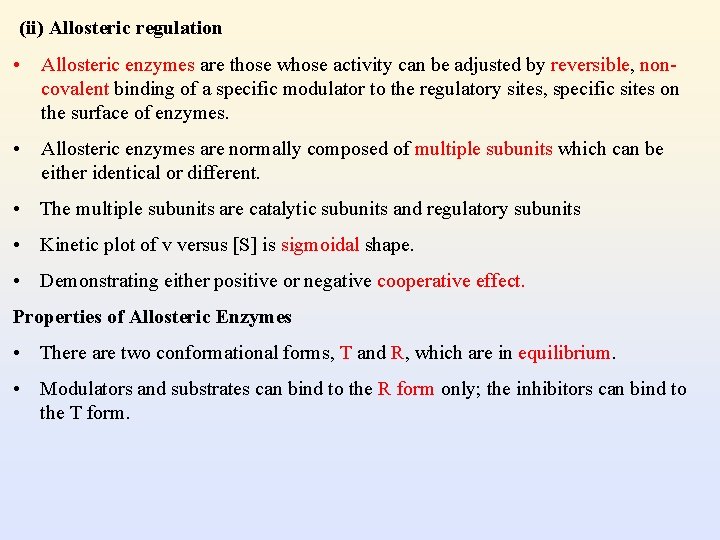
(ii) Allosteric regulation • Allosteric enzymes are those whose activity can be adjusted by reversible, noncovalent binding of a specific modulator to the regulatory sites, specific sites on the surface of enzymes. • Allosteric enzymes are normally composed of multiple subunits which can be either identical or different. • The multiple subunits are catalytic subunits and regulatory subunits • Kinetic plot of v versus [S] is sigmoidal shape. • Demonstrating either positive or negative cooperative effect. Properties of Allosteric Enzymes • There are two conformational forms, T and R, which are in equilibrium. • Modulators and substrates can bind to the R form only; the inhibitors can bind to the T form.
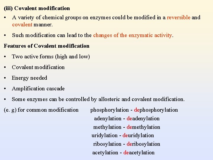
(iii) Covalent modification • A variety of chemical groups on enzymes could be modified in a reversible and covalent manner. • Such modification can lead to the changes of the enzymatic activity. Features of Covalent modification • Two active forms (high and low) • Covalent modification • Energy needed • Amplification cascade • Some enzymes can be controlled by allosteric and covalent modification. (e. g) for common modification phosphorylation - dephosphorylation adenylation - deadenylation methylation - demethylation uridylation - deuridylation ribosylation - deribosylation acetylation - deacetylation
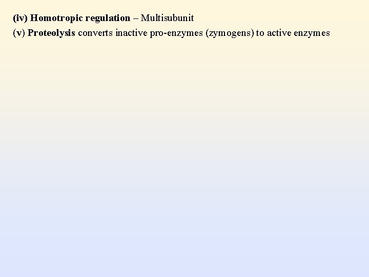
(iv) Homotropic regulation – Multisubunit (v) Proteolysis converts inactive pro-enzymes (zymogens) to active enzymes
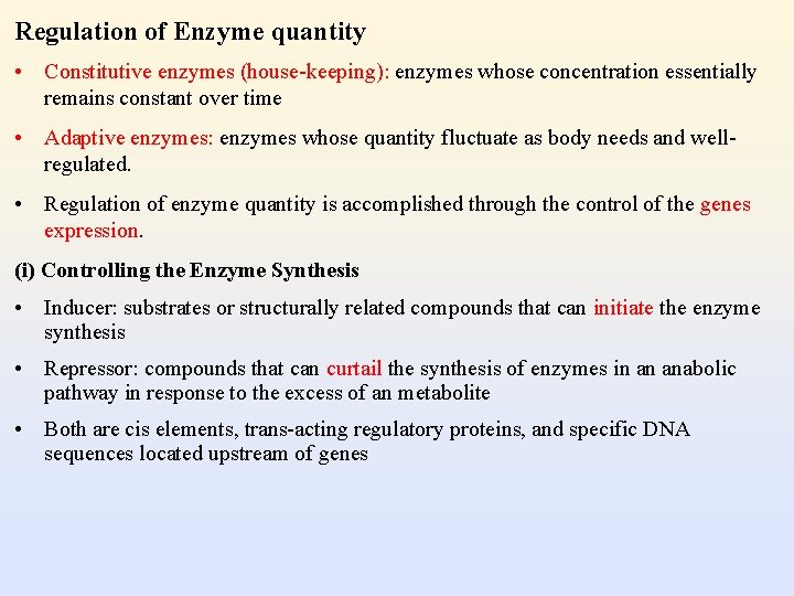
Regulation of Enzyme quantity • Constitutive enzymes (house-keeping): enzymes whose concentration essentially remains constant over time • Adaptive enzymes: enzymes whose quantity fluctuate as body needs and wellregulated. • Regulation of enzyme quantity is accomplished through the control of the genes expression. (i) Controlling the Enzyme Synthesis • Inducer: substrates or structurally related compounds that can initiate the enzyme synthesis • Repressor: compounds that can curtail the synthesis of enzymes in an anabolic pathway in response to the excess of an metabolite • Both are cis elements, trans-acting regulatory proteins, and specific DNA sequences located upstream of genes
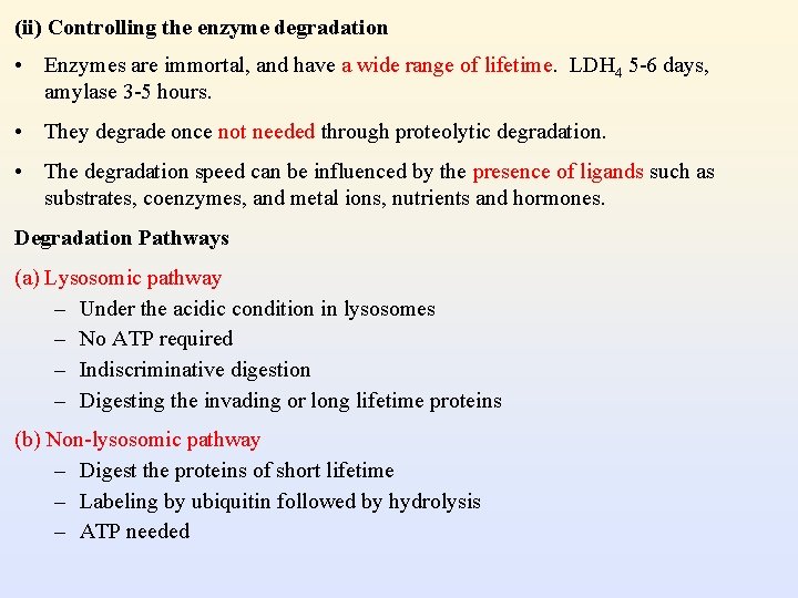
(ii) Controlling the enzyme degradation • Enzymes are immortal, and have a wide range of lifetime. LDH 4 5 -6 days, amylase 3 -5 hours. • They degrade once not needed through proteolytic degradation. • The degradation speed can be influenced by the presence of ligands such as substrates, coenzymes, and metal ions, nutrients and hormones. Degradation Pathways (a) Lysosomic pathway – Under the acidic condition in lysosomes – No ATP required – Indiscriminative digestion – Digesting the invading or long lifetime proteins (b) Non-lysosomic pathway – Digest the proteins of short lifetime – Labeling by ubiquitin followed by hydrolysis – ATP needed
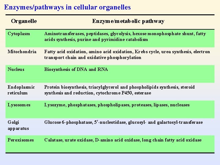
Enzymes/pathways in cellular organelles Organelle Enzyme/metabolic pathway Cytoplasm Aminotransferases, peptidases, glycolysis, hexose monophosphate shunt, fatty acids synthesis, purine and pyrimidine catabolism Mitochondria Fatty acid oxidation, amino acid oxidation, Krebs cycle, urea synthesis, electron transport chain and oxidative phosphorylation Nucleus Biosynthesis of DNA and RNA Endoplasmic reticulum Protein biosynthesis, triacylglycerol and phospholipids synthesis, steroid synthesis and reduction, cytochrome P 450, esterase Lysosomes Lysozyme, phosphatases, phospholipases, proteases, lipases, nucleases Golgi apparatus Glucose 6 -phosphatase, 5’-nucleotidase, glucosyl- and galactosyl-transferase Peroxisomes Calatase, urate oxidase, D-amino acid oxidase, long chain fatty acid oxidase
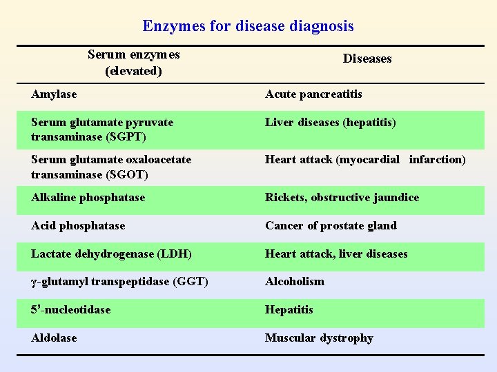
Enzymes for disease diagnosis Serum enzymes (elevated) Diseases Amylase Acute pancreatitis Serum glutamate pyruvate transaminase (SGPT) Liver diseases (hepatitis) Serum glutamate oxaloacetate transaminase (SGOT) Heart attack (myocardial infarction) Alkaline phosphatase Rickets, obstructive jaundice Acid phosphatase Cancer of prostate gland Lactate dehydrogenase (LDH) Heart attack, liver diseases γ-glutamyl transpeptidase (GGT) Alcoholism 5’-nucleotidase Hepatitis Aldolase Muscular dystrophy
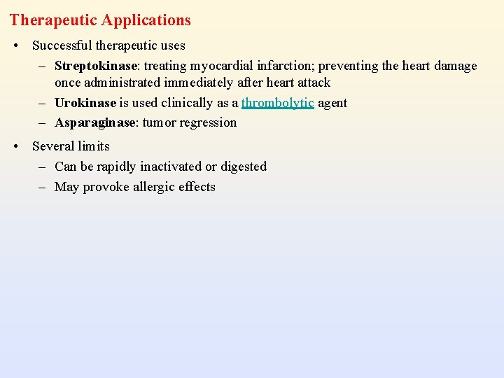
Therapeutic Applications • Successful therapeutic uses – Streptokinase: treating myocardial infarction; preventing the heart damage once administrated immediately after heart attack – Urokinase is used clinically as a thrombolytic agent – Asparaginase: tumor regression • Several limits – Can be rapidly inactivated or digested – May provoke allergic effects
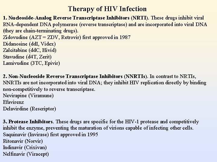
Therapy of HIV Infection 1. Nucleoside-Analog Reverse Transcriptase Inhibitors (NRTI). These drugs inhibit viral RNA-dependent DNA polymerase (reverse transcriptase) and are incorporated into viral DNA (they are chain-terminating drugs). Zidovudine (AZT = ZDV, Retrovir) first approved in 1987 Didanosine (dd. I, Videx) Zalcitabine (dd. C, Hivid) Stavudine (d 4 T, Zerit) Lamivudine (3 TC, Epivir) 2. Non-Nucleoside Reverse Transcriptase Inhibitors (NNRTIs). In contrast to NRTIs, NNRTIs are not incorporated into viral DNA; they inhibit HIV replication directly by binding non-competitively to reverse transcriptase. Nevirapine (Viramune) Efavirenz Delavirdine (Rescriptor) 3. Protease Inhibitors. These drugs are specific for the HIV-1 protease and competitively inhibit the enzyme, preventing the maturation of virions capable of infecting other cells. Saquinavir (Invirase) first approved in 1995 Ritonavir (Norvir) Indinavir (Crixivan) Nelfinavir (Viracept)