Environmental diseases Anthracotic pigment ordinarily is not fibrogenic

Environmental diseases
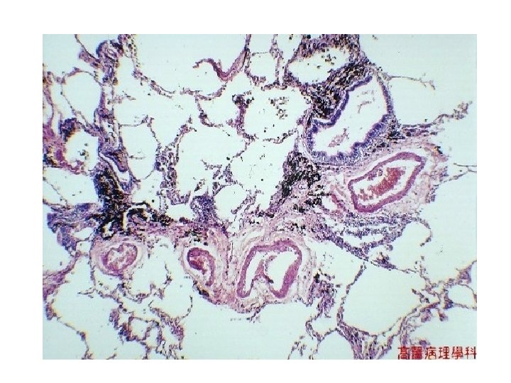

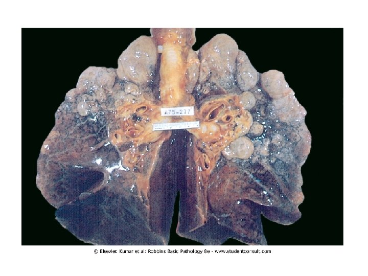
• Anthracotic pigment ordinarily is not fibrogenic, but in massive amounts (as in "black lung disease" in coal miners) a fibrogenic response can be elicited to produce the "coal worker's pneumoconiosis" seen here.

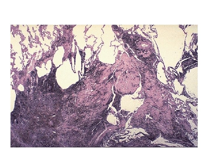
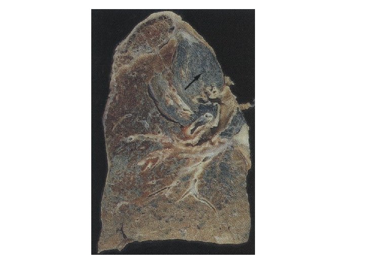
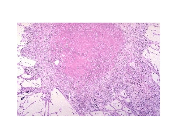
A silicotic nodule within lung parenchyma

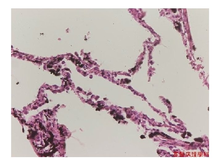
a long, thin asbestos fiber

The asbestos fiber becomes coated with iron and calcium, which is why it is often referred to as a "ferruginous body" as seen here with an iron stain

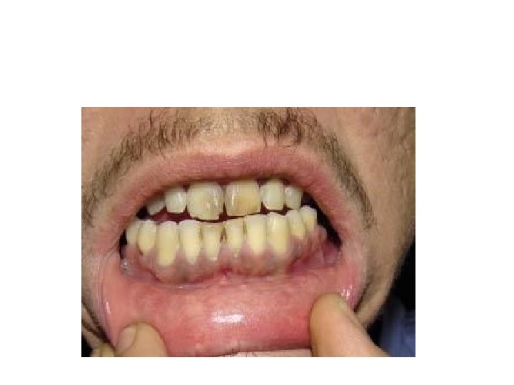
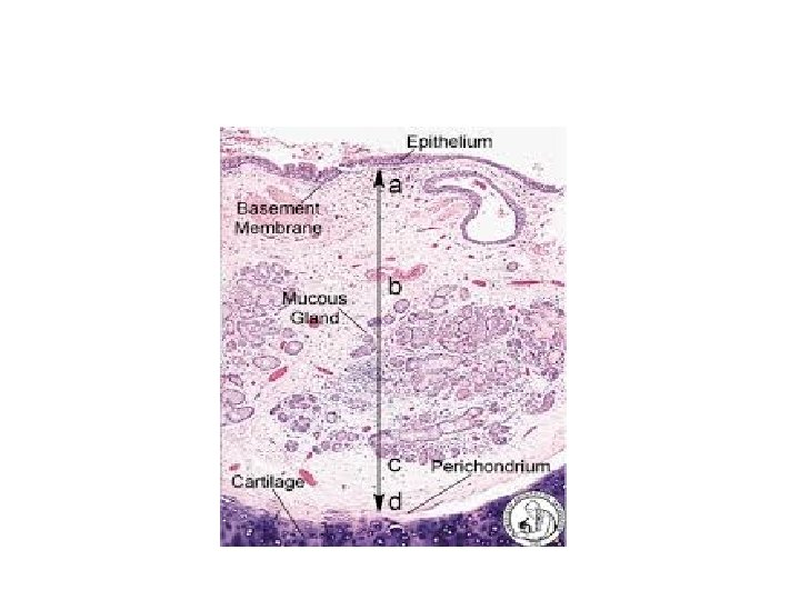
lead line in the gingiva


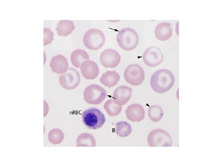
Coarse basophilic stippling
. • Lead inhibits")
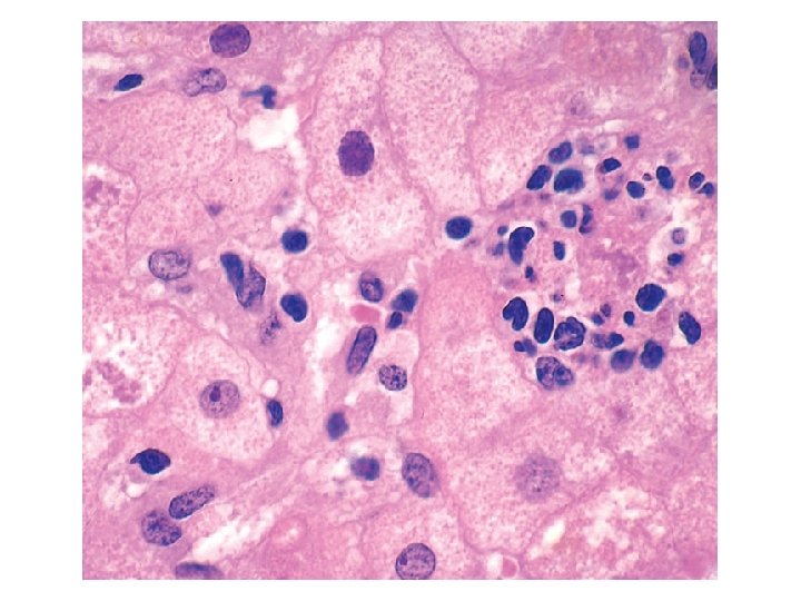
• several hypochromic RBC (which are also target cells, arrowhead). • Lead inhibits the incorporation of iron into heme, so the iron accumulates in mitochondria resulting in the formation of RBC containing iron inclusions or siderocytes (arrows). • A high number of nucleated RBC (n. RBC), disproportionate to the degree of polychromasia and basophilic stippling (lead inhibits the enzyme 5′ nucleotidase which degrades ribosomes) results in basophilic stippling in mature, polychromatophilic RBC (B) and n. RBC

Lead poisoning-

Melanosis of Arsenic

Melanosis, keratosis and amputation of Arsenic

Pulmonary emphysema

Chronic bronchitis

Chronic bronchitis
 and a")
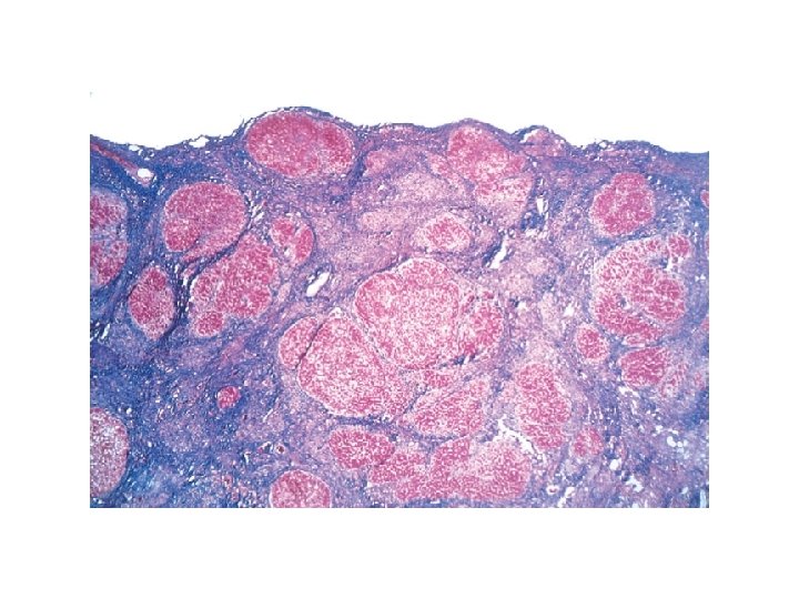
Atherosclerotic plaque in the coronary artery. Overall architecture demonstrating fibrous cap (F) and a central necrotic (largely lipid) core (C); collagen (blue) is stained with Masson trichrome. The lumen (L) is moderately narrowed by this eccentric lesion, which leaves part of the vessel wall unaffected (arrow)
; the internal")
Moderate-power view of the plaque shown in A, stained for elastin (black); the internal and external elastic membranes are attenuated and the media of the artery is thinned under the most advanced plaque (arrow)

Fatty liver

• Alcoholic hepatitis with clustered inflammatory cells marking the site of a necrotic hepatocyte. A Mallory-Denk body is present in another hepatocyte (arrow
 containing prominent Mallory-Denk bodies; clusters of")
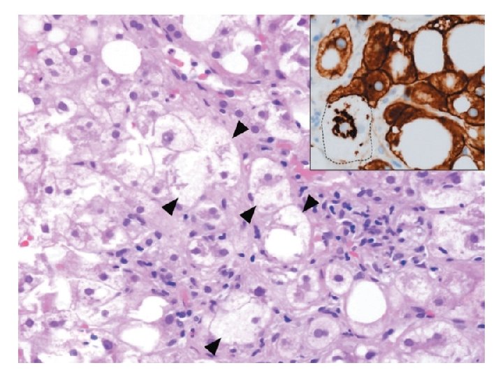
• Steatohepatitis with many ballooned hepatocytes (arrowheads) containing prominent Mallory-Denk bodies; clusters of • inflammatory cells are also seen; inset shows immunostaining for keratins 8 and 18 (brown), with most hepatocytes, including those with fat vacuoles, • showing normal cytoplasmic staining, but in the ballooned cell (dotted line), the keratins are collapsed into the Mallory-Denk body, leaving the cytoplasm • “empty. ”
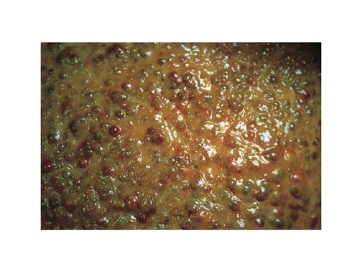

• Alcoholic cirrhosis. The characteristic diffuse nodularity • of the surface is induced by the underlying fibrous scarring. The average • nodule size is 3 mm in this close-up view. The greenish tint is caused by • bile stasis.

• cirrhosis. Small nodules are • entrapped in blue-staining fibrous tissue; fatty accumulation is no longer • seen in this “burned-out” stage. (Masson trichrome stain. )
- Slides: 35