ENTEROCUTANEOUS FISTULA Edited By Basil Smadi DEFINITION A

ENTEROCUTANEOUS FISTULA Edited By : Basil Smadi

DEFINITION A fistula is an abnormal communication between two epithelialized surfaces Enterocutaneous fistula : is an abnormal communication between the small or large bowel and the skin. ECF can arise from the duodenum, jejunum, ileum, colon, or rectum. ECF is an external fistula The ileum was the most common site of ECF

PATHOPHYSIOLOGY * The pathophysiology of all forms of small-bowel fistulas is related to the exposure of nonintestinal tissue to intestinal contents because of the fistula. * The local effect of intestinal fluid can be damaging or corrosive to the nonintestinal tissue, leading to breakdown, erosions, and loss of normal organ or organ system function.

CLASSIFICATION OF FISTULA 1 _Anatomic classifications define the sites of fistula origin, drainage point, and whether the fistula is internal or external 2_ Physiologic classifications rely on fistula output in a 24 -hour period The type of ECF, as based on the output of the enteric contents : 1. Low-output fistula (< 200 m. L/day), 2. Moderate-output fistula (200 -500 m. L/day) 3. High-output fistula (>500 m. L/day) 3 _Etiologic classifications (eg malignancy, Inflammatory bowel disease , or radiation) define the associated disease entity leading to the development of the fistula

ETIOLOGY 3. The etiology of ECFs can be characterized as Postoperative 75 -90 % Traumatic 15 -25 % spontaneous. Postoperative : 1. Disruption of anastomosis Inadvertent enterotomy Inadvertent small-bowel injury 1. 2. 3.

Disruption of anastomosis can result from inadequate blood flow due to an improper vascular supply, especially when extensive mesenteric vessels have to be ligated. Tension anastomosis Minimal leak or infection can lead to perianastomotic abscess formation, resulting in disruption.

Gastroduodenal fistulas are seen most often after surgery for perforated peptic ulcer. In patients with a perforated duodenal ulcer, when the perforation is large, extensive contamination is present. When the duration between perforation and surgery is long, there is a high possibility of a postoperative leak, leading to a lateral duodenal fistula. This problem is difficult to treat, and mortality is high.

Surgery for appendicitis, appendicular perforation at the base, or drainage of an appendicular abscess can also lead to a colocutaneous fistula. Radiation therapy is also another major cause of colonic fistula.

TRAUMATIC Traumatic ECF results from iatrogenic surgical trauma to the bowel that may or may not be recognized. Road traffic accidents with injury to the gut can also lead to an ECF.

SPONTANEOUS Spontaneous causes of ECF : Malignancy Radiation enteritis with perforation Intra-abdominal sepsis IBD (eg, Crohn disease) Diverticulitis (mcc of CCT)
 include postoperative abdominal pain,")
HISTORY &PHYSICAL EXAMINATION Features suggestive of an enterocutaneous fistula (ECF) include postoperative abdominal pain, tenderness, distention, enteric contents from the drain site, and the main abdominal wound. Tachycardia and pyrexia may also be present, as may signs of localized or diffuse peritonitis, including guarding, rigidity, and rebound tenderness.

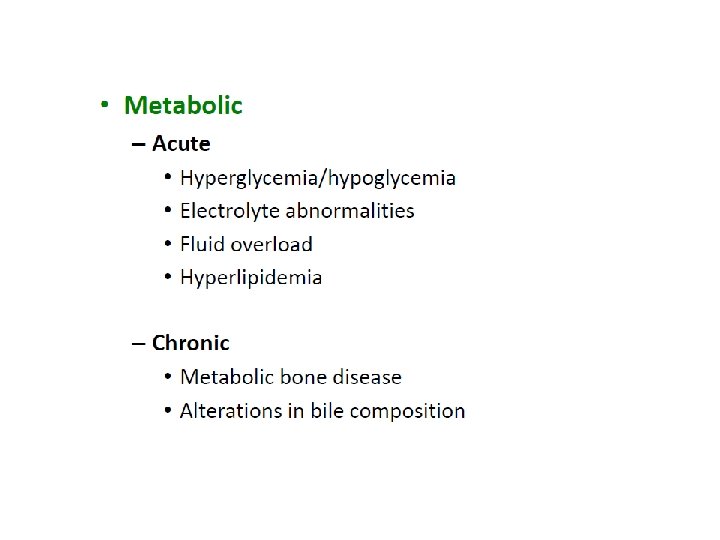
COMPLICATIONS Patients with ECF present with associated complications : Sepsis dehydration Electrolyte and fluid abnormalities Malnutrition Skin excoriation

The degree of sepsis depends on the state of the ECF. If the fistula forms a direct tract through which the bowel contents are draining onto the skin, then the sepsis may be minimal, whereas if the fistula forms an indirect tract through which the bowel contents are draining into an abscess cavity and then onto the skin, the degree of sepsis may be higher. In the presence of extensive peritoneal contamination or generalized peritonitis with ECF, the patient can be toxic due to severe sepsis.

Leakage of protein-rich enteric contents, intraabdominal sepsis, or electrolyte imbalance– related paralytic ileus, as well as a general feeling of ill health, leads to reduced nutritional intake by these patients, resulting in malnutrition. in patients with high-output enterocutaneous fistulas because of intestinal fluid loss. Hypokalemia, hypochloremia, and metabolic alkalosis

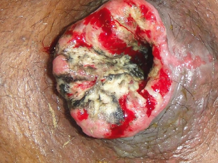
Skin excoriation is one of the complications that can lead to significant morbidity in patients with ECF. When the enteric contents are more fluid than solid, this becomes a difficult problem; the skin excoriation makes it difficult to put a collecting bag or dressings over the fistula, and more leakage leads to an increase in the excoriation.
 count -")
LABORATORY STUDIES 1. 2. 3. 4. 5. Total white blood cell (WBC) count - This is important because sepsis can lead to leukocytosis Serum sodium, potassium, and chloride levels Electrolyte abnormalities can result from fluid and electrolyte loss Complete blood count (CBC), total proteins, serum albumin, and globulin - These can demonstrate the presence of malnutrition-associated anemia/hypoalbuminemia Serum transferrin - Low levels (< 200 mg/d. L) are a predictor of poor healing Serum C-reactive protein (CRP) - Levels may be elevated

PROGNOSIS ECF is a common condition in most general surgical wards. Mortality has fallen significantly since the late 1980 s, from as high as 40 -65% to as low as 5 -20%, largely as a result of advances in intensive care, nutritional support, antimicrobial therapy, wound care, and operative techniques. Even so, the mortality is still high, in the range of 30 -35%, in patients with highoutput ECFs. Once a patient develops an ECF, the morbidity associated with the surgical procedure or the primary disease increases, affecting the patient's quality of life, lengthening the hospital stay, and raising the overall treatment cost. Malnutrition, sepsis, and fluid electrolyte imbalance are the primary causes of mortality in patients with an ECF.

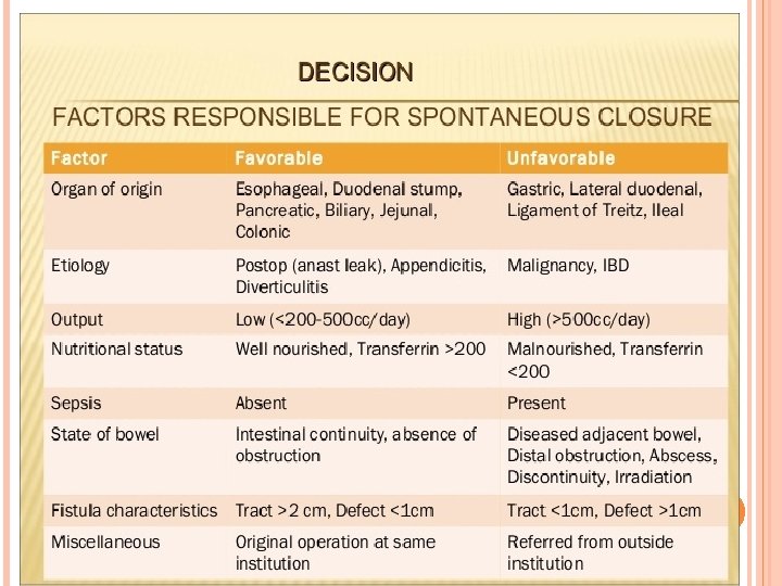
However, patients with an ECF with favorable factors for spontaneous closure have a good prognosis and a lower mortality. Favorable factors for spontaneous closure 1. End fistulas 2. Jejunal fistulas 3. Colonic fistulas 4. Continuity-maintained fistulas 5. Small-defect fistulas

6. 7. Long-tract fistulas Spontaneous closure is also possible if the bowel-wall disruption is partial and other factors are favorable.

UNFAVORABLE FACTORS FOR SPONTANEOUS CLOSURE Factors preventing the spontaneous closure of an ECF can be remembered by using the acronym HIS FRIEND, which represents the following : 1. High output fistula (>500 cc/day) 2. Intestinal destruction ( >50% of circumference) 3. Short segment fistula ( <2. 5 cm) 4. Foreign body 5. Radiation 6. Inflammation/infection/IBD 7. Epithelialization of the fistula tract
 9. Distal obstruction - A distal obstruction prevents the spontaneous closure")
8. Neoplasm (malignancy) 9. Distal obstruction - A distal obstruction prevents the spontaneous closure of an ECF, even in the presence of other favorable factors; if present, surgical intervention is needed to relieve the obstruction 10. Lateral duodenal fitsula

THANK YOU

Suliman Dmor

MANAGEMENT S-S-N-A-P Stabilization and resuscitation Control of Sepsis and appropriate Skin care Nutrition Define underlying Anatomy Plan to deal with the fistula

Stabilization and Resuscitation - Restoration of normal circulating blood volume - electrolyte & acid base imbalance. -hypokalemia and metabolic acidosis, which require correction. - albumin administration.
 - Strict input and output measurements - Urine output should")
- Rehydration --isotonic fluid(NS/RL) - Strict input and output measurements - Urine output should be restored to greater than 0. 5 m. L/kg/hr.

Control Of Sepsis & Fistula Effluent - fever, tachycardia and leukocytosis along with failure to improve adequately points towards possible sepsis or abscesses. - May require surgical drainage of abscess, if any along with adequate antibiotic cover.
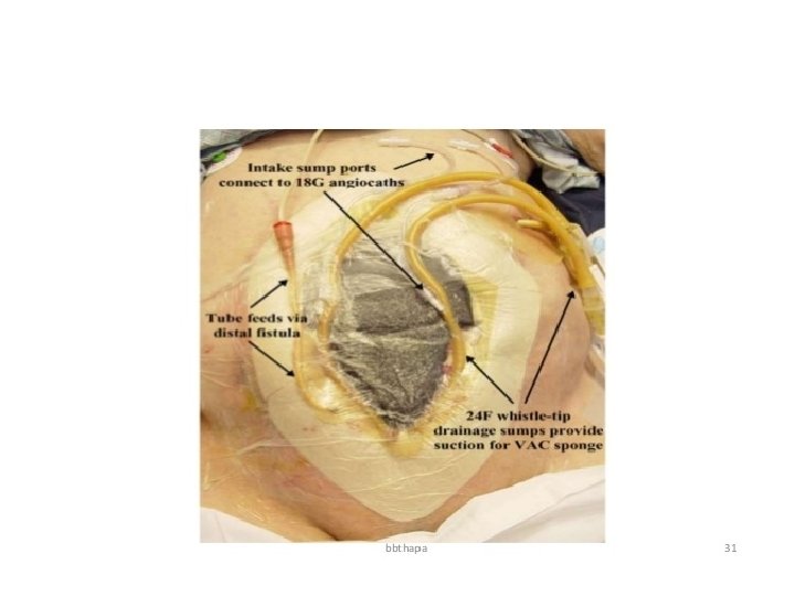
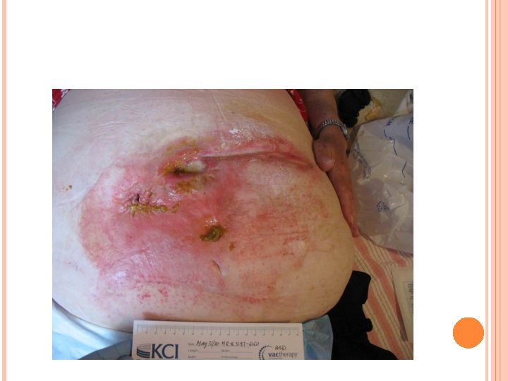
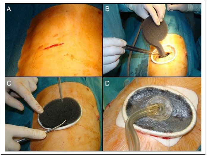
![Vacuum assisted closure [VAC] - device drainage system. - Due to the negative pressure](http://slidetodoc.com/presentation_image_h2/2dbdc6ee84a07c178359dbcd9cae7516/image-34.jpg "Vacuum assisted closure [VAC] - device drainage system. - Due to the negative pressure")
Vacuum assisted closure [VAC] - device drainage system. - Due to the negative pressure application, helps to control drainage, minimizes the size of the abdominal wound, reduces frequency of dressing and protects the skin while helping to promote fistula healing. - Removes chronic edema, leading to increased localized blood flow, and the applied forces result in the enhanced formation of granulation tissue”

Laparotomy may be required for: 1 - Extensive cellulitis/necrotising fascitis 2 - Incomplete percutaneous drainage of collections 3 - Disruption of anastomosis

Skin care management • Problems in skin around the fistula: Wetness Burning pain Discomfort from skin edema

• Goals of skin care: Containing the effluent
: provide a good barrier, before the")
Skin Barriers: 1 - Solid wafers (pectin based): provide a good barrier, before the skin breaks down and ulcerates. 2 - Powders (Pectin/ Karaya based): Powders are preferred over a paste in wet, weepy, perifistular skin when severe skin maceration is present. 3 - Paste 4 - Spray and wipes 5 - Ointments and creams (zinc/petroleum based)

Zinc oxide cream barrier for skin protection.

• Wound pouch dressings

Pharmacological Support SOMATOSTATIN N ANALOGUE - Octreotide Reduces fistula output by 40 – 60% by the end of 24 hrs. - Octreotide and TPN seem to have a synergistic effect on reduction of effluent volume and improvement in fistula closure rates. - Proton-pump inhibitors and H 2 receptor antagonists/antimotality agent also help reduce fistula output especially in proximal fistulas.

• Mode – Inhibit gastrin & cholecystokinin – Reduces splanchic blood flow – Reduces rate gastric emptying – Inhibit gall bladder contraction Plasma half life 1 -2 min - Should be discontinued if ineffective for 48 hrs as it has side effects like hyperglycemia, elevated cholesterol and reduced bowel motility.

Cyclosporine helps to treat refractory fistulae associated with Crohn’s disease. Infliximab administeration helped in partial resolution of 68% of multiple lesions and complete closure in 55% of patients.

- Excessive fistula output can be controlled by: 1. nasogastric tube placement 2. withdrawal of oral feeds 3. initiation of parenteral nutrition

Nutritional management - May be: • Enteral • Parenteral - based on the anatomy of the fistula. - combination of parenteral and enteral nutrition can be used in high-output fistulas
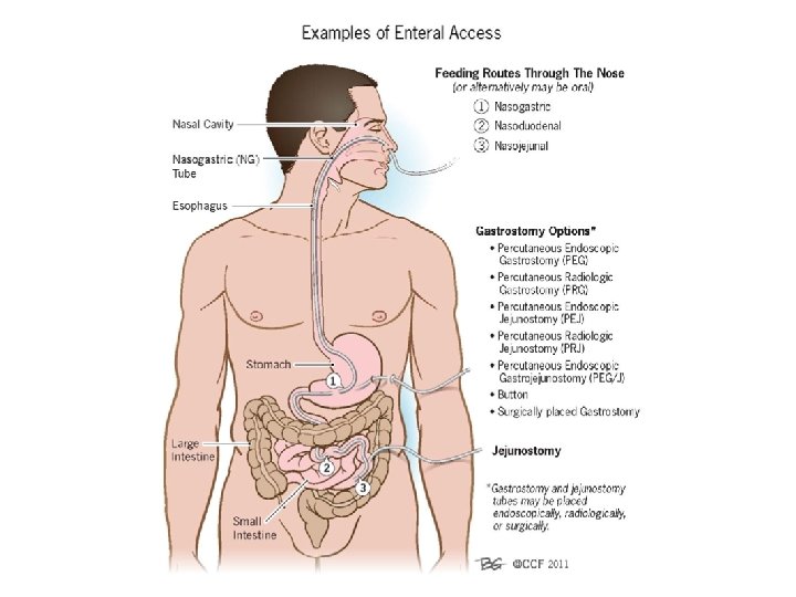

- Nutrition via the enteral route helps in maintaining the intestinal mucosal barrier, more efficacious delivery of nutrients, stimulating hepatic protein synthesis. - The routes of administration via nasogastric/jejunal tubes or a gastrostomy, High rates of feeding should be avoided to prevent hyperosmolar diarrhea. - In proximal fistulae, the enteral feeding tube may be entered beyond the fistula to provide enteral nutrition(fistuloclysis) - In patients with a proximal fistula, if a nasojejunal tube can be introduced beyond the site of the fistula, then these patients can be supported with enteral nutrition, provided that there is at least 4 -5 ft (1. 2 -1. 5 m) of functional bowel distal to it and no distal obstruction. - Patients with chronic small-bowel ECFs may need additional supplementation with copper, folic acid, and vitamin B 12. - During the time the enteral nutrition is increasing, patient should be supplemented with TPN.

- If BW loss> 20%, TPN initiated gradually to avoid refeeding syndrome TPN indications 1 - gastric, duodenal, or small-bowel fistula. 2 - Inability to obtain enteral access 3 - High output fistulas 4 - GI intolerance with enteral nutrition 5 - Multiple unfavorable factors (ileus, obst, ) - Patients should receive 3000 to 5000 non proteins calories per day
 - 80 -100 m. Eq/day")
Requirements for TPN 1 - Electrolyte requirements Sodium (Na) - 80 -100 m. Eq/day Potassium (K) - 75 -100 m. Eq/day Magnesium (Mg) - 15 -20 m. Eq/day Calcium (Ca) - 15 -20 m. Eq/day 2 - Water 1 m. L/kcal/day or 30 ml/kg/day 3 - Calorie and protein requirements are as follows: 25 -30 kcal, 1. 0 -1. 2 g/kg/day 4 - nonprotein calorie-to-nitrogen ratio should be as follows: 200 -300: 1 5 - Glucose, 75 g Amino acids, 20 g Lipids, 30 g/L Conc. dextrose: 500 ml of 20% Dex. (=400 kcal) Fat: 500 ml 10% fat emulsion (=450 kcal) Crystalline Amino Acids: 500 ml 10% Amino acids (=8. 4 g Nitrogen) Daily Vitamin Supplementation ( Vit. K 10 mg/wk)

• Rate of infusion • Starting: 50 – 100 ml/hr • Gradually increased by 25 – 50 ml/hr every second day

Surgical therapy Haneen Al-Hayajneh
 following stabilization. Objectives of investigation plan: To define • Precise")
Imaging (7 -10 days) following stabilization. Objectives of investigation plan: To define • Precise anatomical location • Is the bowel in continuity or is disrupted • Abscess cavity • Condition of adjacent bowel • Is there a distal obstruction

�Often, a combination of studies is necessary to fully appreciate the fistula anatomy. Computed tomography, fistulography, a small bowel followthrough study, and contrast enemas are all useful modalities for defining anatomy depending on the location of the fistula. � Magnetic resonance enterography is another adjunct, particularly useful in patients with IBD. .

Fistulography �During fistulography a water-soluble contrast is injected into the fistulous tract.

�Fistulography provides the following information: 1. Length of the tract 2. Extent of the bowel-wall disruption 3. Location of the fistula 4. Presence of a distal obstruction fistulogram, and percutaneous drainage may decompress a complex fistula and convert it to a simple one.

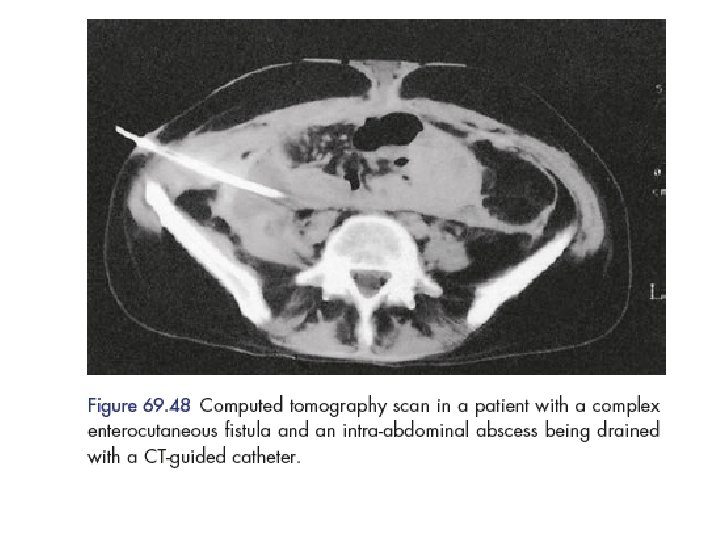
CT scan with oral contrast is considered the single best radiologic test locating the fistula defining its anatomy commenting on the gut surrounding fistula abdominal leaking presence of any intra-abdominal distal obstruction/pathology foreign bodies abscess CT scan is highly recommended for duodenal and pancreatic fistula. CT scan be therapeutic by helping in CTguided aspiration of intra-abdominal abscesses, if any.

Endoscopy may also be used occasionally though its principal use is in internal fistulas. Endoscopy is usually delayed till acute inflammation gets reduced. CT scan

�Water-soluble contrast enema �The different types of tracts that can be seen by using a water-soluble contrast enema (WCE) in patients with ECF with failure of low colorectal anastomosis may be classified as follows : �I – Simple, short blind ending, < 2 cm �II - Continuous linear, long single, >2 cm �III - Continuous complex, multiple linear

�Closure rates without operative intervention in the era of advanced wound care and parenteral nutrition (PN) vary considerably in reports 19 to 92%, with most studies demonstrating closure rates in the 20 to 30% range. �With historical wound care measures, 90% of spontaneous closure occurred in the first month after sepsis resolution, with an additional 10% closing in the second month, and none closing spontaneously after 2 months. � With vacuum assisted closure (VAC) and other negative pressure wound therapies (NPWT) therapy, there are case reports of fistulae closure well into the second and third month �However, the development and management of an ECF remains a chronic, debilitating condition associated with prolonged intensive care unit and hospital length of stay, and hospital costs of over $500, 000

Timing of surgery �Depending on the surgeon, 2 to 3 months of will be attempted before the surgical correction of a fistula is considered. This waiting period gives the fistula an appropriate amount of time to close spontaneously. It also decreases the morbidity and mortality of surgical correction. �The absolute minimal waiting interval after original surgery to return to the operating room is 6 weeks.

� The time interval in which operative intervention is associated with a significantly higher mortality has been outlined by Fazio et al, counting from the day of previous surgery: return to the operating room within 10 days resulted in 13% mortality, operating between 11 and 42 days was associated with 21% mortality, and after 42 days mortality returns to 11% in average patients

�There is a high risk for recurrence. Surgical approach may be difficult due to previous surgeries and adhesions. The bowel must be run carefully, and extreme care must be taken to not cause any accidental enterotomies during lysis of adhesion and bowel mobilization. �As long as the bowel looks healthy, the best option is to excise the fistula tract and resect a small amount of associated bowel followed by an anastomosis to reestablish bowel continuity. �To decrease recurrence rate, one must make sure to close the fascia where the fistula tract was traversing.

�Avoidance and proper repair of any enterotomy is essential since 36% of recurrent fistulas result from inadvertent injury to the bowel. Operative success for definitive ECF resolution ranges from 80 to 95%. �Additionally, following surgery, ECF recurrence is 14 to 34%. Recurrence rates are minimized (18%) when the involved bowel is fully mobilized and resected. Oversewing or wedge resection/bowel repair results in higher rates of recurrence at 33%. �Similarly, Runströmet al reported that ECF failure rate is lower when no anastomosis is constructed and, instead, a stoma is chosen (recurrence rate 14 vs. 34% with anastomosis). � More broadly, one has to balance the risk of ECF recurrence with the morbidity of another operation if anastomosis is avoided.

Indications for surgery �ECF with adverse factors may require earlier surgical intervention �Lateral duodenal or ligament of Treitz fistula �Ileal fistula �High-output fistula �Fistula associated with diseased bowel, distal obstruction, or eversion of mucosa �Enteroatmospheric fistula

Surgery timing 30 -70 per cent need definitive surgerical repair In patients with adverse factors, surgical intervention should be undertaken after a 6 week trial of conservative therapy Pt with no adverse factors Wait atleast 3 months from the initial insult Needs resolution of acute inflammatory response

Fit for surgery �if the patient is stable �optimization of nutrition �eradication of infection

Preoperation �Antibiotic prophylaxis should be performed and �parenteral nutritional supplementation �Enteral feeding should be decreased to allow luminal antibiotic preparation

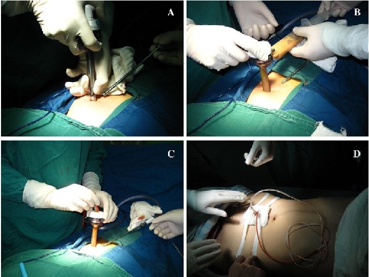
Surgery main steps � 1. Access : The entire bowel from the ligament of Treitz to the rectum is made free of all adhesions. � 2. intestinal repair : resect segment containg fistula + anastomosis �bowel continuity : 2 layers � A : outer non reabsorbale suture � b: inner absorbable suture � 3. abdominal wall closure : primary closure best �Biological mesh second best

�The abdomen should be irrigated any bleeding controlled. �The large abdominal wall defect is closed with the musculocutaneous flap latissimus dorsi flap

Enteroatmospheric fistula �floating stoma �Fistuloclysis in combination with NPWT may also be quite useful in select patients.

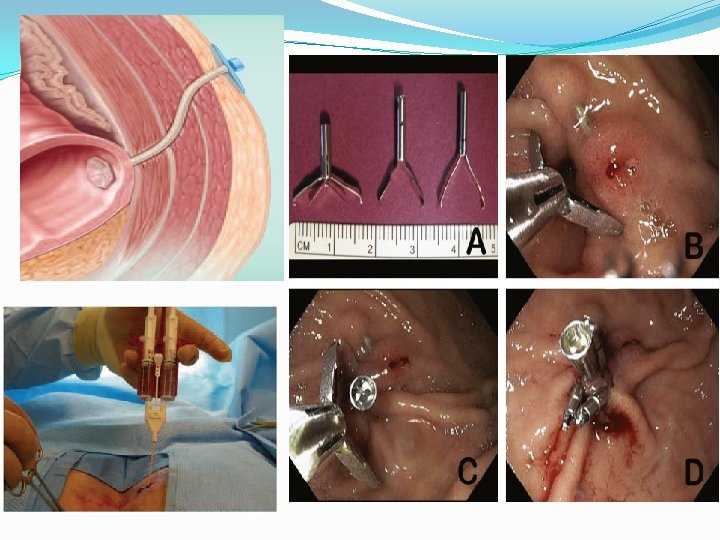
Other Interventions Use of fibrin glue and plugs �Use of fibrin glue and plugs: Once a fistula had been endoscopically located, fibrin glue injected through a catheter �May expedite fistula closure �Usually used with endoclip �Endoclips (for acute fistulas) �Fistula plug

- Slides: 76