Endotracheal Intubation BY JUNAEDI STUDENT POST GRADUATE OF

Endotracheal Intubation BY JUNAEDI STUDENT POST GRADUATE OF NURSING BRAWIJAYA UNIVERSITY- INDONESIAN
 into the trachea to secure")
Definition: Introducing a tube through the mouth (or nose) into the trachea to secure open airways. Advantages: Cuffed E. T tubes protect the airway from aspiration. E. T tube provides access to the tracheobronchial tree for suctioning of secretions. E. T tube does not cause gastric distention and associated danger of regurgitation. E. T tube maintains a patent airway and assists in avoiding further obstruction. E. T tube enables delivery of aerosolized medication. the mouth (or nose) into the trachea to secure open airways.

Indication Endotracheal Intubation: Respiratory Failure: Hypoxia, Hypercapnia, tachypnea, or apnea ; ie. ARDS, asthma, pulmonary edema, infection, COPD exacerbation Inability to ventilate unconscious patient Maintenance or protection of an intact airway Cardiac Arrest Medication administration

Contraindication : Inability of patient to extend head Moderate to severe trauma to the cervical spine or anterior neck Infection in the epiglottal area Mandibular fracture or trismus Mild hypoxia Uncontrolled oropharyngeal hemorrhage Intact tracheostomy Basilar skull fracture (during nasal intubation)

Complications: Ø Ø Ø Hypoxia (Long duration of procedure, Intubation of a bronchus ( right more common, Failure to recognize misplacement of tube, Aspiration) Pneumothorax (resulting from over ventilating with a BVM without a pressure release valve) Trauma (to the teeth, vocal cords, soft tissues of the larynx and related structures) Hypertension and tachycardia (can occur from the intense stimulation of intubation. This is potentially life-threatening in the cardiac patien) Gastric distention and regurgitasi (Failure to secure the placement into esophagus). Cardiac arrhythmias (related to vagal stimulation or sympathetic nerve stimulation may occur)
 : Mask Seal : Small Hands,")
Difficult to intubation: 1. Difficult to bag (MOANS) : Mask Seal : Small Hands, Wrong Mask Size, Oddly Shaped Face, Bushy Beard, Blood/Vomit, and Facial Trauma Obesity or Obstruction: Heavy chest, Abdominal contents inhibit movement of the diaphragm, Increased supra glottic airway resistance, Billowing cheeks, Difficult mask seal, Quicker desaturation Age > 55: Associated with BVM difficulty, possibly due to loss of tone in the upper airway No Teeth: Face tends to “cave in”, Consider leaving dentures in for BVM and remove for intubation. Stiff : Refers to Poor Compliance, Reactive Airway Disease, COPD, Pulmonary Edema/Advance Pneumonia, History of Snoring/Sleep Apnea, Also predicts a higher Mallampati score

Difficult to Laringoscopy and intubation: 1. LEMONS: Look Externally : Beards or facial hair, Short, fat neck, Morbidly obese patients, Facial or neck trauma, Broken teeth (can lacerate balloons), Dentures (should be removed), Large teeth, Protruding tongue, A narrow or abnormally shaped face. Evaluate 3 -3 -2 : Bottom of Jaw/Chin to Neck > 3 fingers, Jaw/Palate > 3 fingers wide, Mouth opens > 2 fingers wide.

Mallampati Obstruction Score : : Anatomy, Trauma, Foreign body obstruction, Grade 1 Edema (burns). Best view grade 1

Neck Mobility approximately : Ideally the neck should be able to extend back 35° Problems: Cervical Spine Immobilization, Ankylosing Spondylitis, Rheumatoid fixation Scene Do and Situation : Scene safety and Environment you have a reasonable chance to get the tube? Space, positioning, access Egress Will A you be able to ventilate during egress? respiratory rate of 4 is better than a rate of 0! Enough meds for a long extrication? Arthritis, Halo

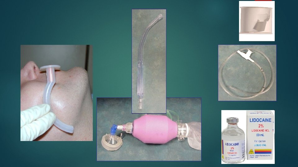
Oral Intubation With local anesthesia: It is also practical to apply surface anesthesia: vagal excitation is less, the patient may tolerate the tube better, arrhytmias and laryngospasm after extubation are rare. Apply 10% Lidocain spray (2 or 3 spurts - 1 spurt=4. 8 mg) If the distal end of tube is also sprayed with Lidocain before intubation, the patient will also tolerate the tube after recovering consciousness. Except : Reserved for the completely unconscious, unresponsive, and apneic, and Arrest situations only (without drug).

Equipment
 and straight (Miller) Endotracheal tubes of")
Equipment Endotracheal Intubation: Laryngoscope Blades: curved (Mac. Intosh) and straight (Miller) Endotracheal tubes of various sizes: q. Neonates and full term infants: no. 0 and 1, q. Adult women: 7. 0 mm i. d. , q. Adult men: 7. 0 to 8. 5 mm i. d. q Pediatric size: (age in years/4) + 4 or width of fingernail of the fifth digit
 Oxygen and manual")
Lubricant, Malleable stylet 10 -ml syringe (to inflate ET cuff) Oxygen and manual bag valve mask Suction apparatus Stethoscope Sterile gloves and goggles Oropharyngeal airway CO 2 Detector
 Blade tipe Macintosh (curve blade) Blade tipe Miller (straight blade)")
Handle and Blade (Laryngoscope) Blade tipe Macintosh (curve blade) Blade tipe Miller (straight blade)

Engaging laryngoscope blade and handle

ETT, Stylet, and Syringe

High volume Low pressure cuff High pressure cuff

Magil Forceps, sterile gloves and goggle

Procedure

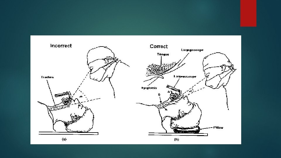
Position patien’s head - Position yourself at the patient’s head - Inspect the oral cavity for secretions or foreign material. - Suction if necessary
")
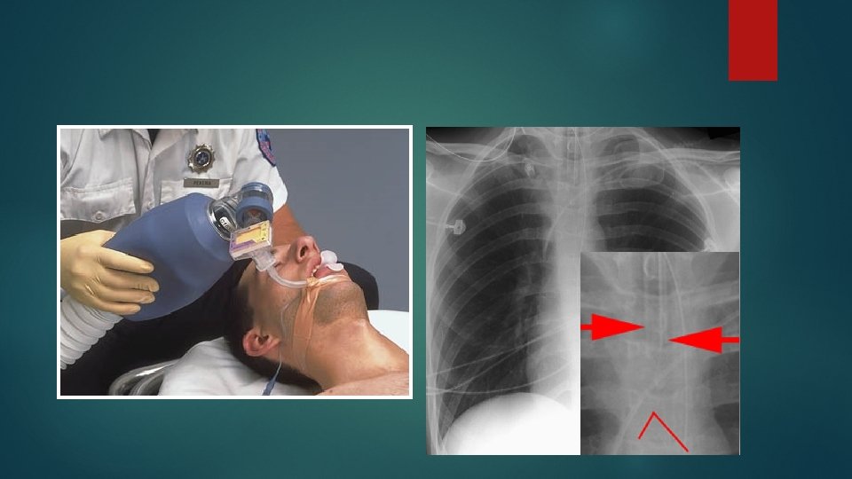
Hiper ventilate with 100 % oxygen for approximately 1 min (prior 2 minutes)

Intubation Technique 22 cm

Sellick Manuver § Helps prevent regurgitation and reduces gastric distention. § Locate the cricoid cartilage by palpating the thyroid cartilage and the feel the depression just below it (cricothyroid membrane). § Using your thumb and index finger of one hand, apply pressure to the anterior and lateral aspects of the cricoid cartilage just next to the midline.

Laringoscopic View

CO 2 exhaled from the lungs: color change to MELLO YELLOW
, ETT easily")
NEVER let go of the tube until secured (Tape, Commercial tube holder), ETT easily displaced so requires ongoing assessment Oro Pharyngeal Airways (OPA)
___depth___cm Post ET lung sounds")
Documentation ET Tube Placement On patient care report: ET (size)___depth___cm Post ET lung sounds ET Attempt (x___) Capnography Checked Suction Boxes used to indicate crew member activity 30

Thank you
- Slides: 31