Endokrinoloji Anamnez FM Dr Mustafa Sahin Ankara Universitesi


 • Görme alanı")


 ATP III criteria : Aşağıdakilerden ≥ 3 • Abdominal obezite :")


 acromegaly. org. uk Picture of")


. Graves")


 The great")


 Graves Disease Both Bushes were")


- Slides: 63
Endokrinoloji – Anamnez / FM Dr. Mustafa Sahin Ankara Universitesi Tıp Fakültesi Endokrinoloji ve Metabolizma Hastalıkları Bilim Dalı
• Sens clinique…. sadece görerek tanı
Nasıl prezente olur ? • Çoğu zaman insidental saptanır • Çoğu asemptomatik
Bası belirtileri: • Başağrısı (Frontal, duranın gerilmesi , bazen hidrosefali…. kusma) • Görme alanı defekti • (Konfrantasyon ile perimetrik inceleme…. bitemporal hemianopsi) • Diplopi (3. sinir basısı) • Rinore (sella tabanı) • Nöbet • İnme • Kişilik değişiklikleri
• Tiroid bezi muayenesi hasta ile aynı seviyede ve hastanın boynu hafif fleksiyon halindeyken yapılır. Hasta yutkunurken ve normal pozisyonda iken boyundaki görülebilir kitle ve guatr için ins pekte edilir. Tiroid dışı kitleler • yutkunmakla yerdeğiştirmezler, • Tiroglossal kistler orta hat • kitleler olup dilin öne çekilmesi ile yer değiştirirler. Tiroid palpasyonu önden ve ya arkadan yapılabilir.
• Önden muayenede hastanın başıı ha if fleksiyonda iken bir elin baş parmağı ile bir loba hafifçe bastırılarak karşı lobun öne doğru belirginleşmesi sağla nır ve diğer elin başparmağı ile öne çıkan lob palpe edilir. • Arkadan muayenede hasta oturur pozis yon da iken arkasına yerleşilir, • 2, 3 ve 4. parmakların uçları ile tiroid lob larıpalpe edilir.
12
ANATOMIC RELATIONSHIPS OF THE THYROID
ANATOMIC RELATIONSHIPS OF THE THYROID
Classical Goiter Classification • Ia -thyroid not visible, lateral lobes smaller than the distal phalanx of the thumb • Ib - thyroid visible with neck in extended position • II - thyroid visible with the neck in normal position • III - thyroid visible at distance
• Palpasyonda ele gelen nodüllerin yeri, boyutları, • kıvamı, fikse ve ağrılı olup olmadığı be lirlenir. • Ağrılı : subakut tiroidit, nodül içine kanama, akut süppüratif tiroidit, tiroid kanserleri • Boyunda lenfadenopati muayenesi
• Plonjan guatr: Semptomatik • olgularda dispne ve disfaji olur. • Plonjan guatrı olanlarda kolların yukarı kaldırılması ile yüzde pletore gelişir (Pemberton belirtisi)
• • Thyroid Enlargement Diffuse Enlargement: isthmus and lateral lobes, no nodules. Grave’s disease, Hashimoto’s thyroiditis, endemic goiter • Single node: Cyst, benign tumor, false positive (only one nodule of multinodular goiter detected). Elevates index of suspicion for malignancy. • Assess for risk factors: radiation exposure, hardness, rapid growth, fixation to surrounding tissue, cervical LAD, male, others. Multinodular Goiter (iodine deficiency) • • • Soft in Graves Disease and may have bruit. Firm in Hashimoto’s thyroiditis, malignancy, & benign and malignant nodules. Tender in thyroiditis. Systolic or continuous bruit may be heard over lateral lobes in hyperthyroidism.
Metabolik Sendrom Obezite Dislipidemi NAFLD Diyabet Hipertansiyon
Metabolik Sendrom (Sendrom-X) ATP III criteria : Aşağıdakilerden ≥ 3 • Abdominal obezite : Etnik farklılık • (Bel çevresi > 102 cm ♂, > 88 cm ♀) • Trigliserid > 150 mg/dl • HDL < 40 mg/dl ♂, < 50 mg/dl ♀ • KB ≥ 130/85 mm. Hg • AKŞ ≥ 110 mg/dl ? ? 100 mg/dl
25
Cushing’s Syndrome • Due to excessive adrenocortical activity or corticosteroid medications • Women between the ages of 20 and 40 years are five times more likely than men to develop Cushing's syndrome. 31
Cushing’s Syndrome 32
Hipokalsemi • Trousseau belirtisi: Tansiyon aleti man şonu üst kola yerleştirilir ve sistolik kan basıncı nın 10 -20 mm. Hg üstün de şişirilir ve 3 -5 dk beklenir. Ebe eli şek • Chvostek belirtisi: Kulak tragusun 1 cm önüne parmakla veya refleks çekici ile vurulma sı ipsilateral fasial kaslar da kontraksiyo na yol açar.
3 4
Endocrine Images: Acromegaly Andre the Giant by EKavet (Flickr) acromegaly. org. uk Picture of wrestling star Andre the Giant and Skull X-ray of man with acromegaly. Notice the characteristic prominent supraorbital ridge (“frontal bossing”), large jaw, and dental malocclusion with underbite (x-ray).
Endocrine Images: Acromegaly 28 yrs 49 yrs 55 yrs 65 yrs Greenspan & Strewler, Basic & Clinical Endocrinology, 5 th Ed. , 1997 From Reichlin S. Acromegaly. Med Grand Rounds 1982; 1: 9 Individual with acromegaly photographed over a 37 -year span. Ages in years are in lower left corner of each photograph. Note that the changes occurring with acromegaly may be very gradual and go completely undetected by the patient or his or her family for many years. It is often only thorough the comparison with old photographs or complaints involving complications of acromegaly, such as sleep apnea, diabetes or dental problems that acromegaly is suspected.
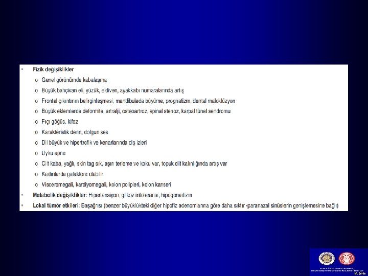
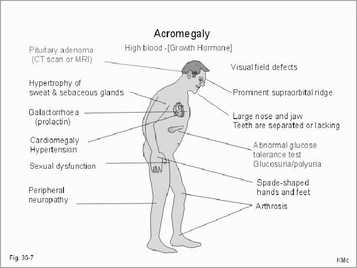
Typical clinical features Acromegaly
Endocrine Images: Acromegaly University of Iowa Dept. of Dermatology Hands of individual with acromegaly (left) compared to hand of non-acromegalic adult (far right).
Endocrine Images: Acromegaly Amilcare Gentili, M. D. Foot X-ray of Patient with Acromegaly. Notice the unusually thick “pad” of soft tissue overlying the calcaneus (double arrow). It is said that a good clinical sign of acromegaly is the inability to feel the calcaneus when pressing on the heel.
Endocrine Images: Acromegaly Clinical Findings in Acromegaly. Symptoms & Signs: • Excessive sweating, snoring. • Arthalgias, carpal tunnel syndrome. • Change in ring/glove or shoe size. Signs: • Dental malocclusion and widely spaced teeth. • Macroglossia. • Large hands and feet. • Large heart (may see signs of heart failure). Laboratory results: • Impaired glucose tolerance or diabetes. • Elevated IGF-1. • Enlarged cardiac silhouette on chest x-ray. Greenspan & Strewler, Basic & Clinical Endocrinology, 5 th Ed. , 1997 From Reichlin S. Acromegaly. Med Grand Rounds 1982; 1: 9
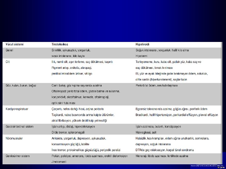
Typical clinical features Hyperthyroidism Graves ophthalmopathy
Endocrine Images: Graves Disease The Handbook of Ocular Disease Management. Graves Ophthalmopathy (Exophthalmos). Graves ophthalmopathy is due to autoimmune-mediated inflammation and edema of the extraocular muscles. Graves eye disease may be asymmetrical and often progresses independently of hyperthyroidism and may lead to diplopia, corneal dryness, ulceration, and blindness. Severe cases may require surgical decompression. Exophthalmos is specific to Graves disease. On the other hand, “lid lag, ” in which the eyelids do not closely follow downward gaze, may be seen in all forms of hyperthyroidism and is due to hyperstimulation of the orbicularis occuli muscles.
Endocrine Images: Graves Disease This photo was taken from Dr. Koenig’s thyroid lecture and is meant to highlight the eye findings in Graves disease: the classic “stare” of hyperthyroidism and a prominent goiter. Notice in Graves that the thyroid is symmetrically enlarged and “plump. ” This is because the entire thyroid is being stimulated by thyroid stimulating immunoglobulin (TSI), which causes constitutive activation of the TSH receptor in the absence of TSH. Auscultation of the goiter of an individual with active Graves disease may reveal a thyroid bruit, due to the hypervascularity of the overactive gland. This bruit must be distinguished from cardiac (or carotid) bruits by localizing its source over the thyroid. Source Undetermined
Endocrine Images: Graves Disease Dermnet Graves Dermopathy Graves dermopathy is also known as “pretibial myxedema, ” which is an unfortunate term, since “myxedema” usually refers to hypothyroidism. The term “myxedema” describes the “doughy” or “peau d’orange” texture of the skin. Graves dermopathy involves inflammation and mucopolysaccharide deposition most prominently in the pretibial regions of the legs. It is a relatively uncommon --albeit, classic-- finding in Graves disease and affects approximately 5% of patients with Graves.
Endocrine Images: Graves Disease Clinical Findings in Graves Disease. Symptoms & Signs: • Heat intolerance, excessive sweating. • Anxiety, “hyperkinesis. ” • Sleep disturbances. • Weight loss despite increased appetite. • Hyperdefecation (not diarrhea). Source Undetermined Signs: • Tachycardia, wide pulse pressure. • Warm, moist skin. • Exophthalmos may be present. • Symmetrical, “plump” goiter. • Fine tremor of outstretched hands. • Brisk reflexes.
Endocrine Images: Hypothyroidism Greenspan & Strewler, Basic & Clinical Endocrinology, 5 th Ed. , 1997 University of Missouri Health Systems Child with Congenital Hypothyroidism (cretinism) Woman with Severe Hypothyroidism This pair of photographs illustrates some general physical features of congenital hypothyroidism and severe hypothyroidism in an adult. The face is has a puffy, “doughy” appearance (hence, the term “myxedematous”). Periorbital edema may be present. The skin is dry and cool, and the hair is coarse. The affect is blunted and apathetic. The child is short and has mental retardation.
Typical clinical features Hypothyroidism
Endocrine Images: Hypothyroidism Clinical Findings in Hypothyroidism. Symptoms & Signs: • Depression. • Cold intolerance. • Weight gain despite unchanged appetite. • Constipation. Woman with Severe Hypothyroidism Signs: • Bradycardia, diastolic hypertension. • “Myxedematous facies” with coarse hair. • Distant heart sounds. • Delayed relaxation phase of achilles reflex. Laboratory results: • Anemia: either macrocytic or normocytic. • Hyponatremia (due to decreased free water clearance by the kidney). • Elevated TSH, low free T 4 (primary hypothyroidism). [Note: since free T 3 may remain normal until hypothyroidism is severe it is useless in the diagnosis of hypothyroidism. ] Greenspan & Strewler, Basic & Clinical Endocrinology, 5 th Ed. , 1997
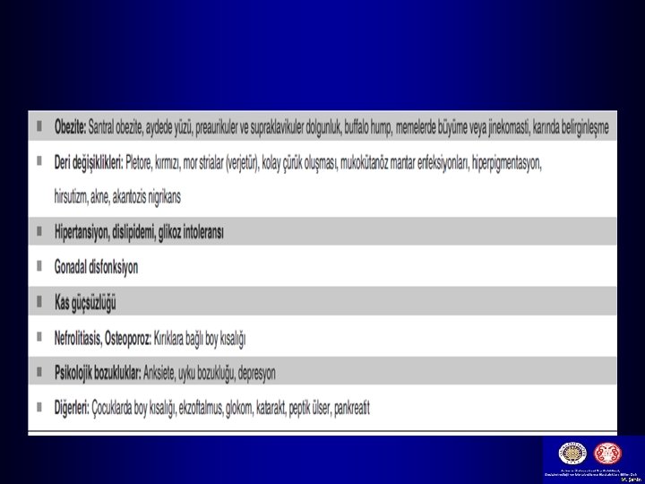
Endocrine Images: Cushings Syndrome Mt. Zion-UCSF Source Undetermined Prominent physical findings in Cushings syndrome include round “moon facies, ” supraclavicular and supracervical fat pads (“buffalo hump”), central obesity and purple abdominal striae. If the result of a pituitary adenoma (Cushings Disease), hyperpigmentation may be present. If an adrenal cortical carcinoma is the cause, there may be hirsuitism and virulization. (Adrenal carcinomas may produce DHEA sulfate, a potent adrenal androgen. ) Adrenal carcinomas also grow more rapidly than adrenal adenomas and tend to be larger: almost always > 5 cm in diameter on an abdominal CT scan.
Endocrine Images: Cushings Syndrome Abdominal Striae in Cushings Syndrome. Classically, these striae are purplish in color and appear on the abdomen, thighs, upper arms and axillae. They are distinguished from silver striae seen in postpartum women or pink striae seen with significant weight loss. Excessive steroid action on skin also may lead to skin fragility and easy bruising during routine activities. G. Hammer, MD, Ph. D University of Michigan (Both images)
Typical clinical features Cushing´s syndrome Moon face Facio-truncal obesity
Endocrine Images: Adrenal Insufficiency NEJM 337: 1666, 1997 This slide of identical twins is from Dr. Hammer’s lecture and is meant to emphasize the hyperpigmentation and thin body habitus that is often seen in primary adrenal insufficiency (the woman with adrenal insufficiency is on the right). Hyperpigmentation may also be seen in the extensor surfaces of the limbs (knuckles, elbows, knees), in newly formed scars and in palmar creases and buccal mucosa. (What’s the cause? )
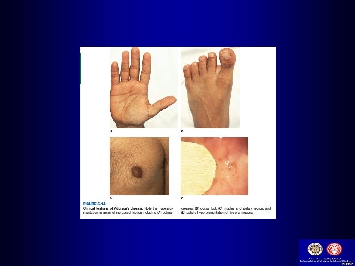
Endocrine Images: Addison’s Disease Clinical Findings in Addison’s Disease. Symptoms & Signs: • General malaise, fatigue. • Weakness and difficulty climbing stairs, arising from sitting, combing or shampooing hair. • Salt craving. NEJM 337: 1666, 1997 Signs: • Orthostatic hypotension. • Hyperpigmentation of extensor surfaces of skin, buccal mucosa, palmar creases. • Weakness of proximal muscle groups. Pertinent routine laboratory results: • Normocytic anemia • Neutropenia (mild) with eosinophilia. • Hyponatremia, hypokalemia and “non-gap” metabolic acidosis. • Mild hypoglycemia (may be pronounced in infants).
Endocrine Images: Addison’s Disease Williams Textbook of Endocrinology, 8 th Ed, 1996. Hyperpigmentation in Addison’s Disease. In primary Addisons disease, one often sees hyperpigmentation of extensor surfaces of the limbs (knuckles, elbows, knees), of the areolae of the breasts, of newly formed scars, and of the buccal mucosa. In this photograph, one may see darkening of the face, fingertips and gingiva as well. (What’s the mechanism? )
Endocrine Images: Addison’s Disease T. Addison “On the constitutional and local effects of disease of the suprarenal capsules” 1855 Hyperpigmentation in Addison’s Disease. This is a (presumably) postmortem drawing from Addison’s original paper of an individual with primary adrenal insufficiency. In Addison’s day, the primary cause was not autoimmune adrenalitis, but tuberculosis.
Endocrine Images: Addison’s Disease James Andrews of Maidenhead Jane Austin (1775 -1817) The great British novelist Jane Austin also suffered from Addison’s disease and died prematurely of its complications. If you look closely, you can see areas of hyperpigmentation on her cheeks…but again, this might be the product of an over-worked endocrinologist’s imagination…
Endocrine Images: Addison’s Disease 1960 Presidential Debate: John F. Kennedy vs. Richard M. Nixon, Chicago, Ill. , September 21, 1960 The Kennedy-Nixon debate in 1960 by scriptingnews (flickr) In the first-ever televised presidential debate, John F. Kennedy was the apparent winner over Richard M. Nixon, a win which helped him in his narrow victory over Nixon in the presidential election of November, 1960. Many observers attributed Kennedy’s “telegenic” character to his youthful, dynamic, tanned (i. e. , “hyperpigmented”) appearance. . .
Endocrine Disorders and World History Marshall Josip “Broz” Tito of Yugoslavia and US President John F. Kennedy, 1962. Yugoslav Government Endocrine disease has clearly affected the course of world history. Kennedy’s year-around “tan” (from his Addison’s disease) helped him win the presidency and lent a youthful air to “Camelot, ” as the Kennedy White House was known, whereas Tito’s death from complications from type 2 diabetes in 1980 eventually led to the break-up of the Yugoslavian federation and the bloody Balkan wars and “ethnic cleansing” of the 1990’s.
Famous Names in Endocrinology U. S. Navy, (wikimedia commons) Graves Disease Both Bushes were diagnosed with Graves disease: Barbara had Graves ophthalmopathy and George presented with atrial fibrillation. Their dog, Millie, had lupus. No kidding. Must be the water. . .
Famous Names in Endocrinology Cecil Stoughton, White House James Andrews of Maidenhead, 1870 Addison’s Disease We now all know about John F. Kennedy, but the novelist Jane Austin was also afflicted with adrenal insufficiency. Her Addison’s disease worsened as she grew older, and she finally succumbed to it at the age of 41 in Winchester, in Central Hamshire (UK) in 1817.
Teşekkürler