
ENDODONTIC DIAGNOSIS BY DR NITHIN S PROFESSOR DEPARTMENT
ENDODONTIC DIAGNOSIS BY DR NITHIN. S, PROFESSOR DEPARTMENT OF CONSERVATIVE DENTISTRY AND ENDODNTICS, YENEPOYA DENTAL COLLEGE
CONTENTS • • • INTRODUCTION DEFINITION HISTORY TAKING EXAMINATION COMMON CLINICAL TESTS SPECIAL METHODS DIAGNOSIS CONCLUSION REFERENCES
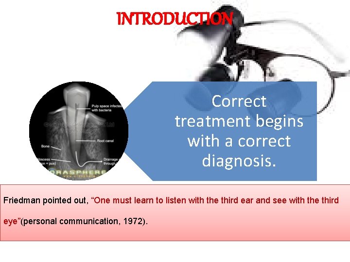
INTRODUCTION Correct treatment begins with a correct diagnosis. Friedman pointed out, “One must learn to listen with the third ear and see with the third eye”(personal communication, 1972).
WHAT IS MEANT BY DIAGNOSIS? “It is a procedure of accepting a patient , recognizing that he has problem , determining the cause of the problem and developing a treatment plan that will alleviate or solve the problem”. - ingle. “Is the process of identifying a disease by its signs, symptoms and results of various diagnostic procedures” - Grossman. DIFFERENTIAL DIAGNOSIS “ Is a process of identifying a condition by differentiating it from all pathologic process that may produce similar lesion” process that may produce similar lesion
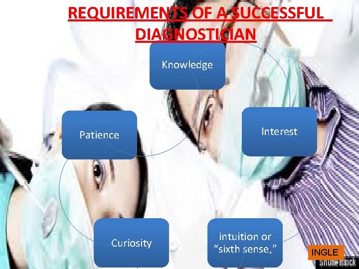
REQUIREMENTS OF A SUCCESSFUL DIAGNOSTICIAN Knowledge Patience Curiosity Interest intuition or “sixth sense, ” INGLE
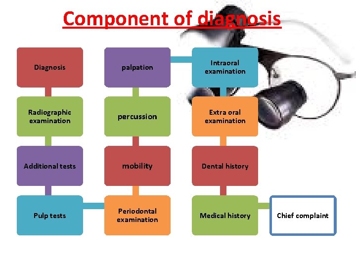
Component of diagnosis Diagnosis palpation Intraoral examination Radiographic examination percussion Extra oral examination Additional tests mobility Dental history Pulp tests Periodontal examination Medical history Chief complaint
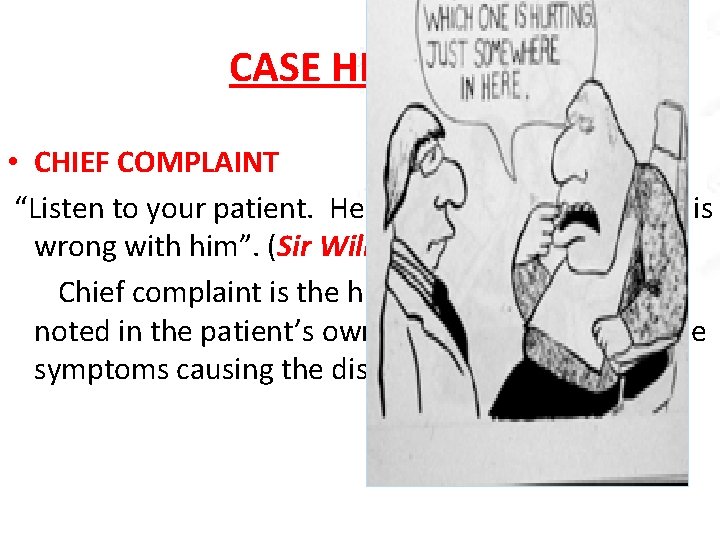
CASE HISTORY • CHIEF COMPLAINT “Listen to your patient. He is trying to tell you what is wrong with him”. (Sir William Osler) Chief complaint is the history of the symptoms noted in the patient’s own words that describes the symptoms causing the discomfort.
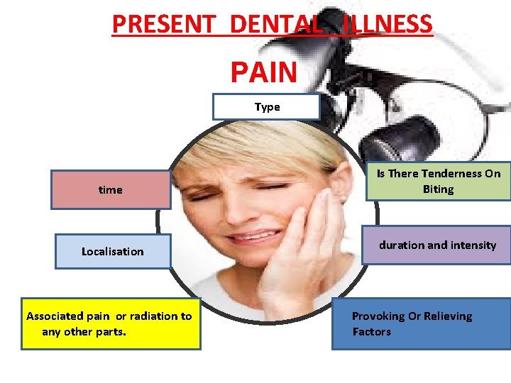
PRESENT DENTAL ILLNESS PAIN Type time Is There Tenderness On Biting Localisation duration and intensity Associated pain or radiation to any other parts. Provoking Or Relieving Factors
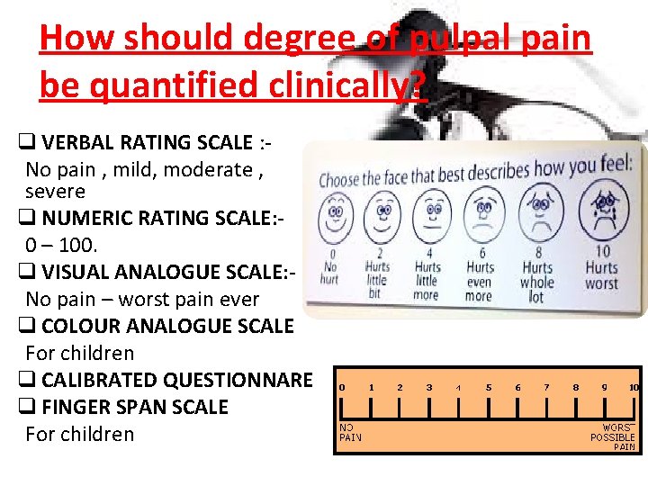
How should degree of pulpal pain be quantified clinically? q VERBAL RATING SCALE : No pain , mild, moderate , severe q NUMERIC RATING SCALE: 0 – 100. q VISUAL ANALOGUE SCALE: No pain – worst pain ever q COLOUR ANALOGUE SCALE For children q CALIBRATED QUESTIONNARE q FINGER SPAN SCALE For children
Past dental history – Reveals information about past dental problems and treatment; reference to chief complaint. – Important to know the date and type of available radiographs to the need for additional radiographs and minimize the patients exposure to radiation
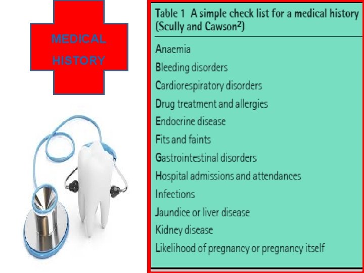
MEDICAL HISTORY
ASA Classification I. A normal healthy patient II. Mild to moderate systemic disease III. Severe systemic disease that limits activity but is not incapacitating IV. Severe systemic disease and is a common threat to life V. A moribund patient not expected to survive 12
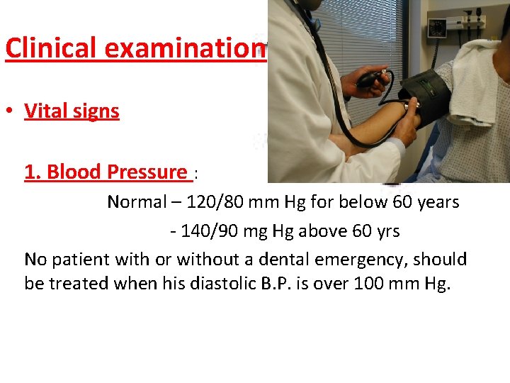
Clinical examination • Vital signs 1. Blood Pressure : Normal – 120/80 mm Hg for below 60 years - 140/90 mg Hg above 60 yrs No patient with or without a dental emergency, should be treated when his diastolic B. P. is over 100 mm Hg.
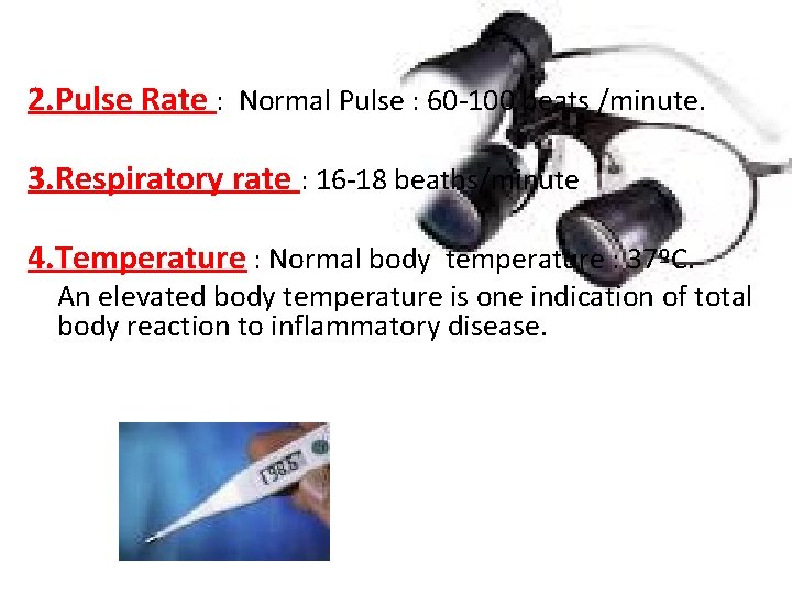
2. Pulse Rate : Normal Pulse : 60 -100 beats /minute. 3. Respiratory rate : 16 -18 beaths/minute 4. Temperature : Normal body temperature : 37ºC. An elevated body temperature is one indication of total body reaction to inflammatory disease.
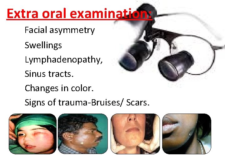
Extra oral examination: Facial asymmetry Swellings Lymphadenopathy, Sinus tracts. Changes in color. Signs of trauma-Bruises/ Scars.
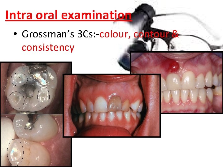
Intra oral examination • Grossman’s 3 Cs: -colour, contour & consistency
Diagnostic tests
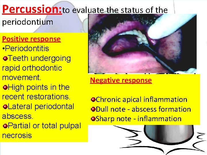
Percussion: to evaluate the status of the periodontium Positive response • Periodontitis Teeth undergoing rapid orthodontic movement. High points in the recent restorations. Lateral periodontal abscess. Partial or total pulpal necrosis Negative response Chronic apical inflammation Dull note - abscess formation Sharp note - inflammation
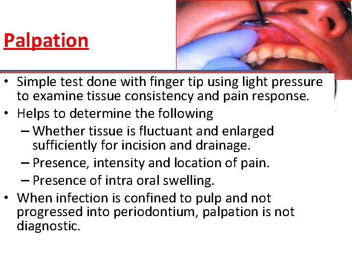
Palpation • Simple test done with finger tip using light pressure to examine tissue consistency and pain response. • Helps to determine the following – Whether tissue is fluctuant and enlarged sufficiently for incision and drainage. – Presence, intensity and location of pain. – Presence of intra oral swelling. • When infection is confined to pulp and not progressed into periodontium, palpation is not diagnostic.
Mobility and Depressibility Tests Rationale – To evaluate the integrity of the attachment apparatus surrounding the teeth. MOBILO METERS Teeth laterally the electronic socket using handles of two instruments or more Theseinare devices /gadgets ; which preferably using two index fingers. aid in determining tooth mobility. Depressibility is performed by applying pressure in an apical direction on the occlusal / incisal aspect of teeth and observing vertical movement if any. GRADES OF MOBILITY Grade I : noticeable movement within the socket Grade II: lateral movement within 1 mm range Grade III : more than 1 mm or Depressibility
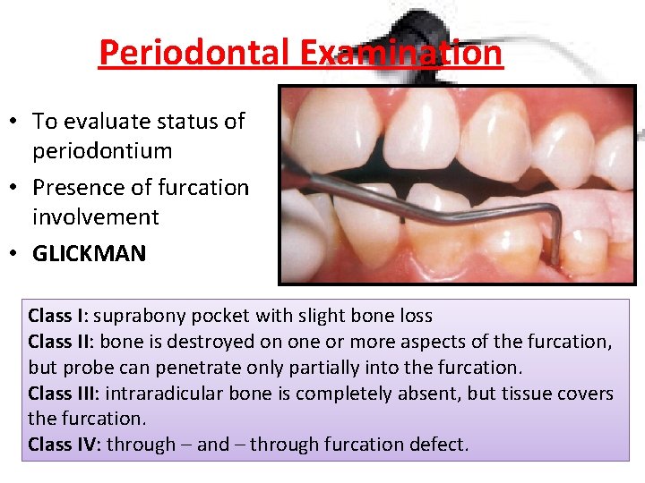
Periodontal Examination • To evaluate status of periodontium • Presence of furcation involvement • GLICKMAN Class I: suprabony pocket with slight bone loss Class II: bone is destroyed on one or more aspects of the furcation, but probe can penetrate only partially into the furcation. Class III: intraradicular bone is completely absent, but tissue covers the furcation. Class IV: through – and – through furcation defect.
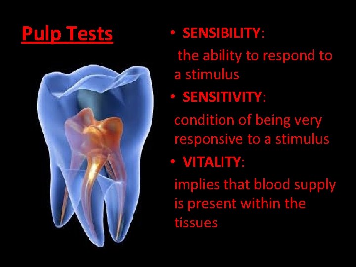
Pulp Tests • SENSIBILITY: the ability to respond to a stimulus • SENSITIVITY: condition of being very responsive to a stimulus • VITALITY: implies that blood supply is present within the tissues
Thermal Tests Cold stimulation • Stimulates fast conducting A delta fibres • Decreases intrapulpal pressure • Sharp localized pain Ingle & Bakland; Endodontics 2002 Heat stimulation • Stimulates the slower conducting C fibres(continued) • Increases intrapulpal pressure • Dull pain of longer duration Bender ; JOE 2000; 26, 175 -9
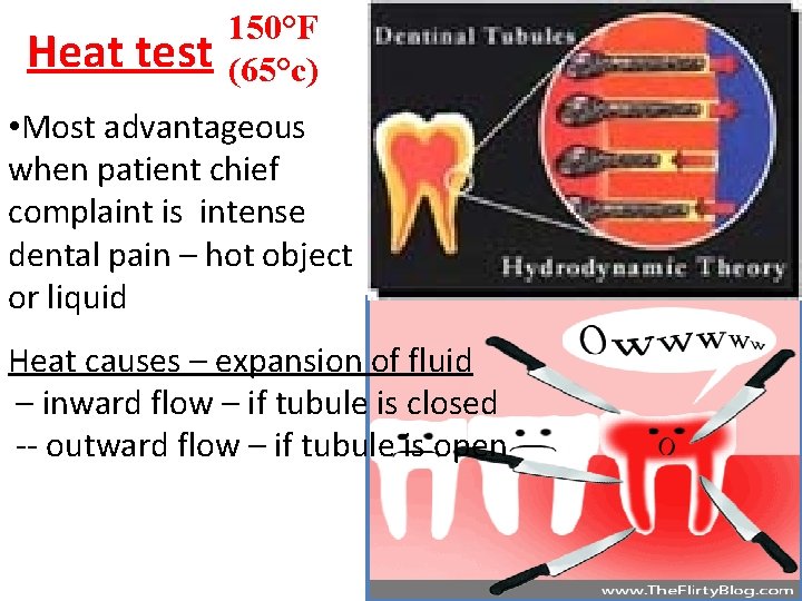
Heat test 150°F (65°c) • Most advantageous when patient chief complaint is intense dental pain – hot object or liquid Heat causes – expansion of fluid – inward flow – if tubule is closed -- outward flow – if tubule is open
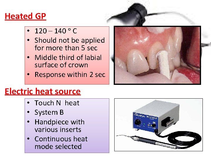
Heated GP • 120 – 140 ᵒ C • Should not be applied for more than 5 sec • Middle third of labial surface of crown • Response within 2 sec Electric heat source • Touch N heat • System B • Handpiece with various inserts • Continuous heat mode selected
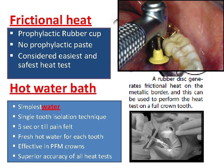
Frictional heat § Prophylactic Rubber cup § No prophylactic paste § Considered easiest and safest heat test Hot water bath § § § Simplestwater Single tooth isolation technique 5 sec or till pain felt Fresh hot water for each tooth Effective in PFM crowns Superior accuracy of all heat tests
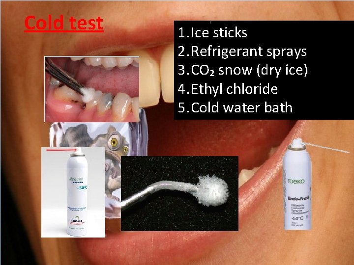
Cold test 1. Ice sticks 2. Refrigerant sprays 3. CO₂ snow (dry ice) 4. Ethyl chloride 5. Cold water bath
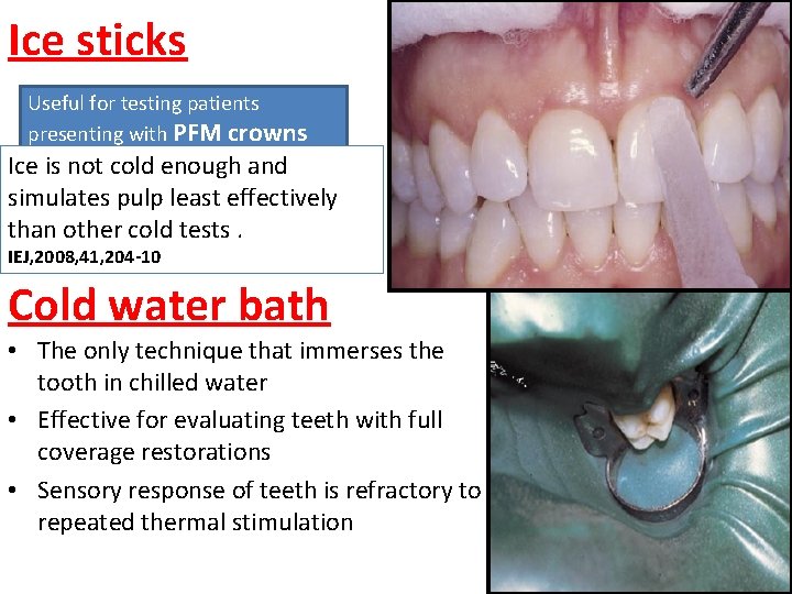
Ice sticks Useful for testing patients presenting with PFM crowns Ice is not cold enough and simulates pulp least effectively than other cold tests. IEJ, 2008, 41, 204 -10 Cold water bath • The only technique that immerses the tooth in chilled water • Effective for evaluating teeth with full coverage restorations • Sensory response of teeth is refractory to repeated thermal stimulation
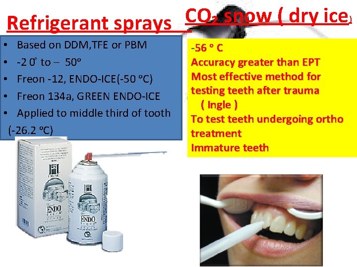
CO₂ snow ( dry ice) Refrigerant sprays • • • Based on DDM, TFE or PBM -2 0 to – 50ᵒ Freon -12, ENDO-ICE(-50 ᵒC) Freon 134 a, GREEN ENDO-ICE Applied to middle third of tooth (-26. 2 ᵒC) -56 ᵒ C Accuracy greater than EPT Most effective method for testing teeth after trauma ( Ingle ) To test teeth undergoing ortho treatment Immature teeth
Responses to thermal test – There are 4 possible responses. – I. No response II. Mild to moderate "degree of awareness of slight pain that subsides within 1 -2 sec. After stimulation has been removed. III. Strong, momentary painful response that subsides within 1 to 2 sec. after the stimulus has been removed. IV. Moderate to strong painful response that lingers for several seconds or longer after the stimulus has been removed.
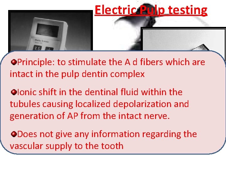
Electric Pulp testing Principle: to stimulate the A d fibers which are intact in the pulp dentin complex Ionic shift in the dentinal fluid within the tubules causing localized depolarization and generation of AP from the intact nerve. Does not give any information regarding the vascular supply to the tooth
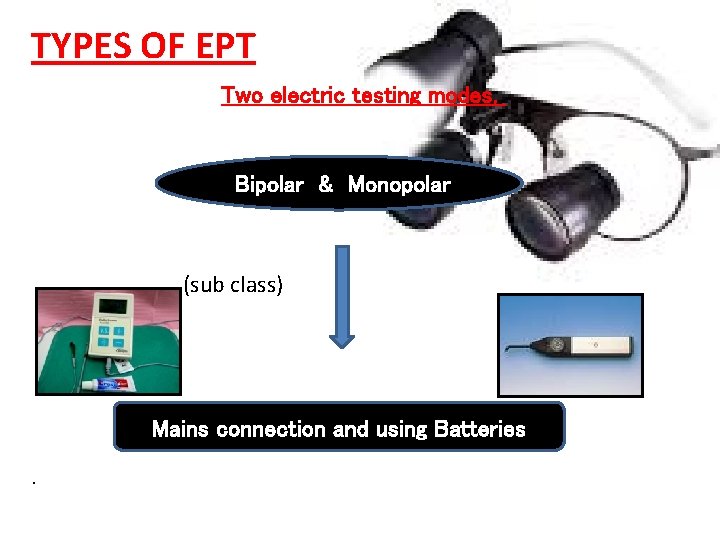
TYPES OF EPT Two electric testing modes, Bipolar & Monopolar (sub class) Mains connection and using Batteries.
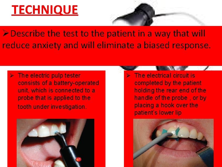
TECHNIQUE ØDescribe the test to the patient in a way that will reduce anxiety and will eliminate a biased response. Ø The electric pulp tester consists of a battery-operated unit, which is connected to a probe that is applied to the tooth under investigation. Ø The electrical circuit is completed by the patient holding the rear end of the handle of the probe , or by placing a hook over the patient’s lower lip
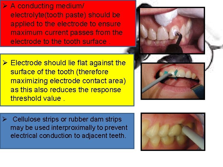
Ø A conducting medium/ electrolyte(tooth paste) should be applied to the electrode to ensure maximum current passes from the electrode to the tooth surface. Ø Electrode should lie flat against the surface of the tooth (therefore maximizing electrode contact area) as this also reduces the response threshold value. Ø Cellulose strips or rubber dam strips may be used interproximally to prevent electrical conduction to adjacent teeth.
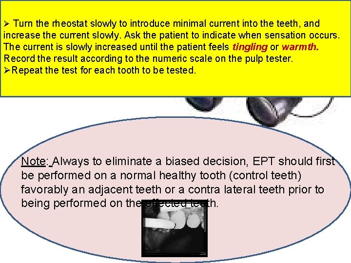
Ø Turn the rheostat slowly to introduce minimal current into the teeth, and increase the current slowly. Ask the patient to indicate when sensation occurs. The current is slowly increased until the patient feels tingling or warmth. Record the result according to the numeric scale on the pulp tester. ØRepeat the test for each tooth to be tested. Note: Always to eliminate a biased decision, EPT should first be performed on a normal healthy tooth (control teeth) favorably an adjacent teeth or a contra lateral teeth prior to being performed on the effected teeth.
PROBLEMS WITH ASSESSING PULP STATUS Ø False-positive results (i. e. non-vital teeth responding positively to testing) Anxious patients Liquefaction necrosis Contact with metal restorations • Vital tissue still present in the partially necrotic root canal system Vital tissue still present in partially necrotic root canal system
Ø False-negative results (i. e. vital teeth responding negatively to testing) Incomplete root development Recently traumatized teeth Sclerosed canals Recent orthodontic activation Patients with psychotic disorder
Anesthetic Test • Pain in the oral cavity is frequently referred from one tooth to an adjacent one or even from one quadrant to the opposing one. • The anesthetic test can help identify the quadrant from where the focus of pain originates. • The suspected tooth should be anesthetized, and, if the diagnosis is correct, the referred pain should disappear, even when it is referred to the opposite arch.
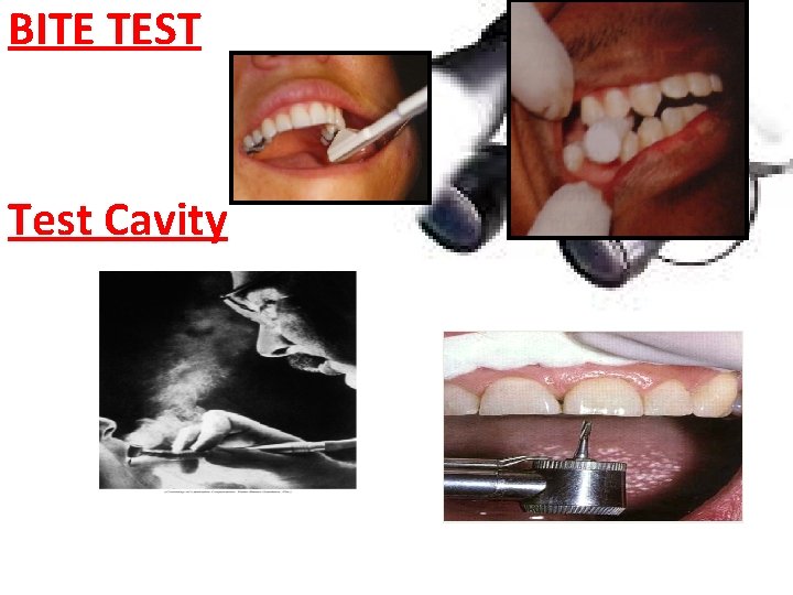
BITE TEST Test Cavity
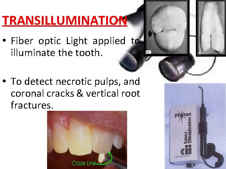
TRANSILLUMINATION • Fiber optic Light applied to illuminate the tooth. • To detect necrotic pulps, and coronal cracks & vertical root fractures.
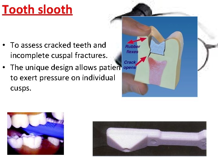
Tooth slooth • To assess cracked teeth and incomplete cuspal fractures. • The unique design allows patient to exert pressure on individual cusps.
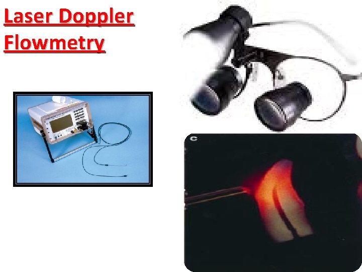
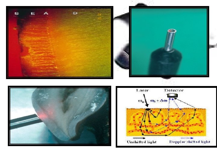
Laser Doppler Flowmetry
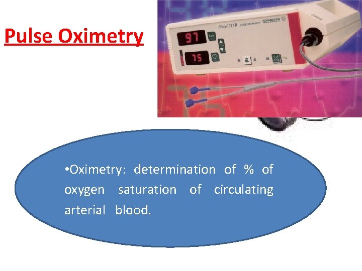
Pulse Oximetry • Oximetry: determination of % of oxygen saturation of circulating arterial blood.
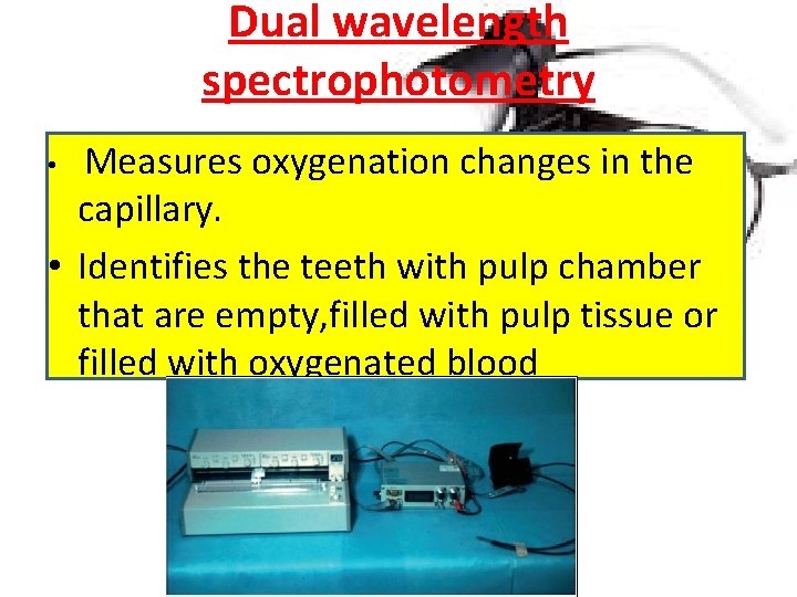
Dual wavelength spectrophotometry Measures oxygenation changes in the capillary. • Identifies the teeth with pulp chamber that are empty, filled with pulp tissue or filled with oxygenated blood •
CONCLUSION • An accurate diagnosis is the key to a successful treatment planning and for restoring the health of the hard tissues and its supporting structures • Determination of the exact pulpal status is complexed as it is dependent on a number of factors like patient history , clinical examination and various diagnostic tests • However with the advent of newer diagnostic techniques, diagnosis can be made more quicker easier and precise.
REFERENCE Ingle’s ENDODONTICS 5 th edition Cohen’s Pathways Of The Pulp 10 th edition ADVANCED ENDODONTICS. R Nageswar Rao Problems in Endodontics- Etiology, Diagnosis and treatment Dental and Maxillofacial radiology-2 nd edition

- Slides: 48