Endocrinology of the Testis John Parrish References Williams


 Mesonephric Tubules Rete Tubules Mullerian Duct Tunica Albuginea")


 Testis")
 Testis pulled")


 Sertolic Cell Precursor")
 Sertolic Cell Precursor Proliferate")
 PGC and Stem Cell")


 • FSH causes Sertoli cells to secret factors that")

 Why is testosterone so high in")
 • • Level of testosterone in Sertoli cell")


 Primary spermatocyte Secondary spermatocyte - short lived and undergoes reduction")


 PGC and Stem Cell")
- Slides: 70
Endocrinology of the Testis John Parrish References: Williams Textbook of Endocrinology
Structure of the Testis Caput Epididymis Spermatic Cord Vas Deferens Vas Efferentia 6 -12 tubules Seminiferous Tubule Tunica Albuginea Corpus Epididymis Rete Testis (within the mediastinum) Cauda Epididymis
Mediastinum Rete Testis Seminiferous Tubule The Testis
Seminiferous Tubule Spermatogonia Primary Spermatocyte Sertoli Cells » Support spermatogenesis Myoid Cells Secondary Spermatocyte Leydig Cells » Testosterone synthesis Round Spermatids Spermatozoa Basement Membrane Capillary
Sertoli Cell
Germ Cell Migration begins by the 4 week of gestation in cow and human.
Migration from endoderm through mesoderm.
XY Male Y Chromosome SRY, SOX 9 Testes develop Sertoli Cells Differentiate Leydig Cells Differentiate SF-1 Testosterone AR Development of Wollfian Ducts 5 a-red SF-1 DHT AMH AR Development of penis scrotum and accessory sex glands AMHR Degeneration of Mullerian Duct
Circulating Androgen • • • Sex Hormone Binding Globulin - 44% Albumin - 54% (1000 fold less affinity than SHBG) Free - 2% Bioavailable Testosterone = free + albumin bound SHBG is made in Liver ABP is made in Sertoli Cells Both also bind estradiol
AMH
Undifferentiated Gonad
Differentiated Reproductive Tracts Male Female
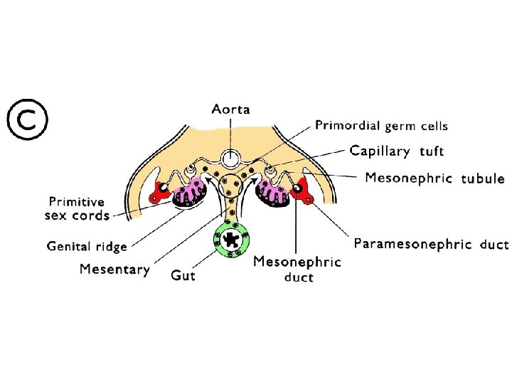
Testicular Development Mesonephric Duct (Wolffian Duct) Mesonephric Tubules Rete Tubules Mullerian Duct Tunica Albuginea Undifferentiated Sex Chords
Mesonephric Tubules Rete Tubules Wolffian Duct Mullerian Duct Primary, Epithelial or Medullary Sex Chords Tunica • Primordial germ cells Albuginea (gonocytes) • Pre-Sertoli Cells
Primary Sex Chords in Fetal Testis Pre-Sertoli - AMH Leydig Cells Testosterone Gonocyte
FSH on Sertoli Cells Hypothalamus Gn. RH • estradiol • inhibin • ABP • tight junctions • growth factors Ant. Pituitary Negative Feedback of Androgens Negative Feedback of Estradiol and Inhibin LH FSH E 2 Leydig Cells ABP + T Inhibin Sertoli Cells T Germ Cells TJ ABP Seminiferous Tubule To Epid.
Negative Feedback • Hypothalamus • Anterior Pituitary » Testosterone conversion to Estradiol » Androgen receptor is less important » Adrogen receptor involved because DHT has negative feedback as well testosterone » Aromatization of testosterone to estradiol also occurs » Inhibin
Leydig Cell and Sertoli Cell Interactions
Hypothalamus Gn. RH Ant. Pituitary Fetal Leydig Cells E 2 Pre-Sertoli Cells T AMH Gonocytes Primary Sex Chord
Testosterone Levels: Fetus - Adult Plasma Testosterone ng/ml Fetal Neonatal Pubertal Adult 5. 0 Adult Leydig Cells 2. 5 Fetal Leydig Cells Birth
Testicular Descent Fusion of the tunica albuginea and peritoneum to form the visceral tunica vaginalis
Front View Spermatic Artery Cranial Suspensory Ligament Fusion of Peritoneum and Gubernaculum Testis Gubernaculum Peritoneum Inguinal Ring
Rapid growth of gubernaculum Spermatic Artery Peritoneum Visceral Growth Testis Gubernaculum (rapid growth) Testis is pulled down to the inguinal ring. Cranial Suspensory Ligament (disappears) Visceral Growth Inguinal Ring Peritoneum Parietal Tunica Vaginalis Visceral Tunica Vaginalis
Gubernaculum regresses Spermatic Artery Peritoneum Visceral Growth Inguinal Canal Testis Gubernaculum (Regressing) Testis pulled into scrotum Peritoneum Vaginal Process Parietal Tunica Vaginalis Visceral Tunica Vaginalis
Spermatic Artery Continued regression of Gubernaculum Peritoneum Inguinal Canal Testis pulled deeper into Scrotum Peritoneum Vaginal Process attaches to Scrotum Space between Visceral and Parietal T. V. is continuous with Peritoneum Vaginal Process Testis Gubernaculum (Fully Regressed) Parietal Tunica Vaginalis Visceral Tunica Vaginalis
Testicular Descent • • • Cryptorchid - failure of descent Controlling mechanisms » » » Androgen dependent DHT supported Estrogen inhibits » INSL 3 - insulin like growth factor 3 » Great/LGR 8 - receptor for INSL 3 Gene Expression – From leydig cells – In gubernaculum
Timing of Testicular Descent in Mammals Gonad to Adominal Pause Transinguinal Inguinoscrotal testis Translocation Testis in Scrotum Cattle Deer Human Horse Pig Dog Rabbit Mouse 0 20 40 60 80 Birth 120 Interval After Conception (% of Gestation) 140
Normal Dog Seminiferous Tubule
Cryptorchid Dog Seminiferous Tubule Sertoli Cells Increased Chance of Testicular Cancer
Primary Sex Chord to Seminiferous Tubule Primodial Germ Cell (PGC) Sertolic Cell Precursor
Primary Sex Chord to Seminiferous Tubule Primodial Germ Cell (PGC) Sertolic Cell Precursor Proliferate to form Spermatogonial Stem Cells Stem Cell Niche • Along basement membrane
Primary Sex Chord to Seminiferous Tubule Primodial Germ Cell (PGC) PGC and Stem Cell Spermatogonia • Express a 3 -, a 5 -, a. V-, b 1 -integrins • Initially express ckit receptor, then stop Sertolic Cell Precursor Proliferate to form Spermatogonial Stem Cells Stem Cell Niche • Along basement membrane
FSH on Sertoli Cells Hypothalamus Gn. RH • estradiol • inhibin • ABP • tight junctions • growth factors Ant. Pituitary Negative Feedback of Androgens Negative Feedback of Estradiol and Inhibin LH FSH E 2 Leydig Cells ABP + T Inhibin Sertoli Cells T Germ Cells TJ ABP Seminiferous Tubule To Epid.
Sertoli Cell Regulation • Switch from FSH to Testosterone » FSH causes » at puberty, – FSH • increase in c. AMP phosphodiesterase increases less effective » Testosterone takes over regulation Germ cells effect ability of Sertoli cell to respond to testosterone
Sertoli Cell Regulation (cont. ) • FSH causes Sertoli cells to secret factors that increase Leydig cell response to LH » at puberty or start of breeding season – FSH increases first – Sertoli cells trigger cells the development of SER in Leydig
Hormonal Regulation of Spermatogenesis • Level of Testosterone » intratesticular testosterone is higher than in circulation » Seminiferous Tubule Fluid (STF) testosterone » Androgen receptor Kd for testosterone IF 600 n. M (Leydig cells here) TV 250 n. M SV 150 n. M PV 20 n. M It is usually 30 to 170 times that seen in PV STF 3 n. M 170 n. M
• Hormonal Regulation of Spermatogenesis (Cont. ) Why is testosterone so high in STF? » Does testosterone work by androgen receptor? – Yes, » What DHT is more effective than testosterone is level of free testosterone in STF? – Have not been able – ABP is produced by to measure sertoli cells under influence of FSH and is present at 30 - 40 n. M • Certainly reduces free testosterone but there is still excess testosterone » Artificial testosterone via implants found need 75 n. M to maintain spermatogenesis
Hormonal Regulation of Spermatogenesis (Cont. ) • • Level of testosterone in Sertoli cell » Testosterone is converted to estrogen in sertoli cell » Has not been addressed No good answer
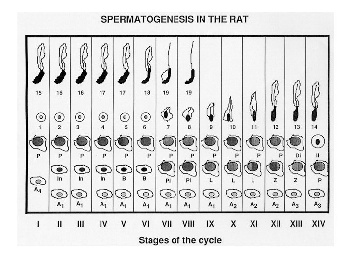
Mitotic Divisions of Spermatogenesis As Apr Aal - stem cells, resting pool, slow dividing A type spermatogonia (1 - 4 types ) Intermediate spermatogonia B type spermatogonia Primary spermatocyte
Mitotic Divisions of Spermatogenesis As Apr Aal - stem cells, resting pool, slow dividing A type spermatogonia (1 - 4 types ) Random Divisions Intermediate spermatogonia B type spermatogonia Primary spermatocyte Stage Specific Divisions
Meiotic Divisions of Spermatogenesis Primary spermatocyte - long interval • preleptotene - DNA synthesis • leptotene - condensation of the chromatin • zygotene - thickening of chromosomes and pairing • pachytene - RNA synthesis, thickening of chromosomes, crossing over • diplotene - chromosomes separate but remain attached at chiasma • diakinesis - cells separate and divide
Meiotic Divisions (cont. ) Primary spermatocyte Secondary spermatocyte - short lived and undergoes reduction division Round spermatid spermiogenesis Spermiation
Sperm Chromatin Sturcture
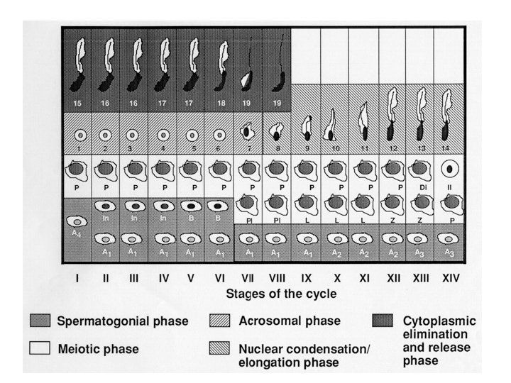
Cytoplasmic Elimination Phase and Spermiation • residual body loss • phagocytosis by sertoli cell • elong. sperm. influences sertoli cell – sem. tubual fluid – ABP – inhibin – interleukin 1
Seminiferous Tubule
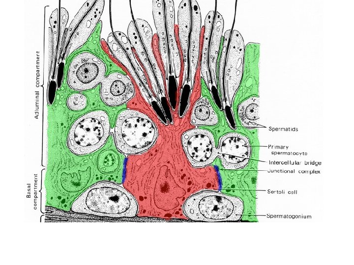
Lumen Amann 1983 Adluminal Spermatids 1° Spermatocyte Basal Interstitial Sertoli Cell Spermatogonium Leydig Cell Blood Vessel
Cytoplasmic Bridges Present Among Daughter Cells All develop surrounded by 1 Sertoli cell ! Degenerating Spermatogonia (Apoptosis, as high as 75%)
Over Population of Spermatogonia Tight Junction Sertoli Formation - FSH Maintenance - T Sertoli SG SG SG Basement Membrane Normal PS Sertoli AP Apoptosis Inhibited Sertoli PS SG Sertoli AP SG Basement Membrane SG Sertoli SG SG SG Basement Membrane
Ap Ad Stem Cell Pool FSH B P Spermatogonial Renewal in the Primate
Spermatogenesis Every 13. 5 Days sperm are released from this point Spermatid Round Spermatid Secondary Spermatocyte Sertoli Cells Primary Spermatocyte Spermatogonia Every 13. 5 Days a new group of cells initiate the cycle Myoid Cells
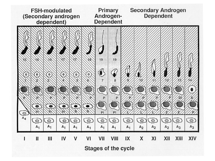
Stages • Specific cellular associations within a small segment of a seminiferous tubule • stages are not the same length in time
Cycle • progression through sequence of all stages
Normal Apoptosis
Effect of Hormone Withdrawal • Experimental Approaches » Hypox. – can’t » Gn. RH restore with testosterone alone agonist – Decreased FSH and LH release – Requires both FSH and Testosterone spematogenesis » Ethane to get normal dimethane sulfonate (destroys leydig cells) – get destruction of spermatogenesis – testosterone can overcome effects of not prevent destruction of Leydig cells EDS except does
Essential Prevents Apoptosis Testosterone Effects
FSH Effects Prevents Apoptosis N-cadherin SCF Activin B to P promoted FSH Receptors Highest SCF Testosterone Effects
N-cadherin loss FSH + T N-cadherin SCF Activin FSH Receptors Highest SCF Testosterone Effects
N-cadherin loss N-cadherin Activin SCF FSH Receptors Highest SCF Testosterone Effects
Normal Dog Seminiferous Tubule
Human Male Reproductive Tract Hormonal Dependence • Testosterone » Wolfian Duct §Vas efferentia §Epididymis §Vas deferens §Seminal Vesicles • DHT » Rest of Male Structures §Prostate §Cowper’s §Penis
Human Testis
Human Male Reproductive Tract and Bladder Seminal Vesicles Prostate Vas Deferens
Hyperplasia of Human Prostate • • • DHT driven via Androgen Receptor 5 -α-reductase inhibitors common treatment » Type 2 enzyme in prostate, hair follicle – No effect on libido – Hair follicles and other DHT dependent tissue affected Estradiol increases androgen receptor in prostate and estradiol increase with age
The End
Primary Sex Chord to Seminiferous Tubule Primodial Germ Cell (PGC) PGC and Stem Cell Spermatogonia • Express a 3 -, a 5 -, a. V-, b 1 -integrins • Initially express ckit receptor, then stop Sertolic Cell Precursor Proliferate to form Spermatogonial Stem Cells Stem Cell Niche • Along basement membrane