
Endocrine System Overview n n n Endocrine system

- Slides: 47
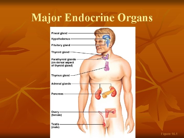
Endocrine System: Overview n n n Endocrine system – the body’s second great controlling system which influences metabolic activities of cells by means of hormones Endocrine glands – pituitary, thyroid, parathyroid, adrenal, pineal, and thymus The pancreas and gonads produce both hormones and exocrine products
Endocrine System: Overview n n The hypothalamus has both neural functions and releases hormones Other tissues and organs that produce hormones – adipose cells, pockets of cells in the walls of the small intestine, stomach, kidneys, and heart
Major Endocrine Organs Figure 16. 1
Autocrines and Paracrines n n n Autocrines – chemicals that exert effects on the same cells that secrete them Paracrines – locally acting chemicals that affect cells other than those that secrete them These are not considered hormones since hormones are long-distance chemical signals
Hormones n Hormones – chemical substances secreted by cells into the extracellular fluids n n n Regulate the metabolic function of other cells Have lag times ranging from seconds to hours Tend to have prolonged effects Are classified as amino acid-based hormones, or steroids Eicosanoids – biologically active lipids with local hormone–like activity
Types of Hormones n Amino acid based n n Steroids – gonadal and adrenocortical hormones, and hormones from the kidneys n n Amines, thyroxine, peptide, and protein hormones Remain in the blood stream longer because they are bound to proteins Eicosanoids – leukotrienes and prostaglandins n Are derived from lipids
Hormone Action n Hormones alter target cell activity by one of two mechanisms n Second messengers: Regulatory G proteins n Amino acid–based hormones n n Direct gene activation n n Steroid hormones The precise response depends on the type of the target cell
Mechanism of Hormone Action n Hormones produce one or more of the following cellular changes in target cells n n n Alter plasma membrane permeability Stimulate protein synthesis Activate or deactivate enzyme systems Induce secretory activity Stimulate mitosis
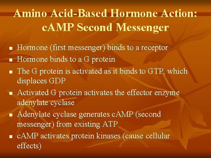
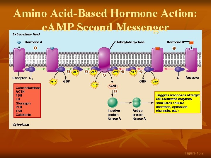
Amino Acid-Based Hormone Action: c. AMP Second Messenger n n n Hormone (first messenger) binds to a receptor Hormone binds to a G protein The G protein is activated as it binds to GTP, which displaces GDP Activated G protein activates the effector enzyme adenylate cyclase Adenylate cyclase generates c. AMP (second messenger) from existing ATP c. AMP activates protein kinases (cause cellular effects)
Amino Acid-Based Hormone Action: c. AMP Second Messenger Extracellular fluid Hormone A Adenylate cyclase Hormone B 1 1 2 Receptor Gs Catecholamines ACTH FSH LH Glucagon PTH TSH Calcitonin GTP GDP GTP 3 GTP ATP 4 GTP 3 GTP 2 GDP GTP Gi Receptor c. AMP 5 Inactive protein kinase A Active protein kinase A Triggers responses of target cell (activates enzymes, stimulates cellular secretion, opens ion channels, etc. ) Cytoplasm Figure 16. 2
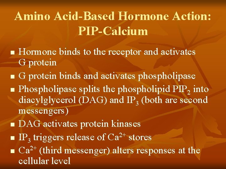
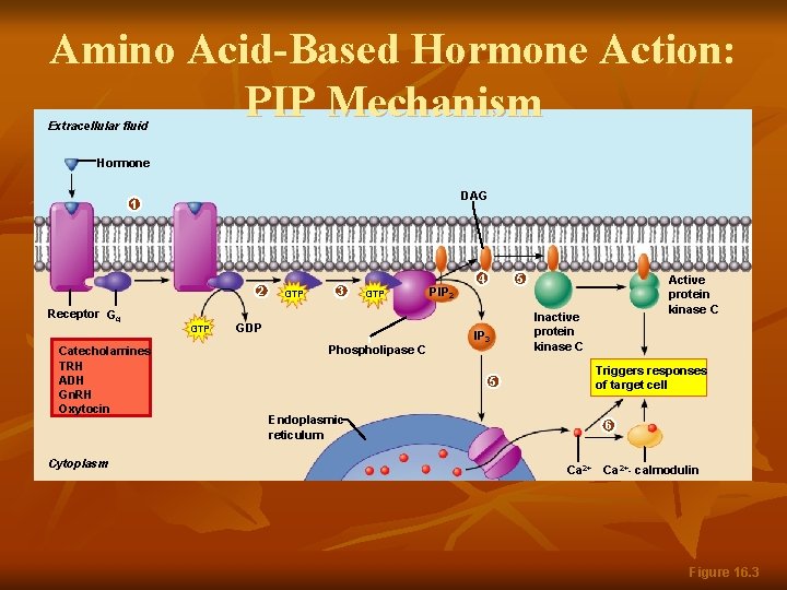
Amino Acid-Based Hormone Action: PIP-Calcium n n n Hormone binds to the receptor and activates G protein binds and activates phospholipase Phospholipase splits the phospholipid PIP 2 into diacylglycerol (DAG) and IP 3 (both are second messengers) DAG activates protein kinases IP 3 triggers release of Ca 2+ stores Ca 2+ (third messenger) alters responses at the cellular level
Amino Acid-Based Hormone Action: PIP Mechanism Extracellular fluid Hormone DAG 1 2 Receptor Gq Catecholamines TRH ADH Gn. RH Oxytocin Cytoplasm GTP 3 GTP GDP Phospholipase C PIP 2 4 5 IP 3 Active protein kinase C Inactive protein kinase C Triggers responses of target cell 5 Endoplasmic reticulum 6 Ca 2+- calmodulin Figure 16. 3
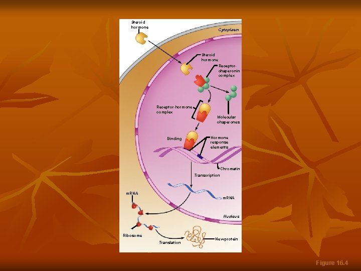
Steroid Hormones n n This interaction prompts DNA transcription to produce m. RNA The m. RNA is translated into proteins, which bring about a cellular effect
Steroid hormone Cytoplasm Steroid hormone Receptorchaperonin complex Receptor-hormone complex Molecular chaperones Binding Hormone response elements Chromatin Transcription m. RNA Nucleus Ribosome Translation New protein Figure 16. 4
Target Cell Specificity n n n Hormones circulate to all tissues but only activate cells referred to as target cells Target cells must have specific receptors to which the hormone binds These receptors may be intracellular or located on the plasma membrane Steroid hormone Figure 16. 4
Target Cell Specificity n Examples of hormone activity n n ACTH receptors are only found on certain cells of the adrenal cortex Thyroxin receptors are found on nearly all cells of the body
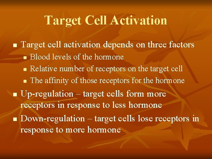
Target Cell Activation n Target cell activation depends on three factors n n n Blood levels of the hormone Relative number of receptors on the target cell The affinity of those receptors for the hormone Up-regulation – target cells form more receptors in response to less hormone Down-regulation – target cells lose receptors in response to more hormone
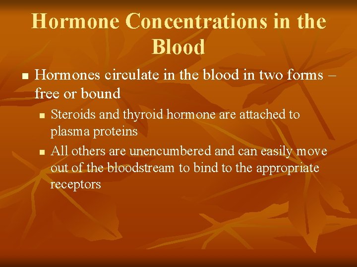
Hormone Concentrations in the Blood n Hormones circulate in the blood in two forms – free or bound n n Steroids and thyroid hormone are attached to plasma proteins All others are unencumbered and can easily move out of the bloodstream to bind to the appropriate receptors
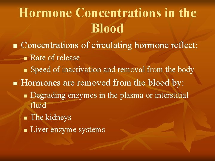
Hormone Concentrations in the Blood n Concentrations of circulating hormone reflect: n n n Rate of release Speed of inactivation and removal from the body Hormones are removed from the blood by: n n n Degrading enzymes in the plasma or interstitial fluid The kidneys Liver enzyme systems
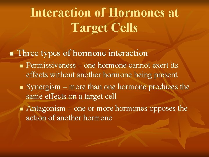
Interaction of Hormones at Target Cells n Three types of hormone interaction n Permissiveness – one hormone cannot exert its effects without another hormone being present Synergism – more than one hormone produces the same effects on a target cell Antagonism – one or more hormones opposes the action of another hormone
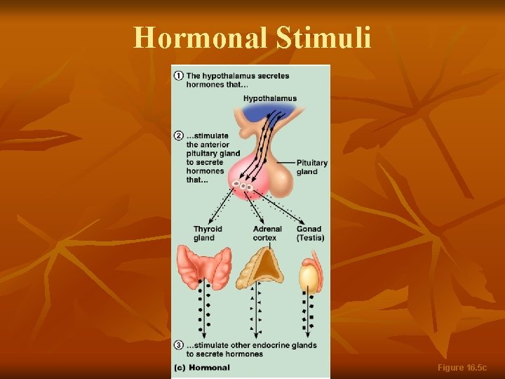
Control of Hormone Release n Blood levels of hormones: n n n Are controlled by negative feedback systems Vary only within a narrow desirable range Hormones are synthesized and released in response to: n n n Humoral stimuli Neural stimuli Hormonal stimuli
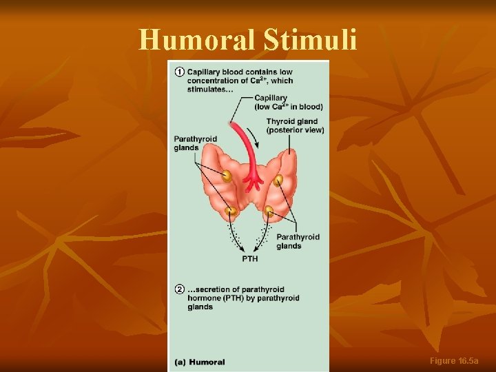
Humoral Stimuli n n Humoral stimuli – secretion of hormones in direct response to changing blood levels of ions and nutrients Example: concentration of calcium ions in the blood n n Declining blood Ca 2+ concentration stimulates the parathyroid glands to secrete PTH (parathyroid hormone) PTH causes Ca 2+ concentrations to rise and the stimulus is removed
Humoral Stimuli Figure 16. 5 a
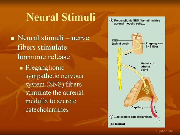
Neural Stimuli n Neural stimuli – nerve fibers stimulate hormone release n Preganglionic sympathetic nervous system (SNS) fibers stimulate the adrenal medulla to secrete catecholamines Figure 16. 5 b
Hormonal Stimuli n Hormonal stimuli – release of hormones in response to hormones produced by other endocrine organs n n The hypothalamic hormones stimulate the anterior pituitary In turn, pituitary hormones stimulate targets to secrete still more hormones
Hormonal Stimuli Figure 16. 5 c
Nervous System Modulation n The nervous system modifies the stimulation of endocrine glands and their negative feedback mechanisms
Nervous System Modulation n The nervous system can override normal endocrine controls n For example, control of blood glucose levels Normally the endocrine system maintains blood glucose n Under stress, the body needs more glucose n The hypothalamus and the sympathetic nervous system are activated to supply ample glucose n
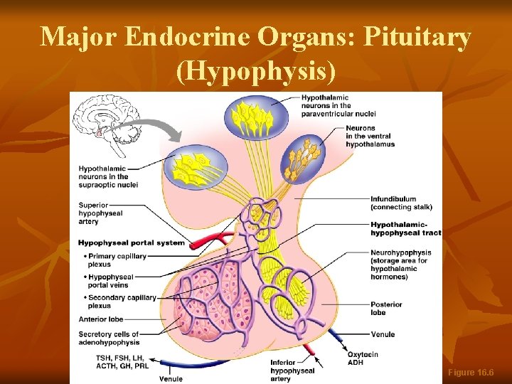
Major Endocrine Organs: Pituitary (Hypophysis) n n Pituitary gland – two-lobed organ that secretes nine major hormones Neurohypophysis – posterior lobe (neural tissue) and the infundibulum n n Receives, stores, and releases hormones from the hypothalamus Adenohypophysis – anterior lobe, made up of glandular tissue n Synthesizes and secretes a number of hormones
Major Endocrine Organs: Pituitary (Hypophysis) Figure 16. 6
Pituitary-Hypothalamic Relationships: Posterior Lobe n n The posterior lobe is a downgrowth of hypothalamic neural tissue Has a neural connection with the hypothalamus (hypothalamic-hypophyseal tract) Nuclei of the hypothalamus synthesize oxytocin and antidiuretic hormone (ADH) These hormones are transported to the posterior pituitary
Pituitary-Hypothalamic Relationships: Anterior Lobe n n The anterior lobe of the pituitary is an outpocketing of the oral mucosa There is no direct neural contact with the hypothalamus
Pituitary-Hypothalamic Relationships: Anterior Lobe n There is a vascular connection, the hypophyseal portal system, consisting of: n n n The primary capillary plexus The hypophyseal portal veins The secondary capillary plexus
Adenophypophyseal Hormones n The six hormones of the adenohypophysis: n n n Abbreviated as GH, TSH, ACTH, FSH, LH, and PRL Regulate the activity of other endocrine glands In addition, pro-opiomelanocortin (POMC): n n Has been isolated from the pituitary Is split into ACTH, opiates, and MSH
Activity of the Adenophypophysis n The hypothalamus sends a chemical stimulus to the anterior pituitary n n n Releasing hormones (RH) stimulate the synthesis and release of hormones Inhibiting hormones (IH) shut off the synthesis and release of hormones Rate of secretion is controlled via negative feedback
Activity of the Adenophypophysis n The tropic hormones that are released are: n n Thyroid-stimulating hormone (TSH) Adrenocorticotropic hormone (ACTH) Follicle-stimulating hormone (FSH) Luteinizing hormone (LH)
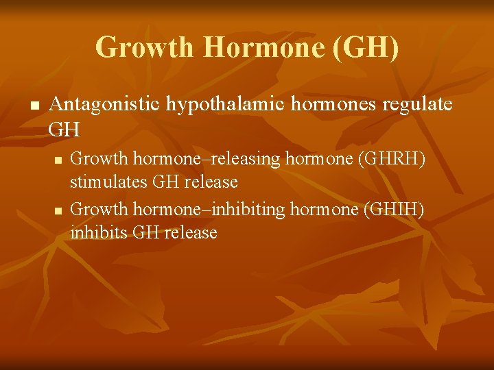
Growth Hormone (GH) n Produced by somatotropic cells of the anterior lobe that: n n n Stimulate most cells, but target bone and skeletal muscle Promote protein synthesis and encourage the use of fats for fuel Most effects are mediated indirectly by somatomedins
Growth Hormone (GH) n Antagonistic hypothalamic hormones regulate GH n n Growth hormone–releasing hormone (GHRH) stimulates GH release Growth hormone–inhibiting hormone (GHIH) inhibits GH release
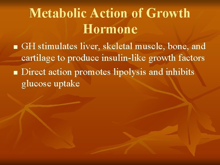
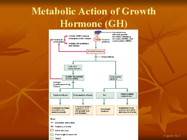
Metabolic Action of Growth Hormone n n GH stimulates liver, skeletal muscle, bone, and cartilage to produce insulin-like growth factors Direct action promotes lipolysis and inhibits glucose uptake
Metabolic Action of Growth Hormone (GH) Figure 16. 7
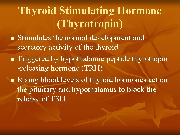
Thyroid Stimulating Hormone (Thyrotropin) n n n Stimulates the normal development and secretory activity of the thyroid Triggered by hypothalamic peptide thyrotropin -releasing hormone (TRH) Rising blood levels of thyroid hormones act on the pituitary and hypothalamus to block the release of TSH
Adrenocorticotropic Hormone (Corticotropin) n n n Stimulates the adrenal cortex to release corticosteroids Triggered by hypothalamic corticotropinreleasing hormone (CRH) in a daily rhythm Internal and external factors such as fever, hypoglycemia, and stressors can trigger the release of CRH
Gonadotropins n Gonadotropins – follicle-stimulating hormone (FSH) and luteinizing hormone (LH) n n Regulate the function of the ovaries and testes FSH stimulates gamete (egg or sperm) production Absent from the blood in prepubertal boys and girls Triggered by the hypothalamic gonadotropinreleasing hormone (Gn. RH) during and after puberty
Functions of Gonadotropins n In females n n n LH works with FSH to cause maturation of the ovarian follicle LH works alone to trigger ovulation (expulsion of the egg from the follicle) LH promotes synthesis and release of estrogens and progesterone
Functions of Gonadotropins n In males n n LH stimulates interstitial cells of the testes to produce testosterone LH is also referred to as interstitial cell-stimulating hormone (ICSH)
Prolactin (PRL) n n n In females, stimulates milk production by the breasts Triggered by the hypothalamic prolactinreleasing hormone (PRH) Inhibited by prolactin-inhibiting hormone (PIH) Blood levels rise toward the end of pregnancy Suckling stimulates PRH release and encourages continued milk production
The Posterior Pituitary and Hypothalamic Hormones n n n Posterior pituitary – made of axons of hypothalamic neurons, stores antidiuretic hormone (ADH) and oxytocin ADH and oxytocin are synthesized in the hypothalamus ADH influences water balance Oxytocin stimulates smooth muscle contraction in breasts and uterus Both use PIP-calcium second-messenger mechanism