Endocrine system Fatima Obeidat MD Department of Pathology

Endocrine system Fatima Obeidat, MD Department of Pathology and Laboratory Medicine

I. Thyroid diseases

I. THYROTOXICOSIS - Most commonly, it is caused by hyperfunction of the thyroid gland ( called hyperthyroidism) Causes of thyrotoxixosis are: I. Associated with hyperthyroidism (Thyroid hyperfunction): 1. Primary a. Diffuse toxic hyperplasia (Graves disease)
 multinodular goiter) c. Hyperfunctioning (toxic ) adenoma Secondary -- TSH-secreting")
. b. Hyperfunctioning (Toxic) multinodular goiter) c. Hyperfunctioning (toxic ) adenoma Secondary -- TSH-secreting pituitary adenoma (rare) II. Not associated with hyperthyroidism - Excessive release of pre-formed hormone in thyroiditis

Clinical manifestations of thyrotoxicosis a. Constitutional symptoms : warm flushed skin, heat intolerance and excessive sweating - Weight loss despite increased appetite. b. Malabsorption, and diarrhea. c. Tachycardia and elderly patients may develop heart failure due to aggravation of pre-existing heart

d. Nervousness, tremor, and irritability e. A wide, staring gaze and lid lag because of sympathetic overstimulation of the levator palpebrae superioris Note: True thyroid ophthalmopathy associated with proptosis is a feature seen only in Graves disease. f. 50% develop proximal muscle weakness (thyroid myopathy).

Note: - The measurement of serum TSH is the most useful single screening test for hyperthyroidism, because TSH levels are decreased even at the earliest stages, when the disease may still be subclinical

- Once the diagnosis of thyrotoxicosis has been confirmed measurement of radioactive iodine uptake by the thyroid gland often is valuable in determining the etiology For example, such scans may show : a. Diffusely increased (whole-gland) uptake in Graves disease, b. Increased uptake in a solitary nodule in toxic adenoma c. Or decreased uptake in thyroiditis.
 Thyroiditis - Is the most common cause of")
II. Thyroiditis 1. Chronic Lymphocytic (Hashimoto) Thyroiditis - Is the most common cause of hypothyroidism in areas of the world where iodine levels are sufficient. - It is characterized by gradual thyroid failure secondary to autoimmune destruction of the thyroid gland

- It is most prevalent between the ages of 45 and 65 years and is more common in women than in men - It can occur in children and is a major cause of non-endemic goiter in children

• Gross : - Diffuse and symmetric enlargement of the thyroid but localized enlargement may be seen in some cases to raise suspicion for neoplasm Microscopic examination reveals 1. Infiltration by small lymphocytes, plasma cells, and well-developed germinal centers

2. The thyroid follicles are atrophic 2. Some follicles are lined by epithelial cells with abundant eosinophilic, cytoplasm, termed Hürthle cells and these Hurthle cells have numerous mitochondria

. Clinically , 1. Painless thyroid enlargement associated with some degree of hypothyroidism, 2. - In the usual clinical course, hypothyroidism develops gradually. ; however, it may be preceded by transient thyrotoxicosis due to disruption of thyroid follicles , and secondary release of thyroid hormones (hashitoxicosis).

Hashimoto thyroiditis

- Patients with Hashimoto thyroiditis often : 1. Have other autoimmune diseases 2. . Are at increased risk for the development of B cell non-Hodgkin lymphomas within the thyroid gland. Note: - The relationship between Hashimoto disease and thyroid epithelial cancers remains controversial, with some morphologic and molecular studies suggesting a predisposition to papillary
 Thyroiditis - Is much less common than Hashimoto disease")
2. Subacute Granulomatous (de Quervain) Thyroiditis - Is much less common than Hashimoto disease - Is most common between the ages of 30 and 50 and, - More frequently in women than in men. - Is believed to be caused by a viral infection and a majority of patients have a history of an upper respiratory infection just before the onset of thyroiditis.

Gross-The gland has intact capsule, and may be unilaterally or bilaterally enlarged. Histologic examination reveals 1. Disruption of thyroid follicles, with extravasation of colloid

leading to a neutrophilic infiltrate, which is replaced by lymphocytes, plasma cells, and macrophages. 2. The extravasated colloid provokes a granulomatous reaction with giant cells that contain fragments of colloid. 3. Healing occurs by resolution of inflammation and fibrosis
 -")
. Clinical Features : -Acute onset characterized by neck pain ( with swallowing) - Fever, malaise, and variable enlargement of the thyroid. - Transient hyperthyroidism may occur as a result of disruption of follicles and release of excessive hormones. - The leukocyte count is increased.

De. Quervain THYROIDITIS

De. Quervain THYROIDITIS

- With progression of disease and gland destruction, a transient hypothyroid phase may ensue. - The condition typically is selflimited, with most patients returning to a euthyroid state within 6 to 8 weeks

3. . Subacute Lymphocytic Thyroiditis : - Also is known as silent or painless thyroiditis; - And in a subset of patients the onset of disease follows - pregnancy (postpartum thyroiditis). - Most likely to be autoimmune because circulating antithyroid antibodies are found in a majority of patients - It mostly affects middle-aged women, who present with a- painless neck mass or features of thyrotoxicosis

- The initial phase of thyrotoxicosis (which is likely to be secondary to thyroid tissue damage) is followed by return To euthyroid state within a few months. - In a minority of affected persons the condition eventually progresses to hypothyroidism. - The histologic features consist of lymphocytic infiltration

4. Riedel thyroiditis, : -A rare disorder of unknown etiology, - Characterized by extensive fibrosis involving the thyroid and contiguous structures simulating a thyroid neoplasm - May be associated with idiopathic fibrosis in other parts of the body, such as the retroperitoneum - The presence of circulating antithyroid antibodies in most patients suggests an autoimmune etiology

Riedel thyroiditis, :

Riedel thyroiditis, :

. IV. GRAVES DISEASE LIs the most common cause of endogenous hyperthyroidism with a peak incidence in women between the ages of 20 and 40 - It is characterized by a triad of manifestations: A. Thyrotoxicosis, caused by a diffusely enlarged, hyper-functional thyroid, is present in all cases.

B. Infiltrative ophthalmopathy -with resultant exophthalmos is noted in 40% of patients as a result of increased volume of the retro-orbital connective tissues by 1. Marked infiltration T cells with inflammatory edema 2. Accumulation of glycosaminoglycans 3. Increased numbers of adipocytes (fatty infiltration)

Exophthalmos

. - These changes displace the eyeball forward, potentially interfering with the function of the extraocular muscles - Exophthalmos may persist after successful treatment of the thyrotoxicosis, and may result in corneal injury. C. A localized, infiltrative dermopathy ( pretibial myxedema) is seen in a minority of cases and involves the skin overlying the shins, and manifests as scaly thickening

Pretibial myxedema

. Gross: Symmetrical enlargement of the thyroid gland with intact capsule, On microscopic examination, a. The follicular cells in untreated cases are tall, and more crowded and may result in formation of small papillae b. Lymphoid infiltrates, consisting predominantly of T cells, with few B cells and plasma cells are present throughout the interstitium; with formation of germinal centers

Laboratory findings and radiologic findings - Elevated serum free T 4 and T 3 and depressed serum TSH - Because of ongoing stimulation of the thyroid follicles by antibodies, radioactive iodine uptake is increased, and radionucleotide scans show a diffuse uptake of iodine

Graves Disease

V. MULTINODULAR GOITER - Enlargement of the thyroid, or goiter, is the most common manifestation of thyroid disease Mechanism : - The goiters reflect impaired synthesis of thyroid hormone often caused by dietary iodine deficiency and this leads to a compensatory rise in the serum TSH, which in turn causes hyperplasia of the follicular cells and, ultimately, gross enlargement of the thyroid gland. ,

- The compensatory increase in functional mass of the gland is enough to overcome the hormone deficiency, ensuring a euthyroid state in the vast majority of persons - If the underlying disorder is sufficiently severe (congenital biosynthetic defect), the compensatory responses may be inadequate to overcome the impairment in hormone synthesis, resulting in goitrous hypothyroidism. - The degree of thyroid enlargement is proportional to the level and duration of thyroid hormone deficiency

Goiters can be endemic or sporadic. I. Endemic goiter : Occurs in geographic areas where the soil, water, and food supply contain little iodine. - The term endemic is used when goiters are present in more than 10% of the population in a given region. - Such conditions are common in mountainous areas of the world, including the Himalayas and the Andes but with increasing availability of iodine supplementation, the frequency and severity of endemic goiter have declined

II. Sporadic goiter : Less common than endemic goiter. - The condition is more common in females than in males, with a peak incidence in puberty or young adulthood, when there is an increased physiologic demand for T 4. - It may be caused by several conditions, including the:

a. Ingestion of substances that interfere with thyroid hormone synthesis , such as excessive calcium and vegetables such as cabbage, cauliflowers, . b. Hereditary enzymatic defects that interfere with thyroid hormone synthesis (dyshormonogenetic goiter). -In most cases, the cause of sporadic goiter is not apparent

Multinodular Goiter

- Multinodular goiters cause multilobulated, asymmetrically enlarged glands which attain massive size and old lesions often show fibrosis, hemorrhage, calcification - Multinodular goiters are typically are hormonally silent, - 10% of patients can manifest with thyrotoxicosis due to the development of autonomous nodules producing hormone independent of TSH stimulation and this condition, called

Clinical Features : a. The dominant features are mass effects of the goiter b. In addition to the cosmetic problem of a large neck mass goiters also may cause airway obstruction, dysphagia, and compression of large vessels in the neck and upper thorax (socalled superior vena cava syndrome). c. The incidence of malignancy in long-standing multinodular goiters is low (less than 5%) but not zero and concern for malignancy arises with goiters that demonstrate sudden changes in size or associated symptoms ( hoarseness)

VI. Thyroid tumors : - From a clinical standpoint, the possibility of a cancer is of major concern in patients who present with thyroid nodules but fortunately, the majority of solitary nodules of the thyroid prove to be either a. Follicular adenomas b. A dominant nodule in multinodular goiter c. Simple cysts or foci of thyroiditis - Carcinomas of the thyroid, are uncommon, accounting for much less than 10% of solitary thyroid nodules.

- Several clinical criteria provide a clue to the nature of a given thyroid nodule: a. Solitary nodules, in general, are more likely to be neoplastic than are multiple nodules. b. Nodules in younger patients are more likely to be neoplastic than are those in older patients. c. Nodules in males are more likely to be neoplastic than are those in females.

d. A history of radiation therapy to the head and neck associated with an increased incidence of thyroid cancer. e. Nodules that take up radioactive iodine in imaging studies (hot nodules) are more likely to be benign than malignant • Note: - It is the morphologic evaluation of a given thyroid nodule by pathological study of surgically resected thyroid tissue that provides the most definitive diagnosis

Carcinomas : - Accounting for about 1. 5% of all cancers - The major subtypes of thyroid carcinoma are 1. Papillary carcinoma ( for more than 85% of cases) 2. Follicular carcinoma (5% to 15% of cases) 3. Anaplastic carcinoma (less than 5% of cases)

1. Environmental Factors. a. The major risk factor to papillary thyroid cancer is exposure to ionizing radiation, during the first 2 decades of life. b. Deficiency of dietary iodine: and by extension, an association with goiter is linked with a higher frequency of follicular carcinomas.

Papillary Carcinoma : Is most the most common form - accounts for the majority of thyroid carcinomas associated with previous exposure to ionizing radiation. - May occur at any age, - - The most common carcinoma in children

Papillary carcinoma

Papillary carcinoma

Clinical Features of papillary carcinomas a. Are nonfunctional tumors manifest as painless mass in the neck, either within the thyroid or as metastasis in a cervical lymph node b. Are indolent lesions, with 10 -year survival rates of 95%. c. The presence of isolated cervical nodal metastases does not have a influence on good prognosis of these lesions. d. In a minority of patients, hematogenous metastases are present at the time of diagnosis, most commonly to lung.

- The bad prognostic factors are: a. Tumors arising in patients older than 40 years b. The presence of extrathyroidal extension c. Presence of distant metastases (stage)

Follicular Carcinoma : - Account for 5% to 15% of primary thyroid cancers. - More common in women and in areas with dietary iodine deficiency (accounting for 25% to 40% of thyroid cancers). - The peak incidence between the ages of 40 and 60 years Clinical Features - Manifest most frequently as solitary cold thyroid nodules. - Tend to metastasize through the bloodstream

. - Regional nodal metastases are uncommon. - As many as half of patients with widely invasive carcinomas succumb to their disease within 10 years, while less than 10% of patients with minimally invasive follicular carcinomas die within the same time span.

Follicular carcinoma

Follicular carcinoma

3. Anaplastic Carcinoma - Are undifferentiated tumors of the thyroid epithelium, - The mean age of 65 years. - They are aggressive, with a mortality rate of 100%. - Metastases to distant sites are common, but death occurs in less than 1 year as a result of aggressive local growth which compromise of vital structures in the neck. .

4. Medullary Carcinoma - Are neuroendocrine neoplasms. - Secrete calcitonin, the measurement of which plays an important role in the diagnosis and postoperative follow-up evaluation of patients. - Are sporadic in about 70% of cases and the remaining 30% are familial cases a. Occurring in the setting of MEN syndrome 2 A or 2 B, b. or familial medullary thyroid carcinoma without an associated MEN syndrome

- Sporadic medullary carcinomas, as well as familial cases without an associated MEN syndrome, occur in adults , with a peak incidence in the fifth and sixth decades.

II. Parathyroid gland

I. HYPERPARATHYROIDISM : 3 categories a. Primary type b. Secondary, c. Less commonly tertiary hyperparathyroidism. a. Primary Hyperparathyroidism - Is a common disorder and important cause of hypercalcemia

-There has been an increase in the detection of cases in as a result of the routine inclusion of serum calcium assays in testing for a variety of clinical conditions Causes of primary hyperparathyrpoidism 1. Parathyroid adenoma (85% to 95%)
 - In more than 95% of")
Primary parathyroid hyperplasia-5% to 10%. 3. Parathyroid carcinoma-(1%) - In more than 95% of cases, primary hyperparathyroidism is caused by a sporadic adenoma or sporadic hyperplasia. 2.

1. The typical adenoma: - lie in close proximity to the thyroid gland or in an ectopic site (the mediastinum) a. Invested by a capsule and is almost invariably confined to single gland , and the remaining glands are normal in size or somewhat shrunken, as a result of feedback inhibition by elevated serum calcium c. Most parathyroid adenomas weigh between 0. 5 and 5 g

Parathyroid adenoma -histology

. 2. Parathyroid hyperplasia - Is a multiglandular process - The combined weight of all glands rarely exceeds 1. 0 g. 3. Parathyroid carcinomas : enlarge one gland - The diagnosis of carcinoma based on cytologic detail is unreliable, and invasion of tissues and metastasis are the only definitive criteria - Local recurrence occurs in one third of cases, - More distant dissemination occurs in another third
")
- Morphologic changes in other organs I. Skeletal changes include: a. Osteitis fibrosa cystica) characterized by 1. Increased osteoclastic activity, resulting in erosion of bone and mobilization of calcium salts, particularly in the metaphyses of long tubular bones.

- Bone resorption is accompanied by increased osteoblastic activity and the formation of new bone. - In more severe cases the cortex is grossly thinned and the marrow contains increased amounts of fibrous tissue accompanied by foci of hemorrhage and cysts
 - Aggregates of osteoclasts, , and hemorrhage occasionally form")
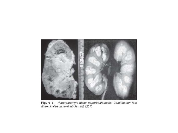
b. Brown tumors of hyperparathyroidism) - Aggregates of osteoclasts, , and hemorrhage occasionally form masses that may be mistaken for neoplasms II. Kidney changes a. PTH-induced hypercalcemia favors the formation ofurinary tract stones (nephrolithiasis) s b. Calcification of the renal interstitium (nephrocalcinosis) III. Metastatic calcification may be seen in the stomach, lungs, myocardium, and blood

Brown tumor of bone

Clinical Features - Primary hyperparathyroidism is a disease of adults and is much more common in women than in men. - The most common manifestation is an increase in serum calcium and is the most common cause of clinically silent hypercalcemia. - The most common cause of clinically apparent hypercalcemia in adults is paraneoplastic syndromes

associated with malignancy and bone metastases - In persons with hypercalcemia caused by parathyroid hyperfunction, serum PTH is inappropriately elevated - Whereas serum PTH is low to undetectable in those with hypercalcemia caused by nonparathyroid diseases, - Clinical Manifestations : - It traditionally has been associated with a constellation of symptoms "painful bones, renal stones, abdominal groans, psychic moans. “

1. Pain was at one time a prominent manifestation of primary hyperparathyroidism and is secondary to a. Fractures of bones b. and resulting from renal stones 2. Gastrointestinal disturbances, including constipation, nausea, peptic ulcers, pancreatitis, and gallstones 3. CNS alterations, - depression, lethargy, and seizures 4. Neuromuscular abnormalities, - weakness and hypotonia

b. Secondary Hyperparathyroidism - Is caused by any condition causing a chronic decreases in the serum calcium level, because low serum calcium leads to compensatory overactivity of the parathyroids. - Renal failure is the most common cause 1. Chronic renal insufficiency causes decreased phosphate excretion, which in turn results in hyperphosphatemia. and the elevated serum phosphate levels depress serum calcium levels and so stimulate parathyroid gland activity

2. Loss of renal substances reduces the availability of α 1 -hydroxylase enzyme necessary for the synthesis of the active form of vitamin D, which in turn reduces intestinal absorption of calcium Gross- The parathyroid glands are hyperplastic. Clinical Features - Are dominated by those related to chronic renal failure
 are less severe than those seen in primary type")
- Bone abnormalities (renal osteodystrophy) are less severe than those seen in primary type - The metastatic calcification of blood vessels (secondary to hyperphosphatemia) occasionally may result in significant ischemic damage to skin and other organs-a process sometimes referred to as calciphylaxis. Note- In a minority of patients, parathyroid activity may become autonomous and excessive, with resultant hypercalcemia-a process sometimes termed tertiary hyperparathyroidism

. HYPOPARATHYROIDISM: is less common than hyperparathyroidism and the major causes are: . a. Surgically induced hypoparathyroidism: inadvertent removal of parathyroids during thyroidectomy. b. Congenital absence: This occurs in conjunction with thymic aplasia (Di George syndrome) and cardiac defects, secondary to deletions on chromosome 22 q 11. 2 c. Autoimmune hypoparathyroidism : This is a hereditary polyglandular deficiency syndrome
. - Mucocutaneous candidiasis are")
arising from autoantibodies to multiple endocrine organs(parathyroid, adrenals, and pancreas). - Mucocutaneous candidiasis are sometimes encountered in affected persons Clinical manifestations - Are secondary to hypocalcemia and include: a. Increased neuromuscular irritability (tingling, muscle spasms, , and sustained carpopedal spasm or tetany), b. Cardiac arrhythmias, c. Seizures.
- Slides: 80