Endocrine block 2016 Anterior Pituitary Disorders Dr Aishah

Endocrine block 2016 Anterior Pituitary Disorders Dr. Aishah Al. Ekhzaimy, MBBS, FRCPC, FACE

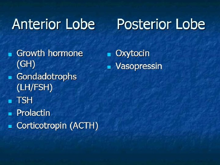
Pituitary Disorders � Anterior pituitary disorders: Non-functional pituitary tumor and mass-effect: hypopituitrism Prolactin secreting cell disorder: prolactinoma Growth hormone secreting cell disorder: acromegaly ACTH secreting cell disorders: cushing’s TSH secreting cell tumor: TSH-secreting adenoma Gonadotropin secreting adenoma � Posterior Pituitary disorders: Diabetes insipidus

Pituitary Development � Anterior pituitary is recognizable by 4 - 5 th wk of gestation � Full maturation by 20 th wk � From Rathke’s pouch, Ectodermal evagination of oropharynx � Migrate to join neurohypophysis � Portion of Rathke’s pouch →→ Intermediate lobe � Remnant of Rathke’s pouch cell in oral cavity →→ pharyngeal pituitary � Lies at the base of the skull as sella turcica � Roof is formed by diaphragma sellae � Floor by the roof of sphenoid sinus

Pituitary Development � Posterior pituitary from neural cells as an outpouching from the floor of 3 rd ventricle � Pituitary stalk in midline joins the pituitary gland with hypothalamus that is below 3 rd ventricle � Development of pituitary cells is controlled by a set of transcription growth factors like pit-1, Prop-1, Pitx 2

Pituitary Development � Pituitary stalk and its blood vessels pass through the diaphragm � Lateral wall by cavernous sinus containing III, IV, VI, V 1, V 2 cranial nerves and internal carotid artery with sympathetic fibers. Both adjacent to temporal lobes � Pituitary gland measures 15 X 10 X 6 mm, weighs 500 mg but about 1 g in women � Optic chiasm lies 10 mm above the gland anterior to the stalk � Blood supply : superior, middle, inferior hypophysial arteries ( internal carotid artery) running in median eminence from hypothalamus � Venous drainage: to superior and inferior petrosal sinsuses to jugular vein

Pituitary Development

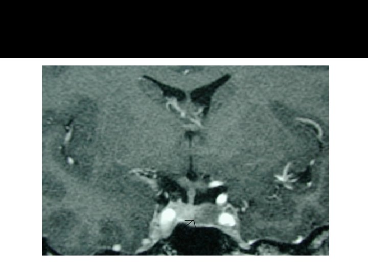
Normal Pituitary Anatomy Modified from Lechan RM. Neuroendocrinology of Pituitary Hormone Regulation. Endocrinology and Metabolism Clinics 16: 475 -501, 1987

Pituitary Development

Endocrine system

Anterior Pituitary Function Hormone Stimulators Corticotroph Gonadotroph Thyrotroph Lactotroph Somatotroph POMC, ACTH FSH, LH TSH Prolactin GH CRH, AVP, gp 130 cytokines Gn. RH, Estrogen TRH Estrogen, TRH GHRH, GHS T 3, T 4, Dopamine, Somatostati n, GH Dopamine Somatostatin, IGF-1, Activins Thyroid Breast and other tissues Liver, bone and other tissues Inhibitors Glucocorticoids Target Gland Adrenals Trophic Effects Steroid production Sex steroids, inhibin Ovary, Testes Sex Steroid, Follicular growth, Germ Cell maturation T 4 synthesis and secretion Adapted from: William’s Textbook of Endocrinology, 10 th ed. , Figure 8 -4, pg 180. Milk Productio n IGF-1 production, Growth induction, Insulin antagonism

Etiology of Pituitary Masses

Etiology of Pituitary-Hypothalamic Lesions � � � � � Non-Functioning Pituitary Adenomas Endocrine active pituitary adenomas Prolactinoma Somatotropinoma Corticotropinoma Thyrotropinoma Other mixed endocrine active adenomas Malignant pituitary tumors: Functional and non-functional pituitary carcinoma Metastases in the pituitary (breast, lung, stomach, kidney) Pituitary cysts: Rathke's cleft cyst, Mucocoeles, Others Empty sella syndrome Pituitary abscess Lymphocytic hypophysitis Carotid aneursym

Disorders of Pituitary Function � Hypopituitarism Central hypoadrenalism, hypogonadism, hypothyroidism or GH deficiency Panhypopituitarism � Hypersecretion of Pituitary Hormones Hyperprolactinemia Acromegaly Cushing’s Disease

Evaluation of Pituitary mass � Pituitary adenoma: 10 % of all pituitary lesions � Genetic-related � MEN-1, Gs-alpha mutation, PTTG gene, FGF receptor-4 � Pituitary incidentaloma: 1. 5 -31% in autopsy ( prevalence) 10 % by MRI most of them < 1 cm

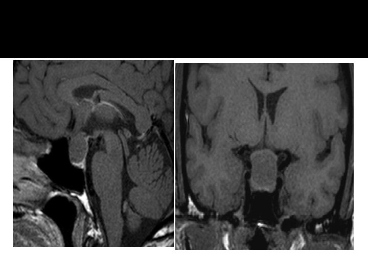
Evaluation of Pituitary lesion

Evaluation of Pituitary lesion �Non-Functional pituitary lesion: � Absence of signs and symptoms of hormonal hypersecretion � 25 % of pituitary tumor � Needs evaluation either micro or macroadenoma � Average 50 – 55 yrs old, more in male

Non- functional pituitary adenoma � Presentation of NFPA: � As incidentaloma by imaging � Symptoms of mass effects ( mechanical pressure) � Hypopituitarism ( mechanism) � Gonadal hypersecretion

Non- functional pituitary adenoma

Non- functional pituitary adenoma � Treatment: � Surgery if indicated - recurrence rate 17 % if gross removal, 40 % with residual tumor - predictors of recurrence: young male, cavernous sinus invasion, extent of suprasellar extention of residual tumor, duration of follow up, marker; Ki-67 � Observation with annual follow up for 5 years and then as needed, visual field exam Q 6 -12 month if close to optic chiasm. Slow growing tumour � Adjunctive therapy: - Radiation therapy - Dopamine agonist - Somatostatin analogue

Functional pituitary mass

Prolactinoma

Prolactin � Human prolactin is a 198 amino acid polypeptide � Primary function is to enhance breast development during pregnancy and to induce lactation � Prolactin also binds to specific receptors in the gonads, lymphoid cells, and liver � Secretion is pulsatile; it increases with sleep, stress, pregnancy, and chest wall stimulation or trauma

Prolactin �Secretion of prolactin is under tonic inhibitory control by dopamine, which acts via D 2 -type receptors located on lactotrophs �Prolactin production can be stimulated by the hypothalamic peptides, thyrotropin-releasing hormone (TRH) and vasoactive intestinal peptide (VIP)

Clinical Features of Hyperprolactinemia/Prolactinoma � Women may present with oligomenorrhea, amenorrhea, galactorrhea or infertility � Men often have less symptoms than women (sexual dysfunction, visual problems, or headache) and are diagnosed later � In both sexes, tumor mass effects may cause visual-field defects or headache

Prolactin �Secretion of prolactin is under tonic inhibitory control by dopamine, which acts via D 2 -type receptors located on lactotrophs �Prolactin production can be stimulated by the hypothalamic peptides, thyrotropin-releasing hormone (TRH) and vasoactive intestinal peptide (VIP)

Causes of Hyperprolactinemia � Hypothalamic Dopamine Deficiency Diseases of the hypothalamus( including tumors, arterio- venous malformations, and inflammatory processes Drugs (e. g. alpha-methyldopa and reserpine) � Defective Transport Mechanisms Section of the pituitary stalk Pituitary or stalk tumors
 � Lactotroph Insensitivity to Dopamine-receptor-blocking agents: phenothiazines (e. g. chlorpromazine),")
Causes of Hyperprolactinemia (continued) � Lactotroph Insensitivity to Dopamine-receptor-blocking agents: phenothiazines (e. g. chlorpromazine), butyrophenones (haloperidol), and benzamides (metoclopramide, sulpiride, and domperidone) � Stimulation of Lactotrophs Hypothyroidism- increased TRH production (acts as a PRF) Estrogens: stimulate lactotrophs Injury to the chest wall: abnormal stimulation of the reflex associated with the rise in prolactin that is seen normally in lactating women during suckling

REMEMBER: Not all hyperprolactinemia is due to a prolactinoma

Work up of Patient with Hyperprolactinemia � In females, pregnancy must always be ruled out � Get a TSH- hypothyroidism is another common cause of elevated prolactin: � Obtain detailed drug history- rule out medication effects � Rule out other common causes including: Nonfasting sample Nipple stimulation or sex Excessive exercise History of chest wall surgery or trauma Renal failure Cirrhosis � If no cause determined or tumor suspected, consider MRI, especially if high prolactin levels (> 100 ng/m. L) http: //www. emedicine. com/Med/topic 1915. htm

Prolactinomas � Most common of functional pituitary adenomas � 25 -30% of all pituitary adenomas � Some growth hormone (GH)–producing tumors also co-secrete PRL � Of women with prolactinomas- 90% present with microprolactinomas � Of men with prolactinomas- up to 60% present with macroprolactinomas http: //www. emedicine. com/Med/topic 1915. htm

Management Medical therapy: Dopamine agonist Surgical resection Radiation therapy

Growth hormone � Pituitary tumor as mass effect →→ Growth hormone deficiency � Hyperfunctioning mass →→ Acromegaly

Growth hormone deficiency �Diagnosis in children and adult

Diagnosis of GH-deficiency and management �GH, IGF-I level �Dynamic testing: clonidine stimulation test, glucagon stimulation, exercise testing, arginine-GHRH, insulin tolerance testing �X-ray of hands: delayed bone age �In Adult: Insulin tolerance testing, MRI pituitary to rule out pituitary adenoma �Management: GH replacement

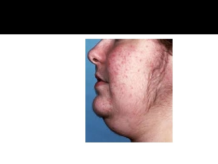
Acromegaly

Growth hormone disorder
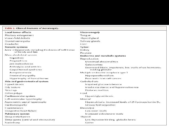

Growth hormone disorder
 � IGF-I")
Acromegaly � Clinical picture and presentation � GH level ( not-reliable, pulsatile) � IGF-I � 75 g OGTT tolerance test for GH suppression � Fasting and random blood sugar, Hb. A 1 c � Lipid profile � Cardiac disease is a major cause of morbidity and mortality � 50 % died before age of 50 � HTN in 40% � LVH in 50% � Diastolic dysfunction as an early sign of cardiomyopathy

Growth hormone disorder. Acromegaly � Medical treatment: Somatostatin analogue � Surgical resection of the tumor

HPA-axis � 2 nd adrenal insufficiency � glucgocorticoid replacement � Circadian rhythm of cortisol secretion � Early morning cortisol between 8 -9 am

ACTH-disorders

ACTH-disorders

Hypoadrenalism � Nausea � Vomiting � Abdominal pain � Diarrhoea � Muscle ache � Dizziness and weakness � Tiredness � Weight loss � Hypotension

Management of hypoadrenalism �Cortisol replacement

ACTH-Adenoma
")
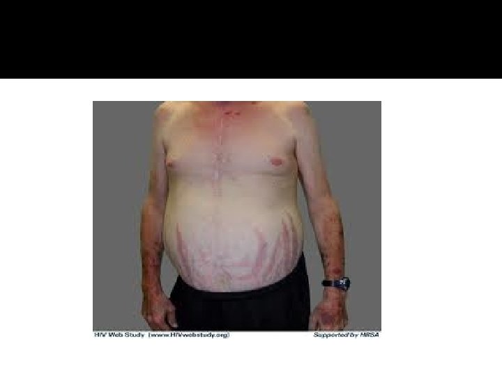
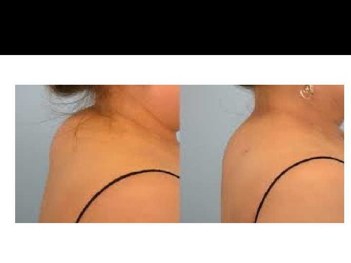
HPA-axis ( excessive cortisol)
 � 80 % HTN � LVH � Diastolic dysfunction, intraventricular")
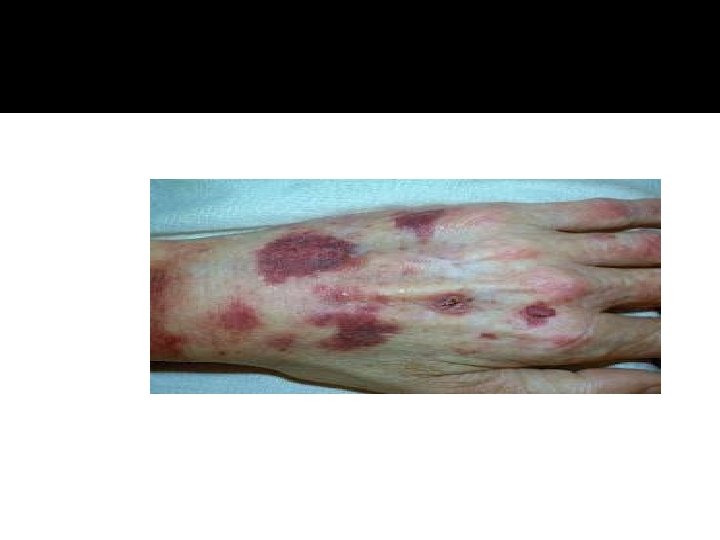
HPA-axis ( excessive cortisol) � 80 % HTN � LVH � Diastolic dysfunction, intraventricular septal hypertrophy � ECG needed: high QRS voltage, inverted T-wave � Echocardiogram preop � OSA: 33% mild, 18% severe. Needs respiratory assessment and careful use of sedative during surgery � Glucose intolerance in 60%, control of hyperglycemia � Osteoporosis with vertebral fracture→→ positioning of patient in OR ( 50 %), 20 % with fracture � thin skin→→ difficult IV cannulation, poor wound healing

Cushing’s-Management �Surgical resection of pituitary �Medical Treatment

TSH-Hypothyroid

Central Hypothyroidism � Low TSH � Low free T 4 and T 3

Central Hypothyroidism � Thyroxine replacement � Surgical removal of pituitary adenoma if large

TSH-hyperthyroid
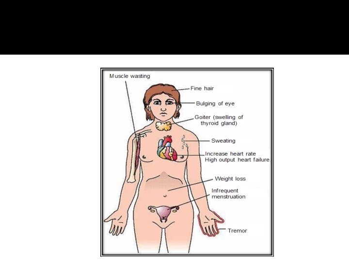

TSH-Producing adenoma � Very rare < 2. 8 % � Signs of hyperthyroidism � High TSH, FT 4, FT 3 � Treatment preop with anti-thyroid meds � Surgical resection of adenoma � Medical therapy: Somatostatin Analogue
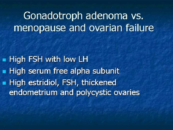

Gonadotroph Adenoma �Surgical resection if large �Radiation therapy

assessment of pituitary function � Baseline: TSH, FT 4, FT 3, LH, FSH, Prolactin, GH, IGF- I, Testosterone, Estradiol � MRI brain � Neuropthalmic evaluation of visual field � Cardiac and respiratory assessment � Anesthesiologist for airway and perioperative monitoring � Neurosurgeon � ENT for Endonasal evaluation for surgical approach � Preop hormonal replacement: all pituitary adenoma should be covered with stress dose of HC
- Slides: 65