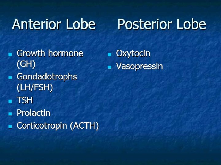
Endocrine block 2011 Anterior pituitary insufficiency Anterior pituitary

Endocrine block 2011 Anterior pituitary insufficiency

Anterior pituitary disorder � Non-functional pituitary tumor mass-effect � Prolactin secreting cell disorder: prolactinoma � Growth hormone secreting cell disorder: acromegaly � ACTH secreting cell disorders: cushing’s � TSH secreting cell tumor: TSHoma � Gonadotropin secreting cell disorder

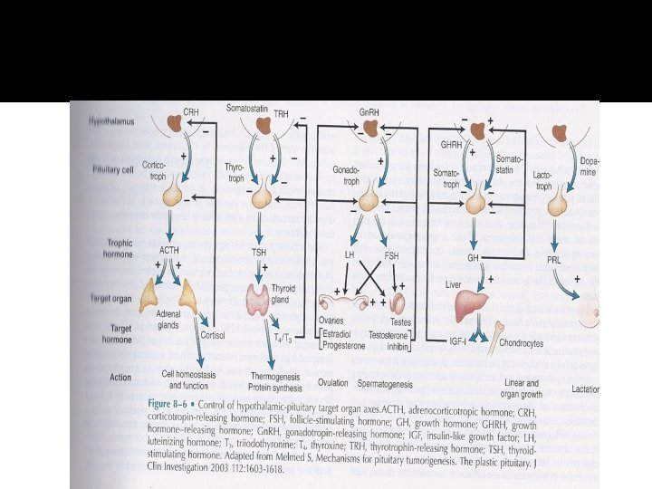
Pituitary Development � Anterior pituitary is recognizable by 4 - 5 th wk of gestation � Full maturation by 20 th wk � From Rathke’s pouch, Ectodermal evagination of oropharynx � Migrate to join neurohypophysis � Portion of Rathke’s pouch →→ Intermediate lobe � Remnant of Rathke’s pouch cell in oral cavity →→ pharyngeal pituitary � Lies at the base of the skull as sella turcica � Roof is formed by diaphragma sellae � Floor by the roof of sphenoid sinus

Pituitary Development � Posterior pituitary from neural cells as an outpouching from the floor of 3 rd ventricle � Pituitary stalk in midline joins the pituitary gland with hypothalamus that is below 3 rd ventricle � Development of pituitary cells is controlled by a set of transcription growth factors like pit-1, Prop-1, Pitx 2

Pituitary Development � Pituitary stalk and its blood vessels pass through the diaphragm � Lateral wall by cavernous sinus containing III, IV, VI, V 1, V 2 cranial nerves and internal carotid artery with sympathetic fibers. Both adjacent to temporal lobes � Pituitary gland measures 15 X 10 X 6 mm, weighs 500 mg but about 1 g in women � Optic chiasm lies 10 mm above the gland anterior to the stalk � Blood supply : superior, middle, inferior hypophysial arteries ( internal carotid artery) running in median eminence from hypothalamus � Venous drainage: to superior and inferior petrosal sinsuses to jugular vein

Pituitary Development

Pituitary Development

Endocrine system

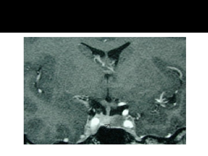
Sellar Mass

Clinical presentations of sellar mass
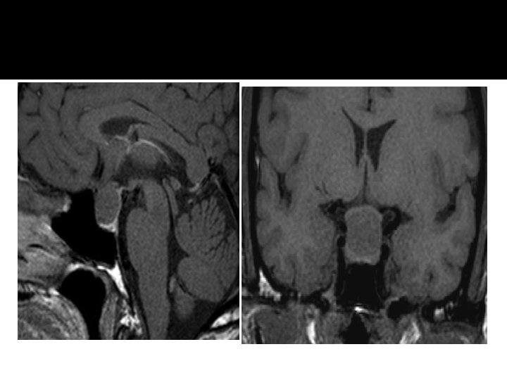

Evaluation of Pituitary mass � Pituitary adenoma: 10 % of all pituitary lesions � Genetic-related � MEN-1, Gs-alpha mutation, PTTG gene, FGF receptor-4 � Pituitary incidentaloma: 1. 5 -31% in autopsy ( prevalence) 10 % by MRI most of them < 1 cm
 �Non-Functional adenoma")
Evaluation of Pituitary lesion �Functional adenoma ( hormonal- secreting) �Non-Functional adenoma

Evaluation of Pituitary lesion

Evaluation of Pituitary lesion �Non-Functional pituitary lesion: � Absence of signs and symptoms of hormonal hypersecretion � 25 % of pituitary tumor � Needs evaluation either micro or macroadenoma � Average 50 – 55 yrs old, more in male

Non- functional pituitary adenoma � Presentation of NFPA: � As incidentaloma by imaging � Symptoms of mass effects ( mechanical pressure) � Hypopituitarism ( mechanism) � Gonadal hypersecretion

Non- functional pituitary adenoma

Non- functional pituitary adenoma � Treatment: � Surgery if indicated - recurrence rate 17 % if gross removal, 40 % with residual tumor - predictors of recurrence: young male, cavernous sinus invasion, extent of suprasellar extention of residual tumor, duration of follow up, marker; Ki-67 � Observation with annual follow up for 5 years and then as needed, visual field exam Q 6 -12 month if close to optic chiasm. Slow growing tumour � Adjunctive therapy: - Radiation therapy - Dopamine agonist - Somatostatin analogue

Effect of pituitary tumor

Prolactinoma

Prolactinoma � Prolactin:

Growth hormone � Pituitary tumor as mass effect →→ Growth hormone deficiency � Hyperfunctioning mass →→ Acromegaly

Growth hormone deficiency �Diagnosis in children and adult


Growth hormone disorder

Growth hormone disorder
 � IGF-I")
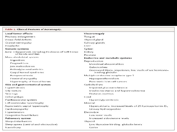
Acromegaly � Clinical picture and presentation � GH level ( not-reliable, pulsatile) � IGF-I � 75 g OGTT tolerance test for GH suppression � Fasting and random blood sugar, Hb. A 1 c � Lipid profile � Cardiac disease is a major cause of morbidity and mortality � 50 % died before age of 50 � HTN in 40% � LVH in 50% � Diastolic dysfunction as an early sign of cardiomyopathy

Growth hormone disorder. Acromegaly � Medical treatment: � Somatostatin analogue � Surgical resection of the tumor

ACTH-disorders

ACTH-disorders

HPA-axis � 2 nd adrenal insufficiency � glucgocorticoid replacement � Circadian rhythm of cortisol secretion � Early morning cortisol between 8 -9 am

Cushing’s
")
HPA-axis ( excessive cortisol)
 � 80 % HTN � LVH � Diastolic dysfunction, intraventricular")
HPA-axis ( excessive cortisol) � 80 % HTN � LVH � Diastolic dysfunction, intraventricular septal hypertrophy � ECG needed: high QRS voltage, inverted T-wave � Echocardiogram preop � OSA: 33% mild, 18% severe. Needs respiratory assessment and careful use of sedative during surgery � Glucose intolerance in 60%, control of hyperglycemia � Osteoporosis with vertebral fracture→→ positioning of patient in OR ( 50 %), 20 % with fracture � thin skin→→ difficult IV cannulation, poor wound healing

TSH-Producing adenoma � Very rare < 2. 8 % � Signs of hyperthyroidism � High TSH, FT 4, FT 3 � Treatment preop with anti-thyroid meds pre-op

assessment of pituitary function � Baseline: TSH, FT 4, FT 3, LH, FSH, Prolactin, GH, IGF- I, Testosterone, Estradiol � MRI brain � Neuropthalmic evaluation of visual field � Cardiac and respiratory assessment � Anesthesiologist for airway and perioperative monitoring � Neurosurgeon � ENT for Endonasal evaluation for surgical approach � Preop hormonal replacement: all pituitary adenoma should be covered with stress dose of HC
- Slides: 41