Encephalon Brain Brain Components Fig 14 1 Brain

Encephalon – Brain

Brain Components Fig 14. 1 Brain Stem Midbrain, Pons, Medulla Oblongata Cerebellum “Little Brain” Diencephalon Thalamus, Hypothalamus, Epithalamus Cerebrum

Protective Coverings of the Brain • Cranial bones • Meninges 1. Dura mater 2. Arachnoid mater 3. Pia mater

Cranial Bones Cranial vault formed by 8 cranial bones Encloses and protects brain Floor divided into 3 fossa: Anterior – front lobes Middle – temporal lobes and base of diencephalon Posterior - cerebellum

Meninges • Surround and protect the brain and spinal cord • Three membranes: 1. Dura mater 2. Arachnoid mater 3. Pia mater

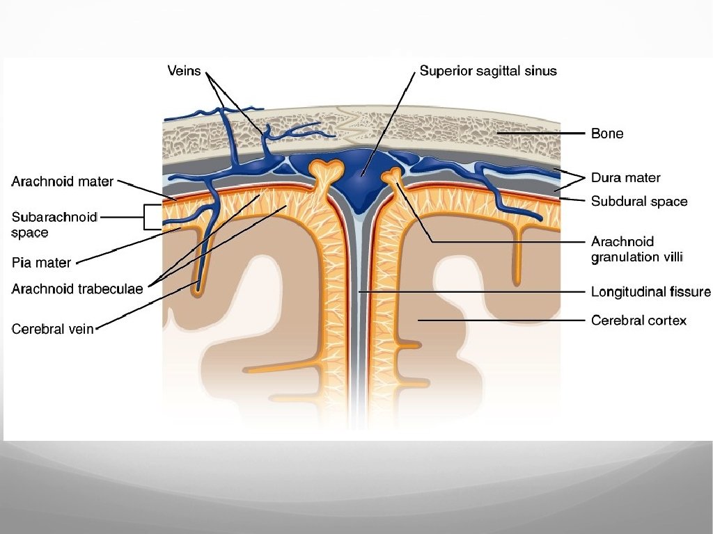
Dura Mater • Tough outermost meninx. • Composed of two layers: • Periosteal • Meningeal • Venous sinuses between the layers.

Dura Mater • Falx cerebri • Falx cerebelli • Tentorium cerebelli Separates cerebrum from cerebellum Separates 2 hemispheres of cerebrum Separates 2 hemispheres of cerebellum

Arachnoid Mater Middlemost meninx Characterized by its filmy, weblike structure Loosely follows contours of cerebral structures but lies over sulci

Pia Mater Thin, delicate innermost meninx Closely adheres to surface of brain and follows sulci and fissures Provides support for blood vessels serving brain tissue Sheath of pia mater
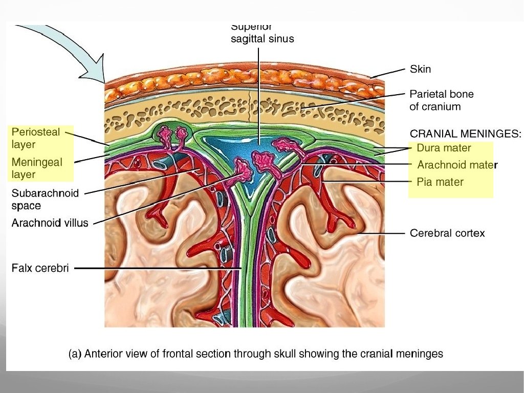

Meningeal Spaces • Real or potential spaces between meningeal layers: • Epidural Space = potential space • Between Skull and Dura • Subdural Space = Real space • Between dura and arachnoid • Small bridging veins (little support) cross the space • Subdural hematoma • Subarachnoid space is a real space • • • Lies between the arachnoid and pia mater Contains cerebrospinal fluid Subarachnoid hemorrhage

Brain Injuries Hemorrhage – active or ongoing bleeding. Hematoma – accumulation of blood within one of the meningeal spaces or surrounding tissues. Contusion – Type of hematoma. Blood escapes ruptured capillaries and enters surrounding tissue.

Brain Blood Flow • Internal carotid arteries • Vertebral arteries • Internal jugular veins

Arterial Brain Blood Flow ANTERIOR: Common Carotid Arteries Internal Carotid Arteries Base of skull Cranium Branch to anterior and middle cerebral arteries

Arterial Brain Blood Flow POSTERIOR: Subclavian arteries Vertebral arteries Transverse foramina of cervical vertebrae foramen magnum Join at junction of pons and medulla Basilar artery Pontine branches Posterior inferior cerebellar arteries (off the vertebral artery) Basilar divides at midbrain anterior inferior cerebellar and superior cerebellar arteries

Arterial Brain Blood Flow Vertebral arteries Subclavian Cervical vertebrae transverse foramina Basilar artery Cerebellum, brainstem and occipital lobes. Pontine arteries Cerebellar arteries: Cerebellum Cerebral arteries Internal Carotid Artery Posterior communicating artery Anterior communicating artery

Arterial Brain Blood Flow Terminal branches of Carotid and Vertebral arteries = circle of Willis Circulatory anastomosis (connection between 2 blood vessels) Backup route in case of blockage Branch into various cerebral arteries to supply brain with blood

Brain Blood Flow Animation https: //www. youtube. com/watch? v=MPc. O 2 ib. O 75 o
- Slides: 19